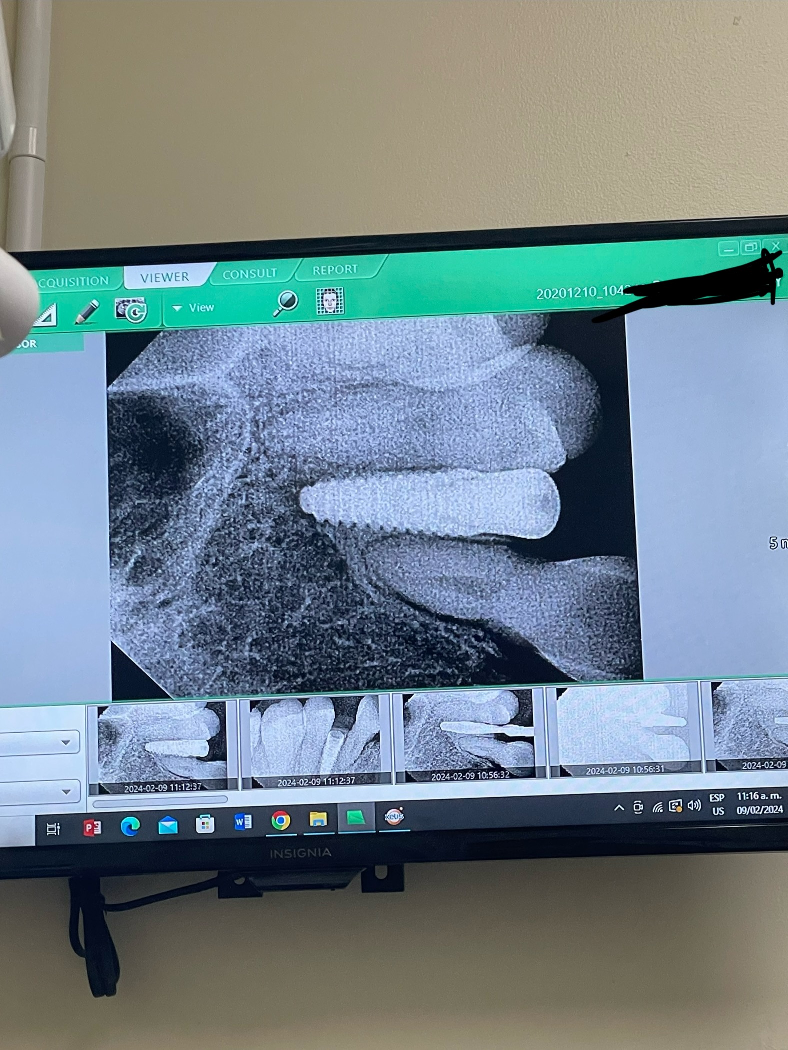
Sinus tract formation after 15 days of implant placement: recommendations?
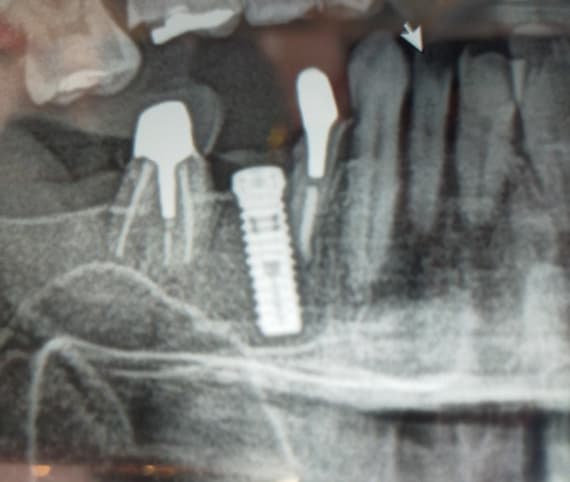
I installed implants in #19 and 21 sites [mandibular left first molar and first premolar; 36, 34]. #20 is vital. A sinus tract has developed around #21 and I traced this with a gutta percha point and periapical radiograph. #21 implant is causing pain. #22 [mandibular left canine;33] is vital. #20 [mandibular left second premolar; 35] had endodontic treatment 10 years prior. I laid a full thickness flap and curetted thoroughly around #21 implant and irrigated with Betadine.. #21 was not mobile. Patient returned 2 weeks later with pain and recurrence of sinus tract. I have placed 9 other implants in this patient without any problems. What do you recommend I do now?
26 Comments on Sinus tract formation after 15 days of implant placement: recommendations?
New comments are currently closed for this post.
CRS
12/9/2013
What is your diagnosis? I can't say without seeing an xray.
Richard Hughes, DDS, FAAI
12/10/2013
CRS make a good point.
I suggest removing the implant , degranulate if necessary and graft with OsteoGen, revisit in 6 months.
Peter Fairbairn
12/10/2013
Yes scant information , but could be some residual issue in the bone in close proximity to the implant from re-existing pathology . the only pain an Implant can experience if still very stable is generallly soft tissue pain .....
There is some confusion as well in the text , 20 is Vital , then later 20 was RCT 10 years ago .
Peter
DrT
12/10/2013
Please show us x-rays and more clinical information. Thank you
Dr Zurita Fernando
12/10/2013
Please take ct scan
Timothy Hacker DDS D-ABOI
12/10/2013
Remove implant. Don't waste time and your patient's confidence by trying to save a failing situation. Degranulate, graft with osteogen or irradiated cancellous bone and place another implant in 6 months.
Dean Licenblat
12/10/2013
I have seen this only once before and despite all efforts to save the implant, I removed it, thoroughly cleaned the site grafted and revisited later.
All measure of antibiotics etc would not work.
Better to bite the bullet than lose more bone and further complicate the scene.
This happened to me in 2008 and even the Maxillofacial surgeon who was with me couldnt explain why it happened.
coryc
12/10/2013
what was the history of #21 (natural tooth) ? if it was a previous rct you're probably out of luck....I've had this happen a few times where the natural tooth failed after rct at some point...even years later and the bone looked good, but there is some residual infection sequestered peri-apically....again, even years after extraction....you can try and irrigate it, antibiotic it, and graft it, but it's like the site has some memory of this infection....the sinus tract just re-forms. strange but true....good luck
Nami Ben Otman
12/11/2013
I have managed one case similar to that, sinus tract developed due to overheat mostly or vibration or over pressure due to blunted or over used drills
x-ray to evaluate extent
surgical apiecectomy of implant with peripical curettage followed by bone augmentation, result was excellant
CRS
12/11/2013
The sinus tract probably developed due to bacteria if you overheated the implant or bone the implant would have most likely failed. What you probably did was remove the bacteria with your "apicoectomy" of the implant and removed contaminated bone. An implant is not a tooth. Please read my next post. Thanks
Nami Ben Otman
12/11/2013
Resistance of bone reduced when exposed to physical factor( overheat, vibration
over pressure...ect) followed by invasion of bacteria cause infection
I notice peiimplant radiolucienies at apex beside crestal radiolucienies so, that is why apeciectomy and curretage of dead infected bone which looks like granuloma, and result was excelant after surgery this my experience in that particlar case
CRS
12/11/2013
That's a real interesting unique theory sinus tracts don't develop from overheating bone. Trauma to bone enough to devitalize it will cause a sequestra to develop as in an osteomyelitis,you probably took care of the bacteria. The implant would have failed due to overheating as I stated before.
Nami Ben Otman
12/14/2013
alternative simple way, to exclude mis-diagnoses if suspect septic foci from adjuscent tooth #20 left second premolar just do re-RCT with calcium hydroxide injection canal and follow up by x-ray if radioluciencies disappear means that source removed and healing will take place near implant if no improvement in the case peri-implant/ periapical curettage with apeiectomy if implant stable and limited radiolucencies to one side only if not removal of implant because during surgery decision of site of sepetic foci
CRS
12/11/2013
I bet it is the ten year old endo tooth, untreated canal or fracture. A non mobile implant usually doesn't have pain unless it is impinging on a nerve. Putting a gutta percha point in a tract is not always diagnostic and can be misleading. What did you find when the flap was elevated what did you currette, that would help in the diagnosis. A recurrent fistula is evidence that the body is trying to discharge fluid or the epithelium was not completely removed. Without a film I'm guessing but unless the endo was done under a microscope there could be an untreated canal or prepping the bone during implant placement could have released some bacteria in privileged sites. Since it is early and the process is active, I would remove the non integrated implant disinfect with the nd-yag laser, graft and have an endodontist evaluate the tooth. Then if it is retreated or fractured the treatment plan will be modified. Better yet get the patient to the endodontist first and take it from there . In the future I advise being aware of placing implants next to old endontically treated teeth as an infection source, it probably is the culprit. With the advent of microscopes I am suspicious of root canals placed without them. Root canals have a finite lifespan of 10-15 years on average. But this is conjecture based on the limited info. Your welcome for the free consult thanks for reading.
Robert J. Miller
12/11/2013
All valid points, but without radiographs very difficult to diagnose. So let's try to deconstruct the case to explain the symptoms. First, was this extraction/immediate placement or a healed site? Second, is the apex of the implant in close proximity to the apex of the adjacent tooth? Third, if a healed site, was it a relatively recent extraction? Last, any radioluscency in the apical part of the osteotomy at time of implant placement?
Retrograde peri-implantitis is almost exclusively caused by reamaining endodontic pathology. Seemingly quiet periapical areas either harbor pathogens in a spore state or are immune responses to untreated necrotic canals. This is why we carefully currette and decontaminate extraction sites prior to grafting or implant placement. Even in the absence of pathogens, granulomatous defects can be self-perpetuating; some of these become apical retention cysts and others just have very slow growing apical areas that are aymptomatic. All it takes, however, is a surgical intervention to activate these sites by causing an exaggerated immune responcse. If you do not completely remove this tissue, it has a high degree of reoccurence. Ablative lasers are excellent as adjunctive treatment and the use of platelet rich fibrin (PRF) can also completely change the host response and clear these areas. But if the implant is well integrated, I always try definitive treatment rather than removal and have an extremely high rate of success.
RJM
CRS
12/12/2013
Retained epithelium in a periapical lesion can form a cyst, sterile granulation tissue can heal, never heard of the spore state of oral bacteria I thought that was fungi, and PRP has nothing specific which will kill bacteria, blood is actually a good nidus for infection. Basically my point is looking for the clinical clues that will give a reasonable diagnosis. Cutting the osteotomy site can release the dormant bacteria in the privileged bone sites that the body has walled off is the theory. Another point is that if there is trouble early prior to osteointegration it is prudent to remove the implant and treat the cause vs the ego. Many of these treatment rationales are based on timing, biology and understanding pathology, it's knowing what to do based on these principles vs making something up. And quite humbly I don't always know exactly what happens sometimes our interventions just work for reasons unknown!
Jaime Ramos
12/12/2013
A thorough vigorous curretagem irrigated with saline is adamant because there exists a difference between infected and affected bacteria but then again without radiographs at least ? Curretagem and irrigate with saline especially in these circumstances . Prevention is easier than the cure . Remember that a radiograph is two dimensional and we working with a 3-dimensional situation .
" Look for the hidden fistulae when there is a endodontic tooth treated nearby and use a periodontal probe on adjacent teeth . You will be surprised what you may discover !
Jaime Ramos
12/12/2013
I meant infected and affected bone .
Robert J. Miller
12/12/2013
Hey CRS; Just a few responses to your comments. Yes, "sterile" granulation tissue does heal spontaneously. But that is the definition of provisional matrix or normal healing tissue in it's earliest stage. We are talking about pathologic granulation tissue that is either pathogen or immune mediated. The pathways and consequences are very different from normal garnulomatous healing.
Several organisms can survive in a spore state, especially spirochetes. A very significant percentage of dental infections are of spirochetal origen and are completely resistant to antibiotics.
I referenced L-PRF, not PRP. PRF contains neutrophils and monocytes, both of which have direct phagocytotic and lysosomal activity. PRF also maintains a neutral pH, interrupting the inflammatory cascade.
Last, we have outstanding success using a combination of laser debridement followed by grafting/autologous biologics in these types of cases. You should try this modality in a few cases; I am sure your paradigm will change with successful outcomes.
RJM
CRS
12/13/2013
Thanks Robert I am starting a protocol with the nd-yag laser on these nasty failing endo cases since the wavelength is appropriate for pigmented bacteria and has enough power to penetrate to get the bacteria hiding in the bone. Can't do this with a erbium or diode wrong wavelength. I like PRGF it works well. I'm hopeful to avoid these early failures since I'm referral based and get the endo when it's pretty bad, I think it's tough to get the patient to have the tooth treated for whatever reason. The white cells can't penetrate the bone but the laser can. When I say sterile I mean clean no pathogens. And the beauty part is that the site is prepared for the replacement implant. This debridement is not as effective without the laser, exciting concept. That's why I got the laser. I also like removing the epithelium with it. Thanks for reading I think we are on the same wavelength , bad pun!
drshalash
12/12/2013
Don't waste your time. U have a failing implant. remove, debride the osteotomy site and graft. revisit in 4-6 months. Good luck
Robert J. Miller
12/12/2013
Without definitive treatment, you will surely have a failed implant.
RJM
Richard Hughes, DDS, FAAI
12/14/2013
CRS, you have mentioned this before about the nd-yag laser wavelength. You got me interested.
CRS
12/14/2013
The difference in this laser is the wavelength and that you can control the amount of joules given for tissue response. It targets hemoglobin and pigmented bacteria which create the niches in the bone to allow the other pathologic bacteria into the site. Diodes just melt tissue they don't have the penetration and erbium lasers target water and hydroxyapatite now since I'm an OMS I really like this laser. I treat the periodontal issues and the failed endo issues in my practice. If you understand the science it really changes the way you treat oral pathogens. It's sort of like early bone grafting and the way extractions were originally taught, a new paradigm. Another way of looking at it is having the right antibiotic specific for a pathogen right laser for the right job. Thanks for reading.
Timothy Hacker DDS D-ABOI
12/14/2013
I have been using the Millennium MPV7 NdYg laser for 8 years with fantastic results. It is a great tool for bleeding control also.
Simon Milbaurr
12/15/2013
I've been using 940nm diode laser for 3 yrs to treat perio along natural teeth and implants results are very good


