Slightly mobile implant with bone loss: How would you treat?

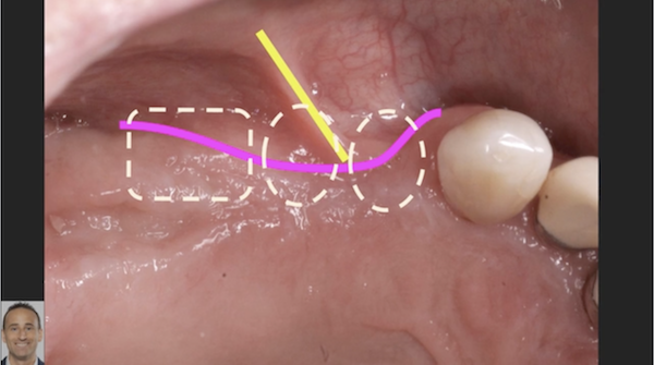
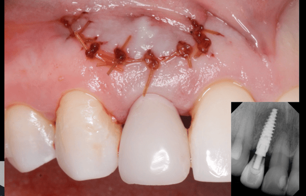
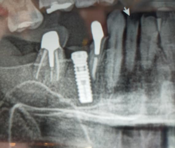
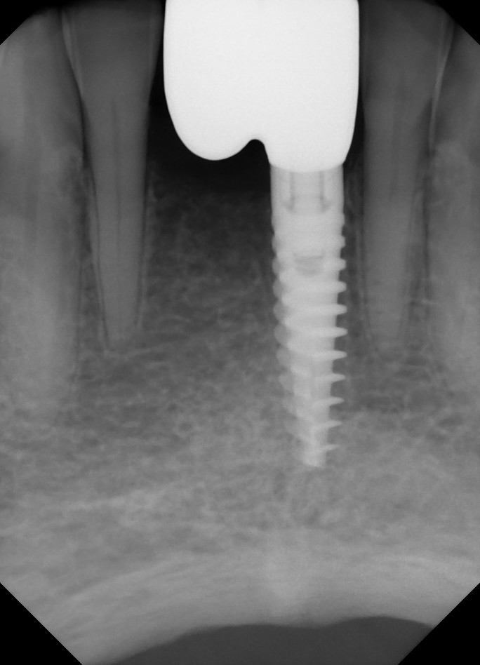
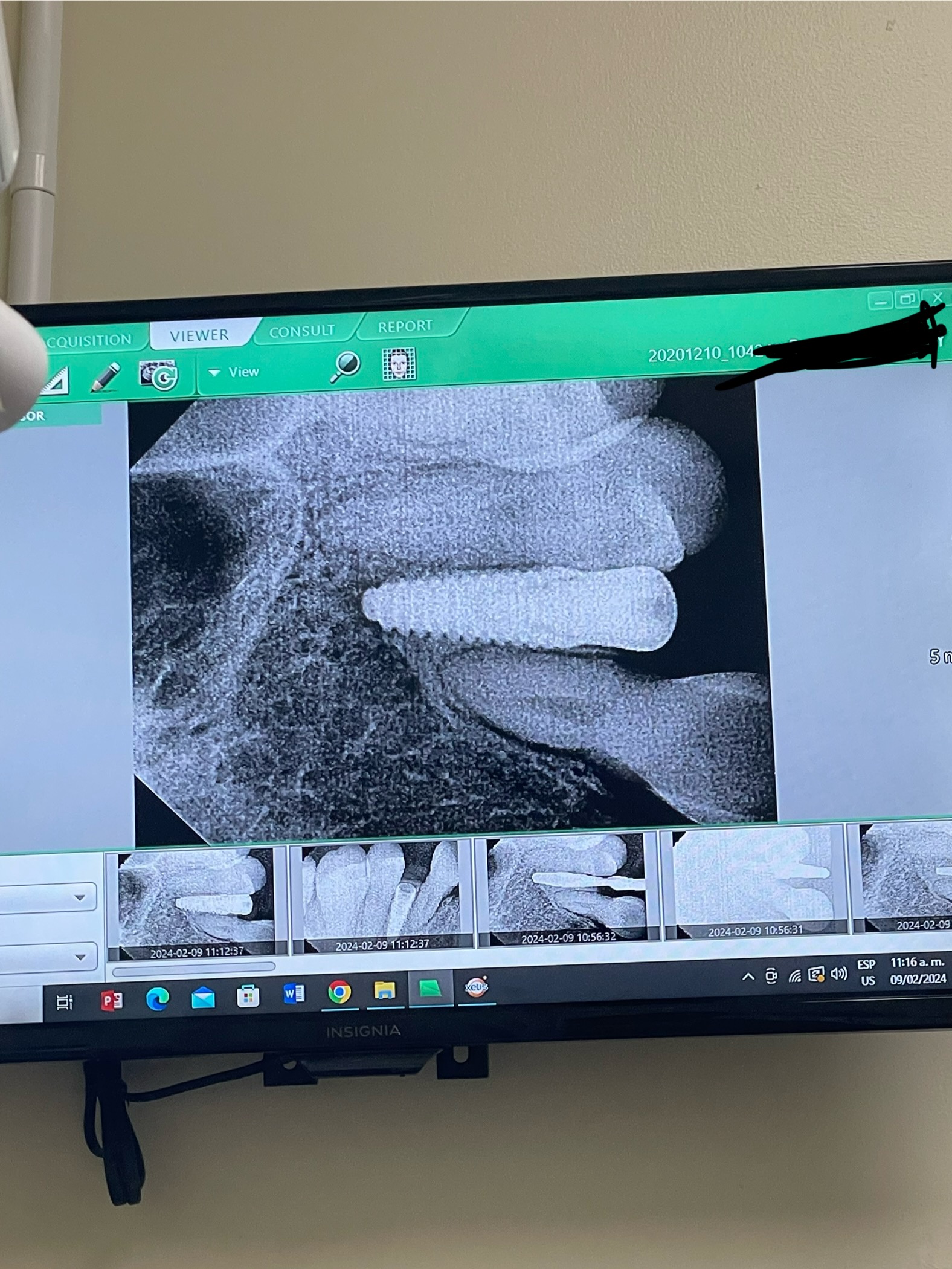
A 60 year old , healthy female presented as a new patient complaining of discomfort on tooth #9(UL central), as well as not being happy with the esthetics of her upper anterior teeth. She reported having had an implant placed 15 years ago and has been having discomfort for 2 years. Her current dentist is a “holistic” dentist who refused to give her antibiotics, as he doesn’t believe in them. On examination the implant appeared slightly mobile and copious pus was draining through a fistula above the crown- see photo. The x-ray shows severe bone loss around the implant (Bicon?), but the crestal bone appears to be intact.

My question has 2 parts. Firstly how would you remove the implant with minimal damage to the remaining crestal bone, what graft materials would you use? And secondly, would you consider a bridge from 8-10 and a crown on #7 as a restorative option rather than another implant? Thanks in advance for your responses.


27 Comments on Slightly mobile implant with bone loss: How would you treat?

New comments are currently closed for this post.
CRS
10/20/2015
Fifteen years is not bad, remove the crown, abutment and use the neobiotech torque wrench, if not take a thin burr or trephine and remove. The crestal bone is most likely what is holding this in. It can be regenerated. I like disinfecting it with the Nd- Yag laser and grafting. See what you get then treatment plan. Resin bonded temp or Essex while graft is healing. There is most likely a perforation in the cortical bone which will need repair. I use particulate with a blood product of choice maybe a membrane depending on the architecture. Good candidate for a connective tissue graft on the anterior teeth and /or veneers for matching in the final restoration.
Bennett Jacoby
10/20/2015
I've never used Biocons but I have used the Neobiotech detorque wrench. My impression, correct me if I'm wrong, is that the Biocons do not have a continuous thread, so that the wrench may break the implant osseointegration, but then it will just be spinning. With the apical section diameter of the implant larger than the coronal section, I don't think the implant would come out easily. I think the bone will need to be trephined. Then again, when the osseointegration is broken, the implant might be able to be "enucleated" from the likely defect in the cortical plate. I agree with all other points CRS.
Justin
10/20/2015
Agreed that this is a failing implant and should be removed. I agree with CRS for surgical protocol which is probably beyond my scope due to my lack of experience in the anterior region and the thin gingival biotype. A new implant/crown #9 or FPD #8-10 will probably not address her chief complaint of poor esthetics. A diagnostic wax up would be a good start. Even if pt decides not to replace implant, graft if for better gingival esthetics with the bridge. Also, check vitality on #10. Difficult case, teeth will be long and may need c.t. graft that CRS mentioned. The patient's smile line doesn't appear to be too high. Good luck and keep us posted!
DrT
10/20/2015
I agree on using an implant removal kit. At the same time I would do a coronally positioned flap with CT graft on teeth #10-12 as well as on the labial of the implant site. I would use a BMP bone putty in the implant exo site after it was thoroughly debrided and disinfected
Peter Hunt
10/20/2015
It would be very difficult to remove this implant without destroying the crestal bone. Any situation without the crestal bone is much more difficult. Think it through another way. It might be better to first remove the crown and the abutment. The labial bone wall already seems to be missing, so then flap the region and open up the lesion. Then the fun part would be to push the implant crestally, and remove it through the labial window. That way you have a chance of preserving the bone crest. It also allows the region to be cleaned out properly and regenerated.
George
10/20/2015
These implants were called "Styker" back when they put this in (now Bicon). I see no indication for the use of these implants, ever. Removal is a problem because middle of implant is bigger than the neck, so trephine will make a really big hole at the crest and you will have a big defect when it heals unless your graft technique is pristine and the patient is a good healer. You have to cut the crown off horizontally at the crest, then trough or trephine around the stump of whatever is left. Sometimes you can wiggle the implant out of the hole in the buccal plate and spare some of the alveolar crest - do this if you can (easier to rebuild buccal plate than to rebuild crest). When titanium junk is gone, debride and graft. This is not a simple case, if you do not have a lot of experience with this kind of thing then I suggest that you send it out and let it be somebody else's problem. Meanwhile, you want to cantilever the central? No thank you, please. If you want to do a conventional FPD and you can't use the lateral (think "interlock" (or tubelock) precision attachment if draw is a problem), then extract the crummy lateral and go to the canine. At this point you might as well do 6-11 so it really looks nice (I would if #7 is too crowded/tilted/whatever). Good luck!
Paul McDonald
10/20/2015
Blimey ! Apart from replacement of the upper left central, there doesn't appear to be much more the patient needs, except a bit of whitening. If the central, the tooth furthest from the jaw elevator muscles, cannot be replaced with a cantilever prosthesis, then what tooth can be ?! Fortunately, resin bonded cantilever bridges perform well, at minimal biological cost, especially if no tooth preparation is performed. Pontic space on protrusion needs to be assessed, and if necessary the opposing incisal edge/s can be reduced. After working impressions, opposing plaster model can be appropriately reduced before wax-up of the bridge, then the localised occlusal reduction replicated clinically at bridge fit, until normal ICP reachieved. I tend to do my own diagnostic mock-ups in composite, very easy, using a familiar material, on mounted study models, so I can see the dynamic occlusion and assess what, if any, localised occlusal reduction may be required, then use to gain consent. There would appear to be little indication to proceed instead directly to a destructive, conventional bridge with a cemented, full crown retainer, especially of a fixed-fixed design.
DrG
10/20/2015
Easy case, I'm not kidding. Vertical release distal 6 and 11, full thickness flap. Your CBCT pre surgery will show you no buccal plate around the implant. (Thank god the holistic dentist let this one soak in its own filth for years). Remove the crown and abutment, then make a small slot apical to the implant and elevate to apical end out buccally. You will be able to easily remove the implant and leave the crestal bone intact. Next completely remove every fiber of tissue present until you see completely clean bone. (Remember there is no buccal plate) then graft, membrane draped over adjacent osseo us architecture 5 mm or so both buccally and palatal to close the entire defect. Over that a CT graft. Release the flap so it's tensionless and suture to primary closure. Honestly, I'd even consider placing and implant same day if possible.... As far as the generalized recession the reload flap can be repositioned to cover those areas. She's got tons of KG present on 6,7,8,10,11 sites so no need for CT graft there when you coronally reposition the flap.
DrT
10/20/2015
One miracle at a time: placing an implant at the same time as you remove the failing one feels to me like pure idio**, or arrogance. Just my opinion. As for increasing the thickness of the gingiva, why not just place a piece of tissue allograft before suturing...no big deal. As for this being an "easy case", hmm..I would be very careful.
DrG
10/20/2015
I should rephrase that, "it's easy for a board certified specialist" Perioderm, mucograft any tissue analog will work. I'm just comfortable with the biocompatible materials myself.
CRS
10/20/2015
I don't have any experience with Bicon implants but I neglected to mention that a buccal flap needs to be raised to access the defect it may be possible to section the implant and push it thru the defect. This needs to be grafted and well healed prior to replacement with another implant.
Robert J. Miller
10/20/2015
As this is a retrograde peri-implantitis, do you understand the mode of failoure? Without understanding the reason for this lesion, I would NOT place another implant at the time of failed implant removal. Is this an encroachment into the nasopalatine foramen? Was there an apical granuloma that was not debrided? Is this primarily related to occlusion? Is there a loss of the facial plate with soft tissue ingrowth through the fenestration? Remove, debride, and attempt to understand the cause of this failure first. Then make a decision on retreatment. RJM
DrG
10/20/2015
I new that would get people commenting... After the first 5,000 implants you start to see typical failure patterns. As well every surgeon needs to know what they are comfortable with. I am willing to guess this failure is due to one of two very common causes. 1. The apex is not on the alveolar housing. Soft tissue and bacteria invaded the implant from the apex down. 2. The original peri-apical lesion was never removed 100% and after years the PA lesion infected the implant, again Apico-coronally. Sound surgical principles should work in redoing this case. A.) complete disinfection of the lesion B.) new implant has primary stability C.) the entire implant when placed is within the alveolar envelope D.) good blood supply to support the CT graft, and osseo is graft E.) tensionless flap without pressure from a prosthesis during the initial healing
DrG
10/20/2015
Actually there is something very interesting in the photos of this case. Any thoughts why the implant is out of occlusion and the surrounding dentition looks like it has supra-erupted since that implant was restored? Hint; I think the tremendous amounts of KG elsewhere is from prior root coverage surgeries.
Raul Mena
10/20/2015
Having placed many of bicon implants and knowing how well thy prform, I will elevate a full mucoperiosteal flap, enter through the buccal plate and with a carbide burr and plenty of irrigation section the implant horizontally right were the abutmenpost end abut the middle of the implant. Debri the area grafted and replace rhe flap. It will function asa short implant and that is about all the implant length that you need. You have nothing to lose.
peterFairbairn
10/21/2015
Agree with Dr G , not a big deal can be removed with a site specific flap at the gingival / mucosal junction after crown removal ( Tap Off ) should be able to remove easily .... Then let heal for three weeks flap and standard SRP again..
Dr Y
10/21/2015
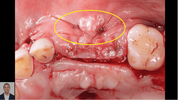
Thanks for all the comments. I removed the implant today, it was not possible to tap the crown off as the implant was mobile and I was concerned about breaking the crestal bone. As it happened, it came out with the crown as I rotated it with my fingers together with the most foul green material I have ever seen. I laid a full thickness flap, curetted the area thoroughly- there was extensive bony destruction but the palate was not breached. The crestal bone was only present on the palatal side. I elected to use dual phase calcium phosphate mixed with B-tcp granules from Steiner. This is the first time I am using this type of material and thought this would be a good case to try it with. I did ask the patient if she had any previous oral surgeries and she said only the implant placement at #9. As far as placing an implant at this time I think it would be highly unadvisable and frankly just not possible due to the amount of bone loss. Peter, in retrospect I think your idea of waiting for healing after implant removal would have been a better idea and is an approach I will be using from now on. One concern I had was the amount of bleeding in the bony defect. It looked like I had hit a blood vessel as the blood was welling up and would not stop. Any ideas on the source of this and ways to stop the flow during surgery. Once the graft was packed it did appear to stop but I was concerned about this. Thanks again for all the tips. Dr. Y
DrG
10/21/2015
Sounds like either you were close to the nasopalantine foramen or you hadn't removed all the granulation tissue down to the bone. If there is any granulation tissue left you won't fully form bone in the defect. When you re-enter it will be mush/type IV bone. However if you have successfully removed all the granulation tissue and it was indeed the nasopalantine artery you hit the regeneration jackpot since that blood supply will be a plentiful source of regenerative cells and growth factors!
Robert J. Miller
10/21/2015
Sounds like there was encroachment into the nasopalatine foramen after all. Probably transected the nasopalatine artery. RJM
PeterFairbairn
10/22/2015
Hi Dr Y , as Dr Millar has suggested there may have been some damage to the artery but curretage but , I think the severe infective state could also have this effect . This is why I suggested allowing to heal for a few weeks prior to grafting .... Greg Steiner makes some great graft materials and has a lot of experience but all of us involved in material development can see the possible issues which may affect the material performance and this excessive bleeding would not help when associated with incomplete closure.. All the best Peter
CRS
10/22/2015
I would have used the Nd-yag laser to disenfect it first going around the implant prior to removal, if there is granulation tissue it helps encapsulate it and gives you a nice margin with good hemostasis Then remove the implant and treat the bed again for disenfection and hemostasis The granuloma most likely was in contact with the nasopalatine contents and may have been torn when blindly curetting the pathology. Bleeding can be controled with digital pressure on the palate, possibly using a suture to keep the pressure on. I would have placed some particulate bone in the bleeder or used Hemcom. This sight most likely has bacteria a few mm of penetration deep to the bone margins. Usually pigmented bacteria seen in perio disease. I doubt you transacted the artery, a pressure suture would have been necessary. With that much pus I would have cultured prior to the laser to see what your are dealing with, I have seen actinomyces which is very difficult to erradicate, at that point I get my infectious disease colleague involved in the management. What most likely happened is that the original tooth may have been an old RCT or fractured with the original bacteria getting a nice head start in the bone. This is a localized osteomyelitis if it has been present chronically. So two issues addressed, chronic bone infection and hemmorhage management. You're welcome☺️
Dr Shyam Mahajan
10/23/2015
Now the implant is removed , granulation tissue removed , flap sutured . Good . Wait for nature do healing. Bleeding must have been due to granulation tissue. Once you currate well , it stops. Does not stop till granulation is completely removed. Many a times pains in spite of good local anesthesia. Would have prefered CBCT.
Carlos
10/27/2015
This might sounds crazy but, why don't you try taking the implant out through the vestibular plate? That will give you access to decontaminate plus maintain the crestal bone and papillae. Allograft and a membrane won't be that difficult after that. I would consider that if I were you. Perhaps you just need to remove a few milimeters of bone in the appical portion since you say it moves already.
Don Rothenberg
10/28/2015
I have used Bicon (DB, Stryker) since 1986. If the implant is mobile and you can feel it moving on the buccal, use a finger, I think it will be easy to remove. Hold the crown or abutment with an extraction forceps...twist and pull. Implant out. If not the abutment will come out. Then use a trephine one size larger then the implant with very cold water and carefully remove as little crestal bone as possible. Once the implant is out curette and graft with whatever you use. I prefer AlloOss with PRF and covering with PRF members. Then treatment plan with the patient. I like Raul's idea...but he is a lot more optimist and creative then I am. Good luck and thanks for posting!
Dr Sandeep Singh
11/4/2015
IMHO do not try to remove that Implant. Expose the buccal Bone plate with modified periosteal flap design preserving the Papilla and attached Gingiva giving a semi-lunar incision. Remove the thin buccal bone, if present and expose the Implant. Remove the Granulation Tissues just the way it is done in Apicecotomy. Disinfect the lesion with Tetracycline / Clindamycin slurry and then with Laser, Diode or Nd yag. After copious Irrigation with normal saline, pack it with CGF Blocks and Graft it with Sticky Bone. Close the bony window with Resorbable Collagen Membrane and then with CGF membrane. Close the flap with pTFE Sutures. Give Antibiotic Prophylaxis of Amoxycillin + Clav 625 mg twice daily x 7 days. I bet you and your patient gonna be the happiest person in this World. God Bless you. Sandeep Singh India
Muhammed Fisal
12/27/2015
• It is amazing to see the crestal bone gain in this case. It is the dream of every implantologist to have bone growing over the crest of implant. This adds to the intrabony length of the implant, improves the crown implant ratio and support the interdental papilla . Almost in every implant case there is some crestal bone loss and literature says that it stops at the first thread. Also the crestal area is the starting point of most of the implant failures. The crestal bone in this case is almost in level with the adjacent natural teeth. This show that there is something admirable in the Bicon design and protocol
Richard Hughes, DDS, FAAI
1/4/2016
One may be able to twist off the crown. And treat the implant as per CRS and Fisal's recommendation. One may not like the Bicon implant but it is still a viable system.

Featured Products

OsteoGen Bone Grafting Plug
Combines bone graft with a collagen plug to yield the easiest and most affordable way to clinically deliver bone graft for socket preservation.
CevOss Bovine Bone Graft
Make the switch to a better xenograft! High volume of interconnected pores promotes new bone. Substantially equivalent to BioOss and NuOss.