Unexpected Anatomic Structure Revealed After CBCT for Implants: Advice?
Dr. T. asks:
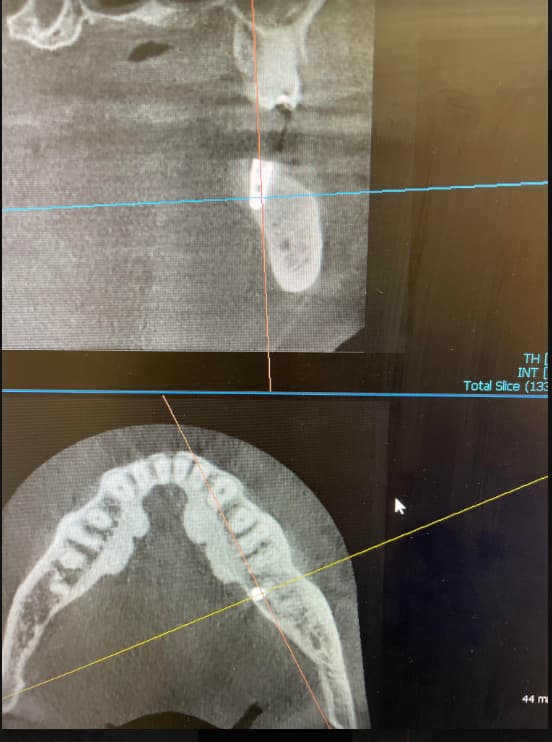
I have treatment planned a patient for installing implants in the mandibular anterior region in the #26, 24 areas [mandibular right lateral incisor, mandibular left central incisor; 42, 31]. A CBVT scan revealed what appears to be an anatomic structure about 1mm diameter in the area. I cannot tell if this is a blood vessel, nerve or some other kind of structure. What do you think this is? What do you think I should do?
CT on lower right later incisor
CT on lower right later incisor
CT on lower left later incisor
17 Comments on Unexpected Anatomic Structure Revealed After CBCT for Implants: Advice?
New comments are currently closed for this post.
Dr. Vaziri
1/9/2012
This is maybe artifact,some fibrotic tissue placed from extracted infected tooth socket or what did you say blood vassel come from jeno-Mussels. Don't worry your are far away from nerve stractur and mental foramen( where you have to do conservative job.
Good lock to you
Dr. Vaziri from Iran
DrShalash
1/10/2012
Did this teeth undergo a socket preservation with a bone graft following extraction? How old is the extraction? Looks to me like an incomplete bone fill within the tooth socket or a fibrous encapsulation around the graft.There are no visible foramina on the lingual cortical plate which is the only anatomical structure u need to worry about in this region. So there no risk of hitting any blood vessels in the region (BTW the actual blood vessels cannot be visible on a CBCT).
Scott D. Ganz
1/10/2012
I would tend to disagree with the comments that there is nothing to worry about in this region. On the contrary, there are vessels, and important ones even in the midline of the symphysis that you can visualize in cross-section CBCT... in fact hemorrhagic have occurred and have been reported in the literature. As to visualization - please check my chapter in Stuart Froum's new textbook on Implant Complications... and other articles that can be found on PubMe. There also appears to be an anterior loop to the mental foramen, but not enough information to tell ... without more slices.
Alejandro Berg
1/10/2012
Seems like an encapsulation of a grat, just remove and good to go
Dr D
1/10/2012
It looks like encapsulation of some grafting material to me...
Could you please clarify...are you placing implants only on sites #24 and #26. I would be concerned if those are the only implants to be placed. The location would create cantelever forces of the overdenture, which will increase as you know, the risk of implant failure. More so, if you have less than ideal bone on those sites...
Richard Hughes, DDS, FAAI
1/10/2012
I would refer the reading to a radiologist that was familiar with head and neck anatomy. Then proceed accordingly.
DREAM DDS
1/10/2012
Dr. T: I think it is great you have noticed and thought about this lucency on the CT. In my opinion, this could be a very important "lesion or structure" and needs to be addressed. You need to have the CT read and reported by a dental/medical radiologist who could be a DDS, MD or dual degree. In the US this report has become mandatory legally. In California at least, CT will soon be mandatory for any implant placement and that means the radiographic report will also be mandatory. Please do not drill into this area before you research this. Understand that our answers are from what we have experienced and what we have researched. I see you have 3 CT images. The clarity of any of our images is not as good when uploaded to be viewed. You need to follow the lucency in your slices. Start from the mental foreman. This could be an anterior n. loop which has been reported up to 13-18mm anterior of the mental foreman. This could be a large cyst but not a residual of bone grafting, it is too far inferior from the crestal ridge. The most important reason I am writing this is it could be a hemangioma bone lesion, previously called an a-v bone shunt. If this is the case, this can be a serious and fatal complication for your patient. There are 25 reported deaths from a dental a-v shunt, mostly in younger patients who had a simple bicuspid extraction. This is nothing to overlook. Ask experienced oral surgeons and they will not pass this off as a benign lesion, it needs to be investigated. Dont be the one who finds out first what this lesion is. A "vascular anamoly" as it may be called, presents as a lucency with pitechial multi focus of densities dotted within the lesion. It is well circumscribed. Other possibilites is that a symphysis foremen carrying artery and vein. Referral to a vascular surgeon may be needed, depends on the radiologist report and you gut. I have one of these going right now, it is very frightening. Sincerely Leonard
Dr. Gerald Rudick
1/10/2012
Dr. T. The timing for the publication of your question was perfect.Tonight, January 10th 2012 from 8:00 -9:00 p.m., Dr. Louis Al-Farage hosted a webinar for the ICOI on complications in implant dentistry.
He specifically addressed your question by showing a case of implants being placed into the symphysis where "an anatomic structure of 1mm in diameter" appeared in this generally harmless area.
Usually the inferior dental nerve exits out through the mental foramena bilaterally; however in addition to aproximately 15% of people having anterior loops of the mental foramen, there are some people who have "incisive canals" which are basically a continuation of the inferior dental canals running bilaterally through into the mid symphysis.
What was previously thought to be the safest area to place implants without any worry of causing nerve damage; turns out to be a region that requires serious consideration........ and you were certainly astute enough to pick it up and took the precaution of having a CT Scan, and not to have proceeded with the surgery.
Scott Ganz was right on the mark with his comments; and is giving all of us who place implants " a wake up call" and to not underestimate the necessity of a CT Scan even in the safest areas.
Dr. T. you have taught us all a valuable lesson, and I thank you for it.
Gerald Rudick dds Montreal, Canada
Richard Hughes, DDS, FAAI
1/10/2012
Dr T: I also thank you for this lesson. I also thank Drs Ganz and Rudick.
Dr.Guy Carnazza DMD
1/11/2012
Dr. Robert Miller just published an article addressing these same types of concerns in the most recent Journal of oral Implantology. Valuable information about what we think is the so called"zone of safety".
Abdusalam Alrmali
1/11/2012
it seems to be an anatomical normal variation, incisive canal may be. there are few cases reported regarding this variation in this save area.
so, if you evaluate the other cuts and see its relation to mental foramen.
if not the second normal landmark prominent in this area [ nutrient canals] but it has different appearance and course.
i think you have to give us more information on the CBCT
thank you
ABDUSALAM, BDS,DDS,MSc
Abdusalam Alrmali
1/11/2012
Considering the incisive canal as anterior extension
of the mandibular canal,the frequency of
incisive canal’s visibility is higher in the inferior canine
area, followed by the inferior lateral incisive and
inferior central incisive areas.
Baker vinci
1/11/2012
Diff. Dx.- Condensing osteitis( not recognized by WHO, exclusionary, typically), hyperdense bone, fib. Dysplasia , several variants of cementum originated lesions, ossifying fibroma, metastasis. Most probable dx, in my opinion, nothing to worry about ;but get a radiologist to look at it. Your copy, probably didn't email well. Again, very poor implant candidate, but that's not what you are asking. Please don't remove grafted material, even if it's a synthetic. Not sure why this suggestion, is so popular! Patient needs good comprehensive dental care. Sorry, I'm sure everyone is tired of hearing this. Bv
Baker vinci
1/11/2012
Transitional stage of periapical dysplasia , has to be considered, as well. Again, just a matter of detail; the "incisive canal" is located just palatal to the maxillary central incisors . The incisive branch, does have it's own canal, however. The 15% anterior loop ,has been questioned , with some pretty strong data. I Might encourage a high definition scan of the area . Bv
Dr Chan
1/11/2012
"1 mm (?) diameter in the area" a bit too vague, Dr T.
Baker vinci
1/12/2012
Yeh, didn't have glasses on , thought I saw 1 cm. Never mind! Bv
Dr Saulo Porto Filho -Bra
1/16/2012
It doesnt looks like a canal. The possibility of the insisor canal cames until the mandibular edge is very short. To me it is a fibrosys. Just open and see.