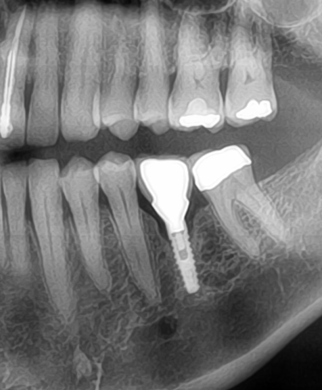
Failing bone graft: any suggestions?
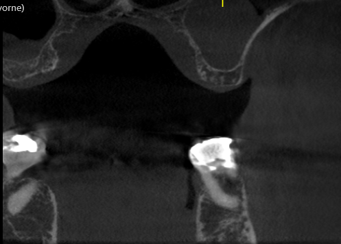
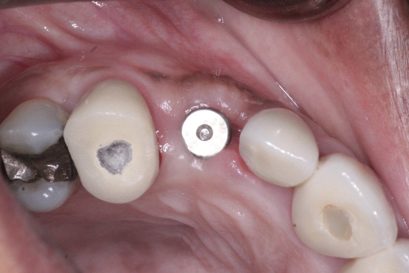
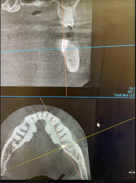
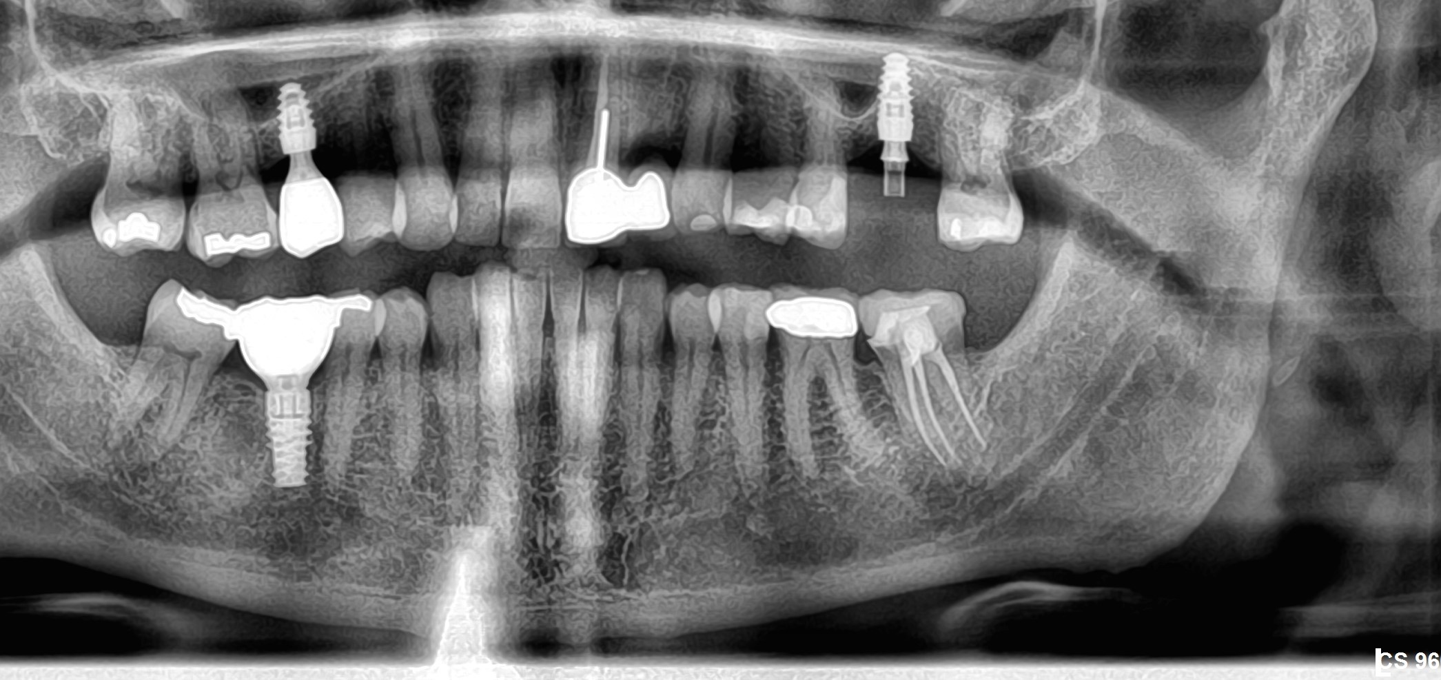
I extracted tooth #19 about 3 mos. ago. It was a failing endo with an attempted apico(by an endodontist) which revealed a root fracture. Here are pre op , post op and 3 months post op. Do you agree with me that the graft encapsulated? If so, now what. Do I enucleate and regraft or enucleate, place the implant and graft around it. I own a diode laser only. Any suggestions for decontaminating the site?


16 Comments on Failing bone graft: any suggestions?
New comments are currently closed for this post.
Justin
1/29/2016
Excellent and well presented case! There are providers here with more extensive training and experience than I - I'll be interested when they chime in. I would probably wait another 3 mo and take a cbct. That is if everything else checks out (no fistula/abscess) with healthy keratinized tissues present.
Thanks for posting!
Justin
PeterFairbairn
1/30/2016
Depends on type of graft and protocol used , so yes more information
Peter
Ezgator
1/30/2016
Beta tri calcium phosphate covered with a titanium ptfe membrane
CRS
1/30/2016
I agree probably some of those nasty residual bacteria and granulation tissue causing a stir. When I have this happen I remove it, disenfect with the Nd-Yag laser and replace bone graft. Peter have started using the tricalcium product so far so good, still use the human demineralized depending on the clinical situation. The diode will just burn the tissue at 910 wavelength and may not go deep enough. Just curetting this small area out may be enough in this case since it is a revision, but I have had consistant results with the Yag. I would culture the area also so that you know what you are dealing with. Lately been seeing cases of localized chronic osteomyelitis if this does not resolve in a few weeks then refer to an OMS, may require a Pic line. P. S. I love how the easy graft handles!
PeterFairbairn
1/31/2016
Agree with CRS , Site preparation is the Key to Success , with a 14 year history with BTcP and thousands of successful grafts I find it is the only particulate to fulfil all the biological needs for true host regeneration . I suspect here the mesial root pathology was not possibly removed leading to an issue .
Here is my protocol and ideas on these materials and their use.
There are lots of BTcP on the market ( there are cars ) and they all perform very differently ......as they can be varied extensively in manufacture ,
Did nice talk here in Athens with Hands on yesterday , to launch a new paradigm shift in regeneration , tomorrow start another study to show just how osteo-inductive CaP products are..
Regards
Peter
ezgator
1/31/2016
So in this case what would you do, especially if you don't own a laser. THANKS
Sb oms
1/31/2016
This is not complicated.
Residual infection and encapsulated bone graft particles.
Flap and debride. Make sure all bone is bleeding. Graft if defect is buccal or lingual, but if it's just a hole- don't graft. The body will re- mineralize the area. Primary closure should by easy. In my hands, I would fill this with PRF.
This is not osteomyelitis and your patient won't require a pic line.
Wait four months, scan and make sure it's healed well, and place your implant.
Best way to avoid is to meticulously clean extraction sockets. Small suction tips to see all the way down into apical regions. Properly shaped currents. Good suction, lots of irrigation. Rotary curretage and perforation of bone surfaces to ensure bleeding and influx of healing cells and mediators.
CRS
2/2/2016
I was just sharing how bone infections can develop from chronic untreated infections. Old school curretage technique may be fine. Yeah I get what an osteomyelitis looks like on a film. If you carefully read my post I said the same thing you did just added some additional information if this does not resolve. Three additional months of residual infection is not a good thing😀
Dr K Gilani
2/2/2016
I would have a different approach to this case. After the extraction and curettage of sockets, i would just wait 2 months , letting the body deal with the infected bone with no disturbance and I would have the gingiva covered the socket for primary closure without coronal displacement of mucosa.. Then I would go in after 2 months and curette out the granulation tissue out of both sockets. Then, all I needed to do was minor drilling in the distal socket and graft the defects with a xeno material, then cover the whole thing with a resorbable membrane. Then just waiting 4 months till exposure. Total time 6 months. This is my standard procedure in this case, which never let me down. I don't trust triphosphate stuff, had many failures in the past.
In this case , I would do what you missed to do from the beginning. Curettage the encapsulated graft, make it bleed. Let body to deal with it for 2 months. Go back and place your implant and graft/membrane around it. It will be cooked after 4 months.
Good luck
seth m black
2/2/2016
If there is no pain or purulence then wait another 3 months. The problem with non autogenous grafting is that there can be delayed resorbtion and therefore delayed replacement with healthy bone.
Are there any possible systematic concerns (AODM, thyroid issues, meds?)....just to name a few.
ezgator
2/2/2016
BTW everyone, I was well aware of this potential and I thought I scraped the heck out of that socket. Maybe next time I will use a rotary round bur as well. Thanks for all your input.
DrG
2/9/2016
Ez, it's not that you didn't scrape the area well enough. The mesial root creates a small domed overhang where the PA granuloma was. No curette or but can get to this area well. To thouroughly clean it (without a 50k laser) you need to remove the Durval bone and curette the area with straight line access. It's like endodontist approach a canal. If your grafting then what's the difference if you ronquer out the mid furcal bone?
pascal valentini
2/3/2016
the problem in that case is : Do we wil still have remaining bacterias in the socket after the graft removal? In such a case I would remove the gaft , curettage and after a 1 week antibiotic treatment I would place an implant with some grafting material around and I would not submerge the implant.
PeterFairbairn
2/3/2016
There are some videos here on Osseonews where I have prepared the site using degranulation Burs ( EK solutions ) placed and grafted .....
As always Ez we all have issues and are not faultless this is biology , things happen....
As for BTcP it is the material of choice as we see in our extensive clinical cases and animal studies , BUT there are plenty cheap not well made products out there as some think it is easy to make , not so .
It is highly Osteo-inductive when made well as seen in research even by Buser.
Regards
Peter
greg steiner
2/4/2016
Hello Peter
I hope your research is going well. You say caP products are osteoinductive. Calcium and phosphate is a natural component of extracellular fluid. How can calcium and phosphate modify the physiology of a progenitor cell to move down the osteogenic pathway? What studies do you have that osteoinduction has occurred? Greg Steiner Steiner Biotechnology
PeterFairbairn
2/10/2016
There are many studies , mainly those of Pamela Habovic ( from about 2004 and a new one out lasy year , ZHao and Watanabe et al in Bone showing genetic up-regulation and even Buser , Sculean , Miron last year in Coir ....
But a well know fact for many years ....bone is grown in muscle tissue.....
I will email the research to you by you can Pubmed it
Regards
Peter


