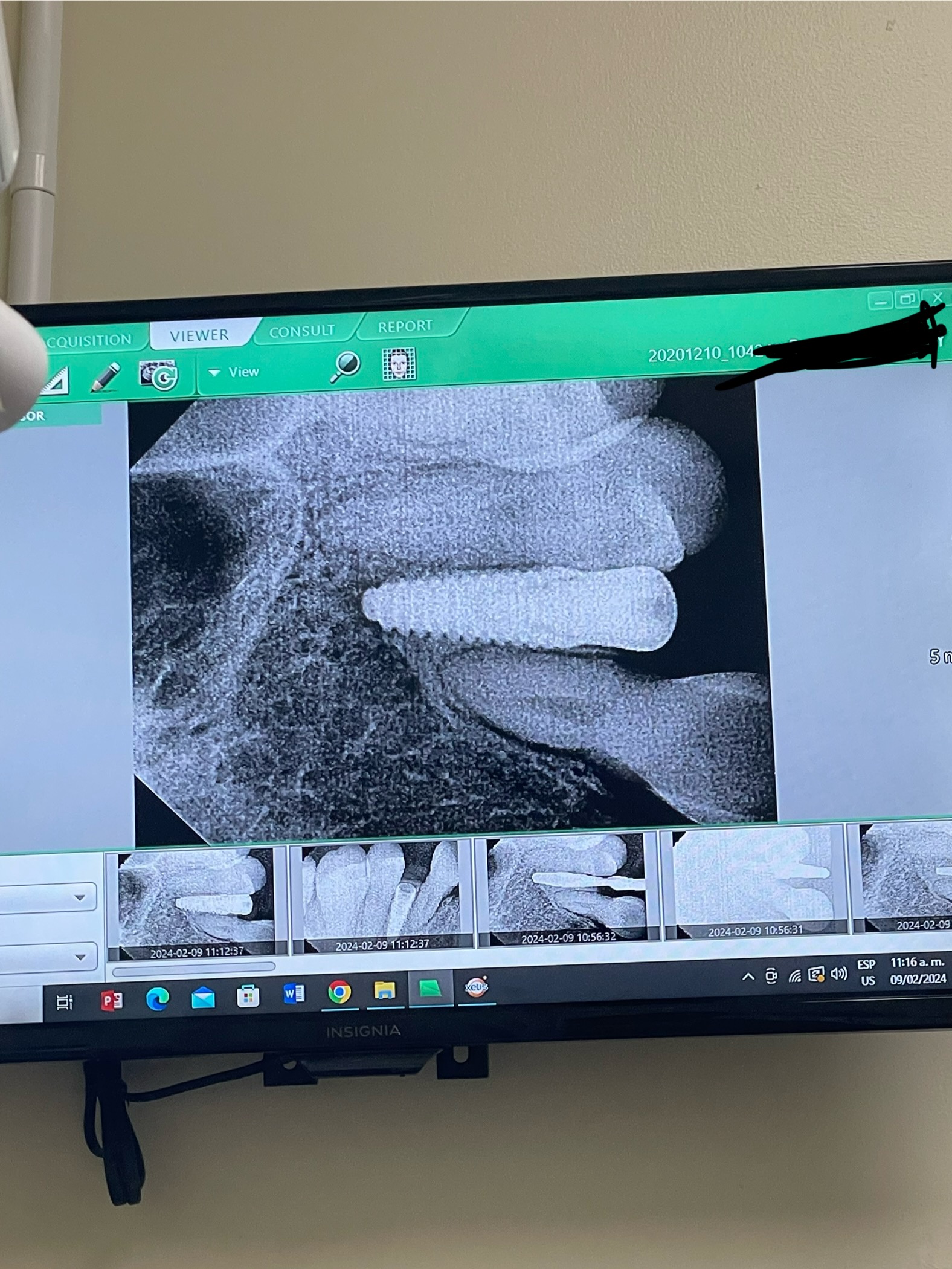
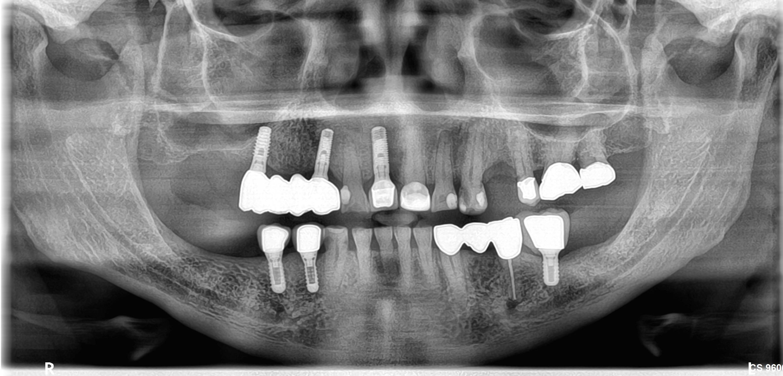
Flap dehiscence in my first implant case: advice?

I placed my first implant on a patient recently under supervision. Patient’s profile: 61 years old, Hepatitis B carrier, no relevant drug allergies or problems. Non-smoker.

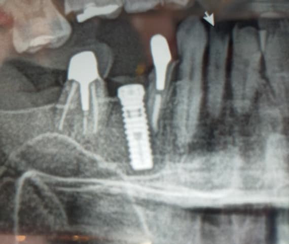
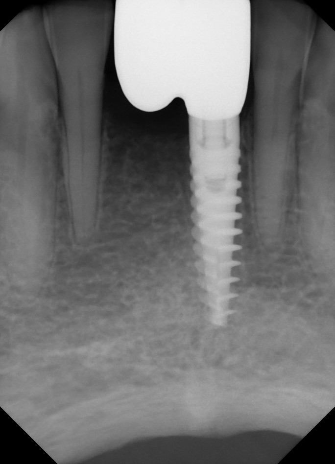
A Straumann 4.1 x 10mm bone level implant was used on the lower left first molar region with a 0.5mm height cover screw. Primary closure was established with simple interrupted 4/0 Vicryl sutures. A similar implant was placed in the region of the lower right first molar. The surgery was uneventful. Standard meds with Augmentin prescribed.

I reviewed him after a week and noticed that the suture on top of the cover screw was gone and the flap was open. Gosh did my heart sink! The lower right implant was healing well.

The surrounding tissue was rather friable and did not look too great. The implant was stable. Patient was advised to continue with Peridex – I wasn’t sure if it would heal up and didn’t intervene.

I reviewed him again at Day 14 and noticed that the flap was still not healing well. The flap tissue is partially healed but the periosteum is still not attaching well to the bone. Implant stability is fine.

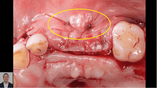
I decided to take action. I flushed the area with Peridex, gently debrided the surface bone and granulation tissue with a spoon excavator and made a small perforation in the distal cortical bone to induce some bleeding. Copious saline irrigation was done, and the flap was sutured. Augmentin was prescribed for another 5 days. I told the patient not to chew on the site for the next few days.

Not sure why the flap broke down but I’m guessing that it may be partly because the patient has thin mucosa on the edentulous ridge and probably ate on it as well?

Another strange thing is that the patient was unwell between days 10 – 13 with some fever and chills. He saw a medical doctor but was not prescribed any antibiotics. He is recovering well at Day 14 but says that he suffered a similar illness when he previously placed 2 implants on his lower anteriors a few years ago. Not sure if it’s related to the surgery or stress?

I really want this case to succeed, but it looks rather worrying especially for a first-timer. What advice would you give? (fingers crossed)


37 Comments on Flap dehiscence in my first implant case: advice?

New comments are currently closed for this post.
Dr Mark Bishara
9/27/2015
Hi , what kind of flap design did you have here? it looks like a crestal release with no vertical distal release. When the area swells up it can pull away on your sutures. How was the thickness of keratinized tissue on buccal , was it lot of moveable mucosa? it looks like the implant is placed to the buccal, I would try and place my implant lingual of the area if permits and make sure to release enough of the tissue so you have passive closure again.
Beginner
9/28/2015
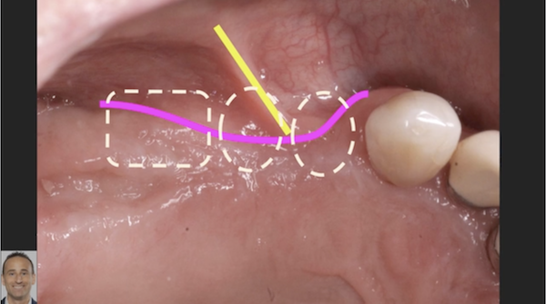
Hi! Thanks for the reply. It was a rather wide crestal release. Not sure if you can see in the first photo but I did try to do a distal buccal release although it was rather diagonal (wonky?) There was quite a lot of moveable mucosa on the lingual. As for positioning, the photo is deceiving but the implant should be quite centred as I used a stent and the supervising surgeon did take a look. How would the prognosis be like any idea?
greg steiner
9/28/2015
This may have nothing to do with your surgery but may be related to the health of the tissue you are working with. One thing for sure is that exposed bone is dead bone it will slough off if it is just surface necrosis but if the necrosis is deeper it will come out in chunks and the implant will likely fail. First was this area grafted prior to implant placement and if so what was it grafted with. In addition we need to see the bone to determine how deep the necrosis is so if you could provide a current digital periapical radiograph this would allow is to do that. Greg Steiner Steiner Biotechnology
Beginner
9/28/2015
Thanks for your reply Greg. No, there was no grafting done at the site. I don't have a new PA at the moment but may take one at a later follow up. In that case, would it be better to attempt to close the flap over the bone or leave it open and let it slough?
CRS
9/28/2015
What was your supervisor's opinion? Mucosa won't attach to necrotic exposed bone. I always thought a releasing incision had to be vertical. Anyway let it granulate in. Peridex is fine to keep it clean. Keep us posted.
Beginner
9/29/2015
Hi CRS. My supervisor just got back to me and told me that there's nothing to be done for now but to monitor and wait even if there is a dehiscence, and that a flap closure is not necessary. He said that he may need to see the patient if the patient has spiking fever or pain at the area. I assume he's referring to osteomyelitis?
CRS
9/29/2015
Fever and chills are most likely not related to implant placement, anecdotal info from a patient needs to be balanced with what you see clinically. Probably some overheated bone or pressure upon placement of the implant. The next 4-6 weeks will tell, keep it clean advise the patient, I would like to see some granulation tissue forming. Now you are gaining clinical experience as a surgeon not a technician. Way too early for an osteo, I suggest you do a little research on pathophysiology of osteomyelitis. The fever of unknown origin if significant can be managed by the physician. It is not all about technique. This is where the real learning and experience begins.
Beginner
9/29/2015
Dear CRS, Thanks for the words of wisdom. Yes, I begin to see how complicated all this further management can be - and I thought implant positioning would be the hardest bit! Will post updates when I get them.
CRS
9/29/2015
It will be fine just let the patient know your concerns worst case scenario you'll do it over, most likely it will be fine.
CRS
9/30/2015
Wish it were that easy, I tell my patients an implant is like a pregnancy and we are watching it, don't want a miscarriage!
Raul Mena
9/29/2015
CRS and others, Nice postings I see that every one is giving positive advice and trying to guide a colleague in the right direction. It may or may not be osteomyelitis. Personally I think is improper flap design and over manipulation of the tissues, both soft and hard. To much suctioning can also produce bone necrosis, due to over cooling and bone dehydration. Chances are that it will heal by second intention. Peridex can be a source of irritation so use it with judgment. Raul
CRS
9/30/2015
It is all in the delivery, if someone truly wants to learn I am ready to give advice. If they want my advice . I don't think there is an understanding of osteomyelitis which is way too early here, it is a chronic condition that takes time to develop, but thank you for your compliment.
Beginner
9/30/2015
Thanks for the feedback Raul. Noted on the flap design!
Carlos Boudet, DDS
9/29/2015
The most common reason that a surgical flap opens post-op is suturing under tension. maybe it needed some periosteal release. Another reason could be taking small needle bites on thin and/or non-keratinized tissue. One more reason could be poor handling of the tissues. When a wound opens in the mouth, it usually will heal by secondary intention (granulation tissue), so by going in at day 14 and removing the granulation tissue, you actually set back the healing response by days, and could have increased the discomfort to the patient. If the implant is stable, supervise the healing and it should eventually be fine unless a lot of bone was exposed. CRS suggestion is valid, but I would start with the basics of bone formation and healing of bone and soft tissues. Good luck!
Beginner
9/30/2015
Dear Carlos, Thank you for your reply. Yes, I think you may be right about the thin tissue. I wasn't sure if I should have removed it or not, but he was having some pain / discomfort at the area and it didn't look too good. I'm glad to say that he reports that the discomfort is much better after the irrigation and debridement was done. Perhaps it was the food debris there causing tissue irritation?
DrG
9/29/2015
Definitely agree with the earlier comments. Exposed bone without vascularized areas is necrotic. Lots of ways to deal with that, but I'd definitely expect the surface bone to come off the implant at some time. Lots of interesting teachable moments in these radiographs. 1. Make big flaps so you can see what you need to see. Vertical releases are fine make sure you miss the ligi artery and the mental nerve branches when you plan your flaps. 2. When you make full thickness flaps make sure you, A) incise the tissue down to bone- no split flaps /especially beginners B) make the incisions in keratinized tissue only 3. The implant is definitely going to be a mesially cantilevered restoration. (I read you used a stent) Think about the orientation of your handpiece and how you press the handpiece with a distal pressure, I can move the center of the osteotomy to the distal as you enlarge the osteotomy. 4. Surgery needs to be reactive and definitive. Waiting in this instance reflects ambiguity on the part of the surgeon and the mentor. Take action at this point. Either decorticate the neurotics bone and do a FGG in the mucosal area or back out the implant, let it heal for 12 weeks and replace the implant and do tissue graft at time of placement. Finally what is the opposing arch like? Does the patient wear a RPD?
CRS
9/30/2015
Actually waiting in this instance is prudent and respectful of the biology vs being technical, each time you intervene the developing blood supply is disturbed.
Beginner
9/30/2015
Dear DrG, thank you for your detailed reply. Yes you're right about the release being insufficient and I should have considered a free gingival graft first to increase the keratinized tissue. Noted on the handpiece angulation too. The implant is likely going to be a single crown as we are planning for a shortened dental arch. I am also unsure if a 4.1mm diameter Straumann can support a mesial cantilever crown - would that work? I would say that the opinions of my supervisors weren't ambiguous - the general consensus was that there was nothing much to be done at this time unless failure occurs and that I would know it. I was anxious to intervene as the patient is my relative and I felt really bad that he was having the discomfort and symptoms mentioned. As for the opposing arch, the plan is to an RPD. He will require bilateral sinus grafting for upper implants and so he's a bit hesitant about proceeding at this time. I have offered him the options of non-intervention, upper and lower RPDs or upper and lower implants. We're proceeding with this plan for now as it leaves his options open for future maxillary implants and that the maxillary RPD is likely to be a lot more comfortable than a mandibular denture for a first time wearer. I have a few questions: 1. By ligi artery do you mean lingual? Besides hitting the books on anatomy, are there easy landmarks you usually take note of to avoid the vital structures especially so on a resorbed ridge? 2. How much surface bone will I expect to lose? Thanks for your time.
DrG
9/30/2015
Yes lingual darn autocorrect.... Glad to hear it's a relative and a RPD opposing. As long as the implant integrates you should have no issue with the slight distal positioning of the implant. Thanks for replying to all of our comments it shows your genuine interest in learning and understanding.
Philip
9/29/2015
I think you did an excellent job here both during surgery and in dealing with the subsequent healing issue. It is well known that mouth ulceration can be strongly stress related and the history of a previous episode points to this. Ulceration will often occur at a trauma site , e.g. injection site. I love what you have done as a first intervention and I would be surprised if this does not resolve well for you. Remember how well extraction sites resolve when even a dry socket occurs. It can look awful for a while. Excellent work and excellent problem solving. Well done!!
Beginner
9/30/2015
Dear Philip, Thank you for your heartening encouragement. Much appreciated!
Dr. Rodger Uchizono
9/29/2015
Regardless of the cause, I feel it's important to get that bone covered with a membrane to protect the implant and control epithelial growth. I would treat it as a guided tissue regeneration case using Renovix membrane by Salvin Dental to protect the implant. Cut a piece large enough to extend past the defect margins and suture passively after removing all granulation tissue. Primary closure is not necessary, and the implant will be saved if the bone was not overheated or over-compressed. This was an ambitious first case, but it will get you to assess the volume and quality of keratinized tissue adjacent to the implant during the planning phase. I hope this helps! -RodgerU
Beginner
9/30/2015
Dear Rodger, Thanks for your input. I've been wondering if I should have placed a membrane from the start. It seems a little bit late now though. Yes, I need to pay more attention to the soft tissue!
Dr. Rodger Uchizono
10/1/2015
It seems that the pre-operative planning didn't account for the volume and quality of attached gingiva in the implant site. Some perform a CT graft either before or during the implant placement, sometimes harvesting from the lingual of the surgical site. You also have the option of moving keratinized tissue from the top of the ridge buccally, placing a healing cap and suturing loosely as needed. Whether or not I place a membrane depends upon the need for a bone graft, presence of a bony dehiscence in an immediate placement case, success in mobilizing the tissue in lower bicuspids, etc. I'm sure other posters on this thread would be happy to share their thoughts on this subject. The use of a surgical guide gave you a sense of security regarding availability of bone, but soft tissue issues are equally important in the planning.
Jafar Kolahi
9/30/2015
I had same experience. Your suture was under pressure and closed. Yet, don't worry. The wound will heal after 2-3 weeks successfully with some minor bone lose.
Beginner
9/30/2015
Thank you Jafar. That's good to hear!
Bashar Kabbani
9/30/2015
Regardless of the cause you can remove the cover screw of the implant and put the healing abutment, Let the tissue heal around it.
Beginner
9/30/2015
Thanks for your comment Bashar. Noted! But most likely we'll wait for the other side to gain better stability as the initial torque there was rather low. This one was about 15Ncm. Also, I'm a little phobic about touching it any more if it's not giving further problems for now.
Dr.Dr.Hossam Barghash
9/30/2015
one of the most common mistake for the beginner, that they think tight suture secure there implants,and dehiscence occur, especially when there is an absence of the keratinized mucosa , there will be more pulling on the suture, and the usual scenario,flap opening. how to handle, back to wound healing physiology, good oral hygiene,gentle mouth wash especially with saline(for neutralizing PH) actually debridment and closure is not commended ,wound edges are friable so you will take another big bite far from wound edges ( compromising the blood supply) plus the area will not be easy to clean. another factor will be systemic condition of the patient. to summarize good oral hygiene, tetracycline capsule dissolve the powder in water in water and use as mouth wash and follow up.
Beginner
9/30/2015
Dear Hossam, You're right about the tissues being friable. They were worse at day 7 when I first reviewed him and removed the other sutures. The tissue below the resorbable Vicryl (polyglactin) sutures looked rather mushy and I did not dare to touch it. Maybe I should have used monofilament nylon instead? I reviewed him again at Day 14 and noted that while the tissue seemed to have firmed up, the flap edges were still not attached to the (necrotic) bone and there was food debris trapped. He also complained of slight pain to the area. I therefore decided to treat it like a pericoronitis case with some cleanup and antibiotics. I don't think he is able to clean the area well by himself, and I was hoping to decrease the size of the gaping wound and deflect food debris away. I tried to use a tension free modified mattress suture if that helps. Glad to say that the symptoms have subsided, but I am not sure how effective the suturing will be given that the necrotic surface bone will not attach to it as I have been taught here. Can the necrotic sequestrum layer be sloughed off beneath the healing gum tissue and would it come off as a big piece or just dissolve away?
Emery Cole, DMD
9/30/2015
Friable non kerotinized tissue can be tricky. Old school advice once given to me, Hibiclens placed topically 4xday with cotton tip applicator (Clear version tastes better if still available), Rinse with a fresh cold pitcher of strong brewed black tea several times per day......hard to beat saline rinse as well.(Pre-Peridex days) Does the patient drink alcohol frequently? Do they have Zerostomia? Surgical field looks extremely dry.
CRS
9/30/2015
Hibicliens is chlohexidine, may be cheaper. Avoid the strong black tea. Like the previous comment about if the patient is wearing a partial often the culprit in wound breakdown.
Beginner
9/30/2015
Dear Emery, Thanks for the reply! The cotton tip applicator sounds like a very good idea. No, I don't think he's a drinker and he doesn't have xerostomia but maybe minor hyposalivation at his age? The surgical handpiece had saline irrigation built in. The tea sounds delicious!
Beginner
9/30/2015
Hi CRS, No, he has never worn dentures before. If he were, would you have recommended the use of a periodontal dressing such as GC Coe-Pak on the area to block out the surgical site? (and make it impossible for him to wear the denture?) Just curious. Thanks!
CRS
10/1/2015
No pack, slows down the healing. Prosthesis need to be carefully relieved and balanced, prefer nothing for 2-3 weeks but not always possible in the esthetic zone. I sometimes use viscogel. I think one of the cruelest things in dentistry is when an ill fitting immediate denture is placed over a newly created surgical site. Painful!
Beginner
10/1/2015
Roger that. Thanks!
Manosteel
12/9/2016
You might want to get in the habit of extending your incisions one tooth over in dimension and use verticle release incisions. If you reflect the flap beyond the mucogingival junction you can pull up a little more for closure and if you incise the periosteum about 1 mm in depth (periosteal release) and spread the flap with Metzenbaum scissors you'll have more than enough for closure. Also if you use horizontal matress sutures they will tend to pull up the flap margins vertically, so you'll have a little more room for constricture as the flap heals. You can also use continuous on top to help closure . As for the flap opening have the pt rinse bid with chlorhexidine gluc. rinses and put them on antibiotics. Monitor express your concerns and sincerity to the patient and reevaluate in 1-2 months.

Featured Products

OsteoGen Bone Grafting Plug
Combines bone graft with a collagen plug to yield the easiest and most affordable way to clinically deliver bone graft for socket preservation.
CevOss Bovine Bone Graft
Make the switch to a better xenograft! High volume of interconnected pores promotes new bone. Substantially equivalent to BioOss and NuOss.