Overcontoured tissue. How to manage?
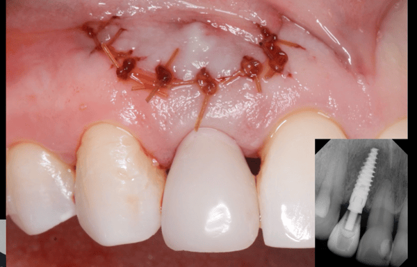
Immediate implant was placed at the site of central incisor which was extracted atraumatically. The implant was placed more labial, so a labial bone resorption was suspected. Three months following its placement, a flap was reflected and a totally resorbed labial bone was detected. Autogenous particulate bone graft mixed with Cerasorb B-TCP Bone Graft were used to regenerate the area and it was covered with PRF and collagen membrane. After additional three months, an overcontoured tissue was detected during follow up visits. How should I manage this case?
![]overcontoured tissue](https://osseonews.nyc3.cdn.digitaloceanspaces.com/wp-content/uploads/2012/07/2012-06-23-11.49.06-e1342443121841.jpg)overcontoured tissue
24 Comments on Overcontoured tissue. How to manage?
New comments are currently closed for this post.
Guy Carnazza DMD
7/16/2012
You may have to consider frenectomy because frenum seems to be inserting at surgical site. Frenectomy, bone graft and re-evaluate.
Baker k. Vinci
7/24/2012
The buccal mucosa maybe the most prominent blood supply, as bad as it may look. A frenectomy or a vestibuloplasty is not what this patient needs. Bv
kais ismail
7/17/2012
Dear dr. I think that the graft is failure and both the grafting material and the implant caused chronic irritation to the gingiva lead to this gingival enlargement
DrT
7/17/2012
With all due respect, why would you want to salvage this implant fixture??? The esthetics of the final case are going to be totally unacceptable. Furthermore, considering that the labial contours of the fixture must be outside the adjacent alveolar housing, there is no way that you are going to be able to regenerate any new buccal bone, no matter how many materials you use. I have to wonder why the implant was placed in such an unfavorable position in the first place?? My recommendation: remove, graft socket, wait at least 3 months and then place another fixture, this time in the proper position. Please, please, lets not have other posters giving their opinions on how to save this fixture. This is just an example, plain and simple of an implant placed in improper position, and there is only one acceptable solution...remove it
DrT
rsdds
7/18/2012
i couln't agree more...
michael w johnson
7/17/2012
couldn't agree more. The anterior region is no place to put bandaids on. Remove, graft and do it right.
Alejandro Berg
7/17/2012
If you were the patient?
Loose the implant and start from scratch witha good graft, also dont forget to release the flap( I´m guessing you didnt). You will also need a soft tissue graft or two.
Perioperry
7/17/2012
Is the implant in a restorable position? Do a transfer impression, pour a study model with implant analog and evaluate the restorability of the implant. Next, tap on the implant healing abutment to determine if the fixture is integrated. Is the keratinized tissue over the facial aspect of the implant moveable or is it firmly fixed in place? If fixed it may be possible to improve the tissue appearance by means of gingivoplasty with electrosurg or laser. If these observations don't predict a favorable end result it would be best to remove the implant, bone graft the defect, contour the hypertrophic soft tissue after at least 4 months of healing, then complete the case.
sherman
7/17/2012
I agree, lose it, regraft, reimplant !
repairing only lead to bigger disaster !
Sb oms
7/18/2012
There are a few things going on here, most of them mentioned in the above well stated posts, but here's my two cents.
1. This is not a labial frenum. This is the result of poorly placed vertical incisions, and a chronic inflammatory tissue reaction. This tissue is telling you something- it doesn't like what is underneath it. The implant / graft/ etc.. is causing an inflammatory reaction, and it all should be removed.
2. I would not graft this case upon removal of the underlying nightmare. I would let this heal on it's own and redevelop a proper blood supply and tissue profile. A graft would not heal well in this site due to the massive chronic inflammatory/infectious nature of the tissue.
3. Your resulting defect will be considerable. As you stated your buccal plate is gone. In my hands, I would scan a few months after removal and most likely plan a block graft. I'm sure I'll get a few moans and groans out of this, but it works. Other options include a conventional fixed bridge.
4. What kind of temporary is the patient wearing? If it's a removable partial, this will only make the problem worse. These sites need to heal free and clear of denture acrylic pressure. Consider a snap on smile type of this without any tissue contact.
5.Consider referral. You've done two surgeries, and the patient is no better then when you started.
6. When you make flaps with vertical incisions, make them distant from your site. By distant I mean 1-2 teeth away. These flaps are easier to release, and have a better blood supply for healing. This is a basic premise of oral surgery which I see beginners forgetting routinely on this site. Look at any basic oral surgery textbook and you will see what I mean. Your flap here was poorly designed, and you were doomed from the start.
Baker vinci
7/18/2012
As sb said, we still have to stick to basic principles . The vestibule is obliterated. Block grafts work, if done correctly. By the way, you can only use autogenous bone, if this is what you choose to do. Refer this out!! Bv
Richard Hughes, DDS, FAAI
7/18/2012
SB and BV are most correct on this casein every way. Prior planning prevents poor performance! The five P's.
Dr Campos FICOI,FADIA
7/18/2012
Soft tissue reminds me of Epulis fissuratum ( maybe cause by temporary appliance ). If you suspect not having enough labial plate ,grafting at the time of placement would be a better course of action to prevent the condition to develop.Depending on the size of the defect particulate bone,cerasorb,membrane might not be enough ,maybe a case for a block graft. Take a CT scan to evaluate what is under,if there is not labial plate you know what to do( remove it) graft and prep for placing another implant at later time. Try to contact some of the local periodontist and /or Oral surgeon the one that you refer patients to.most of them are willing to help you and give you sound advice in some of the issues.Best luck to you
peter fairbairn
7/18/2012
All great points , although we routinely only use site specific flaps retaining the papillae on the adjacent teeth as we do not use traditional membranes a large flap is not required remembering to use only 5.0 or 6.0 sutures.
As Richard has said planning , why so many different materials ? It does look as a local specialist may be needed with removal and a period of healing.
In the aesthetic zone we only use 3.5 or 3,8mm dia Implants and place palatally.
When it comes to soft tissue as after many years we find it unpredictable so it is left to the body to sort that out.
As long as the hard tissue is restored the soft will sort itself out Garber said it .
Good luck but as SB said you may not be able to do it again so time for referral.
Peter
Dr. Alex Zavyalov
7/18/2012
I do not understand how it is possible to give any implant-related recommendations without X ray bone analysis.
Baker vinci
7/19/2012
Because the doctor stated there is no buccal bone. That is better than a panoramic and soft tissue failure is assessed by the naked eye and perio probe. Bv
peter fairbairn
7/19/2012
Alex agreed but here you can see the issue and it is too buccal placement as well as other issues , hence would need a scan to be of any diagnostic use.
Peter
ttmillerjr
7/21/2012
Over-contoured? Really? I hate to say it, but this case is a parade of errors. Decision making errors and surgical errors. It's clear the implant was placed incorrectly, error one. Did you do it flapless? The decision to "regenerate the area", with the implant in it's position, was error two. The surgery itself demonstrates at least a couple more errors. Your flap was not released properly, which resulted in too much tension and the opening of the site, resulting in the healing we see. It is painfully obvious that this case is beyond you now. We have to know our limits and use conservative reliable techniques. I know you don't want to hear this, but you need to start with simple cases. Because you see techniques in a presentation of an accomplished surgeon, doesn't mean it will work out that way for you.
ttmillerjr
7/21/2012
I guess I wasn't very helpful. Okay, a few things to keep in mind; 1) If one draws a line from the facial of #8 to the facial of #10, the implant ideally will be 2mm palatal to the line. 2) When placing your implants don't be afraid to start your osteotomies with a surgical handpiece. It's easy to get pushed too far facially on the upper anteriors. 3) When grafting remember that you can't build bone just anywhere. If one looks "down" at the the upper arch, from the incisal point of view, and draws a line on the facial from healthy bone to healthy bone, across a defect, it's not reasonable to assume you can grow bone outside of this line. When you see your implant interrupts this line, you know grafting isn't going to get it done. Same thing when one looks at the bone on a non-angled PA X-Ray. If one draws a line from the bone level around one tooth, across an edentulous span, to the level of bone on the next tooth, one can't expect to grow bone any higher than this line. Bone grafting is not easy, it takes a clear idea of general principles plus experience.
Richard Hughes, DDS, FAAI
7/21/2012
Perio perry brought up good points. Gingivoplasty may help. It may not get better. It is not a frenum!
ttmillerjr
7/22/2012
If the question is how to correct this esthetically, here are my thoughts. If there is gingiva 360 degrees around the implant simply follow your initial releasing incisions, partial thickness through the gingiva and apically reposition the flap recreating his vestibule.
Paolo Rossetti - Milano
7/24/2012
Correcting mistakes by other mistakes is a game I have learnt not to play. Sometimes doing it all over again so that you can start from scratch is a good idea.
Abdullah
8/7/2012
I think the soft tissue of the flab was over tention, and its treatment need sulcus deepining.
CRS
9/4/2012
Usually the implant is placed slightly palatally so that the implant immerges thru the cingulum of the proposed crown. That way you can have a screw retained crown or a cemented one with room for porcelain. Without an xray it is difficult to judge the size used. I agree that the implant is outside of the arch and should be removed. Place a particulate graft and ct graft. Allow it to heal and for goodness sakes refer it out!


