Ailing Bone Graft Case: Your Thoughts?

Dr. D. asks:
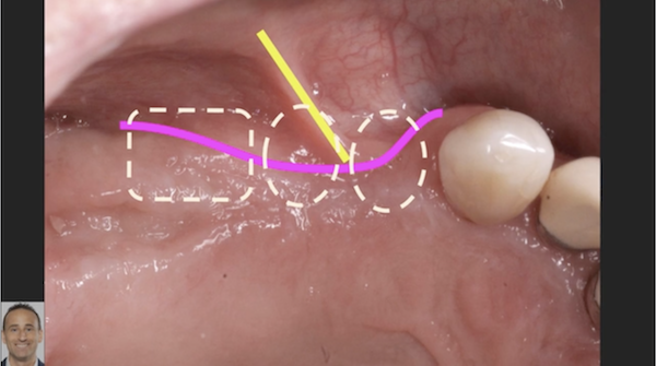
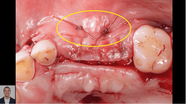
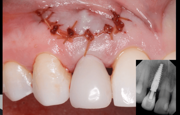
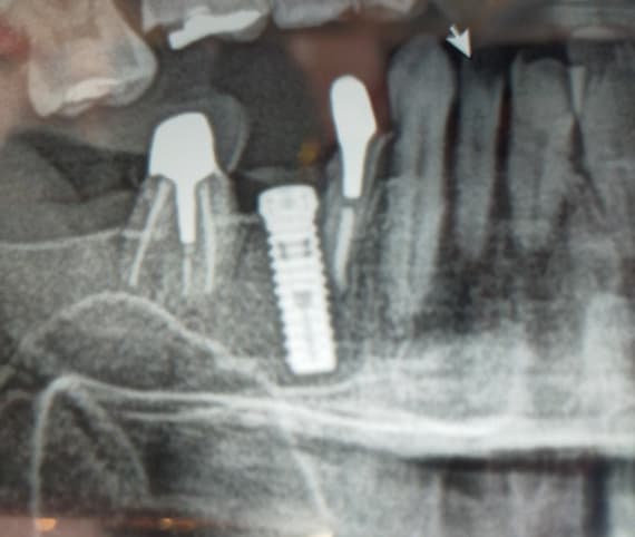
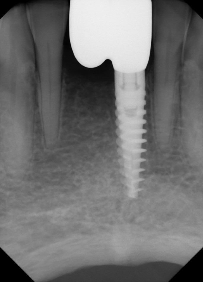
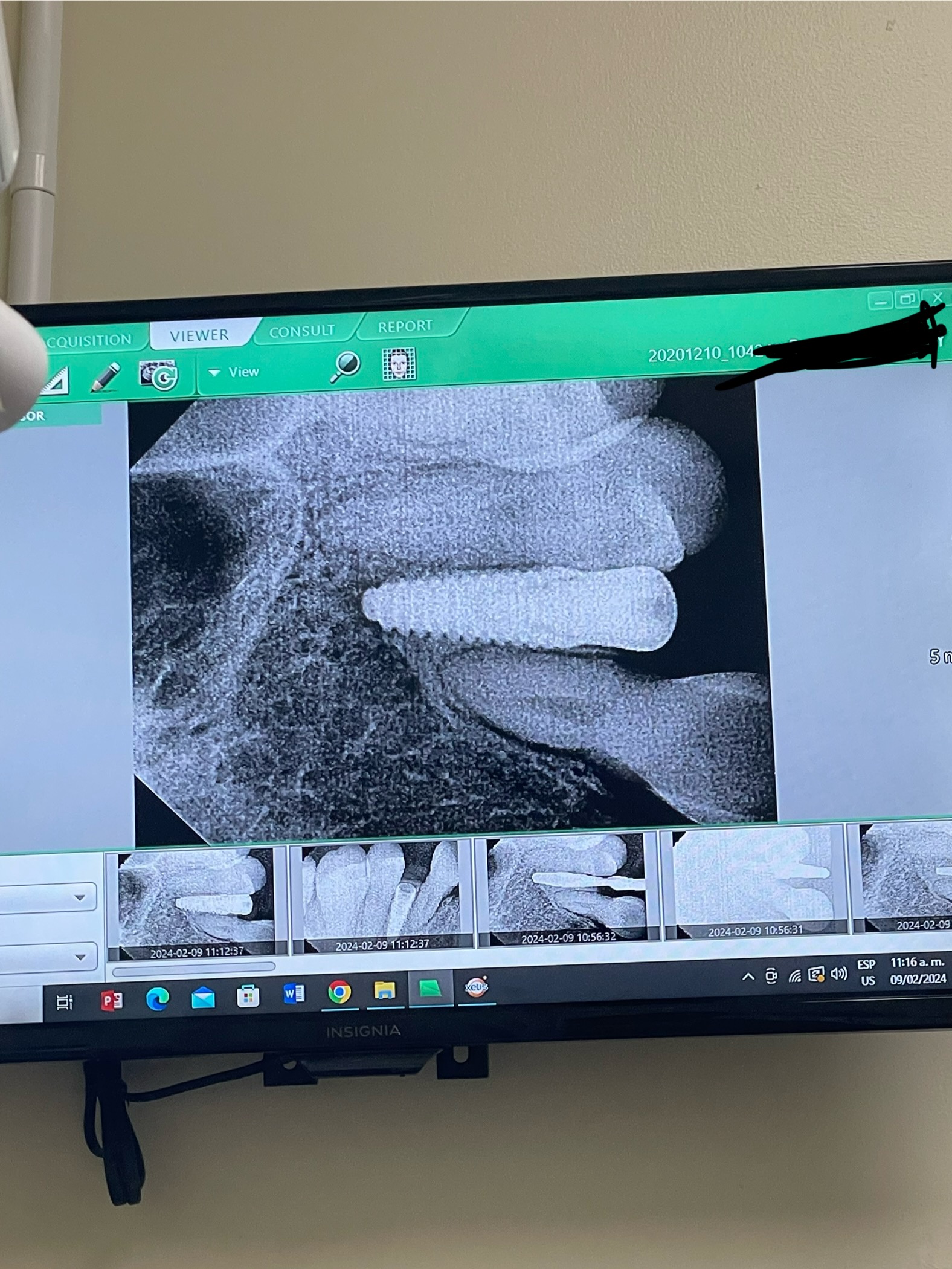
I have a 50 year old male patient in excellent health. I extracted his mandibular incisors #23-26 [mandibular right central and lateral incisors, mandibular left central and lateral incisors; 41, 42, 31, 32] and immediately installed 2 Noble Speedy implant fixtures with a bone graft of Bio-Oss and covered with a Bio-Gide membrane. At 3 weeks post-op, the flap opened and I had to re-suture which probably delayed the healing process. At 2 months post-op, the patient presented with swelling, bleeding and purulence through a hole in the flap. I irrigated with betadine and prescribed antibiotics for 1 week. Have just told patient after 4months that he would probably need to re-open up wound, clean out and possibly redo some GBR, allow to heal for a further 4months and if all goes well complete implant bridge. Patient not too happy with this especially since time is an important factor for him. I haven’t told him that there might be a possibility that the implant may be lost. Your thoughts please. Many thanks.

Pre-Op

Post-Op

22 Comments on Ailing Bone Graft Case: Your Thoughts?

New comments are currently closed for this post.
SBOMS
3/5/2012
"At three weeks post op the flap opened and I had to re-suture..." Why do you think the flap opened? Your site was infected. The flap just didn't open, your site was infected. The body did what it does best and your site fistulated to drain the process. The fistula just came through the path of least resistance - your suture line. And then you re-sutured in an attempt to close the infected site??? At this point, you attempted to lock the infected material back underneath your flap. Is it any surprise that your patient re-fistulated 6 weeks later??? A couple of points to consider here, because your thought process here is completely wrong. 1. Your site has been infected from day one. Your wound opening at three weeks was not a flap dehiscence, it was an infection. 2. Once bone graft material - especially a pourous - non resorbable one like bio-oss is contaminated, it all needs to be removed. 3. Why are you placing bio-oss around peri-implant voids??? What are you trying to accomplish??? Space maintenance or bone growth? 4. The bacteria associated with periodontally involved incisor teeth are extremely virulent and hard to get rid of. When you remove them and place immediate implants, you'd better be ready for stuff like this and know how to handle it. 5. Your restorative dentist will not be too happy about your placement angles here. WHile this is all fixable with customized abutments, it can be difficult in this area due to narrow diameters. (you are obviosuly right handed - your drill trajectories are a dead give-away. 6. I would clean this all out ASAP - implants and graft. Your patient was most likely infected from the start. Let the site heal. At that time refer, or do this with some better judgment. 7. The idea that performing GBR around newly placed and infected implants is psychotic. It clearly shows that you need guidance. So bravo for posting this case.
dr.fadi
3/6/2012
Nice comment
Mario Marcone
3/5/2012
I agree that this case could have been handled differently using accurate diagnostics and a more predictable treatment planning protocol. In addition, less than favorable action was subsequently taken to try to troubleshoot the problem. I agree, with SBOMS, that removing the implants and bone grafting material and allowing to heal would be a possible good solution, but I am not so sure that it would be the right solution for this patient. There may be way to correctly troubleshoot this case and live with a compromise. Open a full thickness flap buccal and lingual, curette out and debride all infected material found till you get bleeding bony tissue in the area. You will see exposed implant threads. Irrigate the area with sterile saline. You must use an aseptic technique. You may use whatever is in vogue these days for implant thread cleansing such as tetracycline or citric acid among others. At this point in the procedure, you have 2 options ... 1. You may choose to close the flaps with primary closure over the implant healing caps. Use appropriate antibiotherapy. This option will lead to exposed implant threads out of bone but covered by soft tissue. With good tissue healing, the prosthetics can be done ... watch and adjust your occlusion, you may have a crown-to-implant ratio issue. 2. This second option is if you want to try to grow bony tissue around the exposed implant threads. After complete debridement under aseptic conditions, irrigate thoroughly with sterile saline, place a mix of Choukroun's L-PRF and a collagenous Irradiated freeze dried cortico-cancellous bone allograft such as MinerOss (BioHorizons) and metronidazole, all around the implant threads and beyond, close the flap, and pray!
Mario Marcone
3/6/2012
The assumption I make is that all knowledge about appropriate required bone and soft tissue morphology and volume is understood and applied. Otherwise, troubleshooting will not work. It requires multidisciplinary expertise. Good luck. This case is interesting because it really tests our knowledge and challenges us to improve ourselves.
carlos boudet
3/5/2012
You should learn a lot from this case. I have always learned more from my mistakes. SBOMS comments although harsh, are likely correct, the opening up of the wound at 3 weeks was a sign of infection, and the placement of the implants would have been better with the aid of a surgical guide. Remember the old saying: "a prosthetic procedure with a surgical component". The bovine graft you probably thought would not resorb and keep the volume of the added bone better but you did not count on it getting infected. Ideally remove everything and graft. If you end up being able to salvage the implants, the case will be a prosthetic compromise. Good luck!
peter fairbairn
3/6/2012
What we must not forget is that the procedure followed by Dr D is the taught route at many Dental Schools and is backed up by extensive "research ". But as we know from experience is that thing can happen and here they have. It is now advised to mix Tetracycline into the xenograft material to help prevent this event . But I prefer to follow the Spinal and Orthopaedic surgeons lead and avoid Xenografts completely , they are my patients and thus my responsibilty. As all the above have said we learn from issues . Peter
Aussi
3/6/2012
Bovine Bone is ceramic, because of the sintering process (heating) and chemical process to “deorganify” the bovine bone (removing the organic components). The product Bio-Oss may resorb in 20 to 30 years or longer, depending on the quantity the doctor placed in your sinus. Find out how much was placed and how much was removed, if you don’t already know. The high elastic modulus (mechanical property) of bovine may tear your Schneiderian membrane because it has sharp corners and it is very strong. The material may come out from your nose because it is very dense; similar to synthetic ceramic hydroxyapatites in this country in 1970-1980. Since the osteoclast cells are not capable of removing the organic foreign material, giant cells and macrophages will transport these small pieces of bovine bone to your larger filter organs (i.e. lymph nodes, lungs and spleen) (Valen, Bollough and El Sharkawy). Your immune system may give up or be compromised because of these cells being overworked trying to remove the Bio-Oss ceramic product over a long periods of time. Due to the product’s inability to resorb under normal conditions, the manufacturer instructs the doctors to pack the Bio-Oss very lightly in the sinus. This method will produce fibrous tissue encapsulation of the material. The reason for fibrous tissue encapsulation is the body’s defense mechanism, due to the materials’ negative mechanical and chemical properties; the body is trying to defend itself from this foreign material. The fibrous tissue results in a problem, as mentioned in a book by Ole T. Jensen, The Sinus Bone Graft, chapter 17 ( of Xenografts for Sinus Augmentation P. Tarnow). Dr. Froum reported “ of relative proportions of vital bone, connective tissue, and residual xenograft to be approximately 25%, 50% and 25% respectively. and incompletely. For example, Piatelli et al 34 retrieved 20 biopsy specimens at time intervals ranging from 6 months to 4 years from sinuses augmented with 100% Bio-Oss. Findings at 6 to 9 months showed them to be composed of about 40% marrow space, about 30% newly formed bone, and about 30% residual Bio-Oss particles. Please look at your white blood count. You will see it is very high. Leukocytes (white blood cells of the immune system) is your body’s first defense mechanism, and they are currently being over worked! You need a resorbable material. How much of the Bio-Oss was removed from your sinus and how was it removed? Do you have a recent x-ray showing Bio-Oss radiopaque, much denser than the host bone? Sincerely, Useby Stuart J. Froum, Stephen S. Wallace, Sang-Choon Cho, and DennisHistomorphometric studies at 6 to 12 months after grafting consistently report findings” He further notes “As previously noted, xenografts have been shown to “resorb” slowly” Corresponding References: SJ Froum, SS Wallace, SC Cho and DP Tarnow. In Jensen OT, ed. Quintessence, 2006: Chp 17; 211-219 Valen M and Ganz SD: Part I: A Synthetic Bioactive Resorbable Graft (SBRG) for predictable implant reconstruction. El Sharkawy HM, Meffert, RM. New Orleans, La: Louisiana State University School of Dentistry; 1987. DiCarlo EF, Bullough PG. Biologic responses to orthopaedic implants and their wear debris. 9:235–260. The Sinus Bone Graft. 2nd ed Chicago, IL:J Oral Implantol, 28(4):167-177, 2002.Biodegradation and Migration of Porous Calcium Phosphate Ceramics [thesis].Clin Mat. 1992; 718 465- Schlegel und Donath [25] konnten bei 126 klinischen Biopsaten mit einem Nachsorgezeitraum bis zu sechs Jahren keine Resorptionszeichen nachweisen. BIO-OSS--a resorbable bone substitute? Department of Oral Maxillofacial Surgery, Ludwig Maximilians University, Munich, Germany. Abstract BIO-OSS is an allergen-free bone substitute material of bovine origin, used to fill bone defects or to reconstruct ridge configurations. Seventy one patients (39 female, 32 male) received 126 BIO-OSS implantations. Some health parameters or habits were documented to eliminate possible risk factors of influence. The diameter of jaw defects filled with BIO-OSS was measured. There was a significant influence of the defect size on the healing result. In X-ray controls, BIO-OSS served to identify the surrounding native bone. The density of the BIO-OSS areas was higher than in control sites. These radiological results were supported by bone biopsies. Histologically, the permanency of the BIO-OSS was still recognizable after 6 years and longer. The ingrowth of newly formed bone in the BIO-OSS scaffold explained the increased density of the implanted regions. There were no clinical signs of BIO-OSS resorption. Therefore, we can assume that form corrections achieved by BIO-OSS insertions will last. PMID: 10186966 [PubMed - indexed for MEDLINE]
SBOMS
3/6/2012
guys, this is not peri-implantitis. Your treatment recomendations to open, disinfect the implants, and graft with primary closure, are completely misleading to the post. These are newly placed, infected, non-integrated implants, they should be removed. most importantly, the patient needs to be told the truth. I'm sure there is a flipper over this site aggravating the situation as well. in my honest opinion, the dentist here has lost control of this case. it should be referred in a politically correct manner. dr fairbaum- i enjoy your posts, and have learned from you. the treatment of infection is also taught in dental schools. i'm not sure if the poster went to class that day!! again, bravo for posting, you should learn a lot from this one.
dr.omar taha
3/9/2012
thanks for your great comment but really i want to ask you in another case if you need to make vertical augmentation in lower second molar area , you need to raise the hight about 9 mm what do you think about using bone block bioteck with pericardial membrane please tell me your opinion thanks
Alejandro Berg
3/6/2012
Why are we still debating... implants and graft out, let it heal and then re do the procedure. Will probably do again GBR and implants, and then a soft tissue graft.
edwin
3/6/2012
"This method will produce fibrous tissue encapsulation of the material. The reason for fibrous tissue encapsulation is the body’s defense mechanism, due to the materials’ negative mechanical and chemical properties; the body is trying to defend itself from this foreign material. The fibrous tissue results in a problem, as mentioned in a book by Ole T. Jensen, The Sinus Bone Graft, chapter 17 ( of Xenografts for Sinus Augmentation P. Tarnow)." Why would you use a material that requires the body to wall off alien tissue? This seems counter to the goal of building bone. Why not use the patient's own bone and Cerasorb? "It is now advised to mix Tetracycline into the xenograft material to help prevent this event ." What is appropriate when the patient is allergic to *cyclines? What if the patient has an allergy and you add this to the wound?
Mario Marcone
3/6/2012
The facts, as presented, are somewhat ambiguous. Before rushing to conclusions about troubleshooting strategy, taking a CBCT may reveal some reality not readily apparent presently. In all fairness, I am of the conviction that some of the opinions expressed here are nothing more than just hot-headed opinions coming from individuals who refuse to think and troubleshoot. It is definitely easier to restart the case from groung zero, but the posts have not convinced me that this should be so.
greg steiner
3/6/2012
All bone grafts fail on occasion but the best you could hope for with Bio-oss in this case is fibrous encapsulation of the graft material. This might look good at first but it would not add any bone support for your implants and would not produce bone integration in the grafted area. The long term result would likely be chronic inflammation due to a fibrous attachment to the implant rather than bone and ultimately failure. Bone grafts behave very differently when they are placed on the surface of bone as opposed to how they behave when they are placed in a hole in bone. I suggest that rather than attempting to retreat this case would be to refer to someone who regularly does ridge augmentation and implant surgery. If the patient is unhappy at this time he will be very unhappy with a second failure. Greg Steiner Steiner Laboratories
Mario Marcone
3/6/2012
Mr Steiner, With all due respect, why should Dr D., as you say, refer to someone who regularly does ridge augmentation and implants? I know such colleagues, in the very city where I practice, who do a very significant amount of ridge augmentation and implants, and, to be quite frank, they're very often off the mark! Dr D. is probably on some kind of learning curve, and he is seeking for some honest advice. There is nothing wrong with that. he should be given a chance to correct his mistakes and it is our role to help because he was honest enough to present the case for our consideration.
Richard Hughes, DDS, FAAI
3/6/2012
SBOMS & Aussie are on target. Aussie, youngot it about BioOss, very well expressed. I have been saying the same thing for years, but not this detailed. Goodmjob.
JJP
3/7/2012
Primary closure is very essential when bone grafts & membranes are used. In a immediate implant case, it is very difficult to achieve the primary closure, without which, there are very high chances of post-op infection. Lack of primary closure may be the cause for infection in this particular case..
Dr G J Berne
3/7/2012
I agree with those comments about the unsuitability of Bioss for this type of augmentation. You end up with a lump of Bioss around the implant which looks lovely on an xray, but is in fact useless. I have been doing some work recently on disinfecting/sterilizing surgical sites with the use of intraoral ozone generators, and to date I have had some excellent results. This case could be one of those that might benefit. Remove all the Xenograft material around the implants and replace preferably with autologous bone after having disinfected/sterilized the implants and surgical site with ozone. Place a non-resorbable membrane over the site and follow up the healing process with ozone gas over the surgical site every 2 days or so till healing. I have had excellent results with this protocol. The ozone unit I use is an "Ozotop" which is made in Switzerland, and I am rapidly finding it a godsend.It is a pity there isn't much research yet on its use for this particular application, although ozone's effectiveness as a sterilizing agent is well documented.
Robert J. Miller
3/7/2012
While I agree with the comments on the unsuitability of BioOss for use in these types of cases, the mode of failure in this case is different than the previous clinicians have posted. There are many papers extolling the virtues of using xenografts, but they all have one thing in common. The sites are infra-bony and free from trauma. This allows bone to form around the graft granules with establishment of periosteal microvasculature to crest. In this case, BioOss has been used to fill a void in contact with periosteum in highly moblie tissue and, highly likely, a prosthesis that comes in contact with the grafted area. This results in contact microtauma and a non-infective inflammatory response. These particles cannot resorb and contribute to resorption of adjacent bone and a breakdown of collagen resulting in wound dehisence. The treatment of this problem is multifactorial: open flap debridement/removal of xenograft, regrafting with a resorbable graft and membrane, connective tissue graft to thicken the biotype, and relief of the prosthesis so there is no contact with tissue. This is a biologic failure that requires a biologic solution. RJM
Mario Marcone
3/7/2012
Absolutely right, RJM. All cases should be treatment planned right from the start with the biology in mind.
Aussie
3/9/2012
Are there any organic risks in Bio-Oss? Do you think that there could be proteins from cows inside? Do you agree that some patients have foreign body reactions with that unresorbable Bio-Oss?
Dr Samir Nayyar
3/10/2012
Many dentists are doing implants without proper diagnosis and treatment planning. More time should be devoted on treatment planning. Infected sites should not be implanted immediately. Waiting for few days after extraction and waiting for the infection to go off should be the best. This competitive society is definitely having negative effects. Prevention is better than cure. Its better to debride the infected site now and do regular check-ups
Aussie
3/11/2012
How can you see the differences between Bio-Oss-Hydroxyapatite and human Hydroxyapatite. With any x-rays? No. Nobody knows where Bio-Oss was placed in the human bone. For patients who have immune reactions/foreign body reactions caused by Bio-Oss it is very dangerous. Where do you want to remove Bio-Oss which recircles from time to time? You can see the difference between the Hydroxylapatites only under the microscope. Please take biopsies. Bio-Oss leads never to human bone. Why do anybody use Bio-Oss for augmentations?

Featured Products

OsteoGen Bone Grafting Plug
Combines bone graft with a collagen plug to yield the easiest and most affordable way to clinically deliver bone graft for socket preservation.
CevOss Bovine Bone Graft
Make the switch to a better xenograft! High volume of interconnected pores promotes new bone. Substantially equivalent to BioOss and NuOss.