Augmentation in the Esthetic Zone with Cerasorb and PRF
Last Updated: Nov 20, 2018
Case presented by: Prof. Dr. Fernando Luiz Almeida (Rio de Janeiro, Brasil)
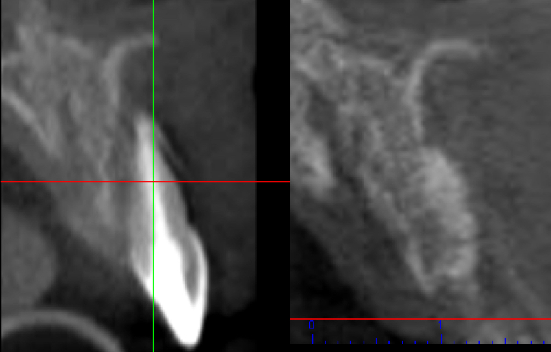
A young male patient visited the clinic with previous endodontic treatment and advanced gingival recession [Fig. 1]. After x-ray scan, it was clear that the buccal lamina is missing and the tooth has a hopeless prognosis [Fig. 2]. First, a full thickness flap was elevated. The massive bucal defect was extending up to the apical third of the extracted tooth. CERASORB M Granules were mixed with plated rich fibrin (PRF). Additionally, two PRF membranes were created to cover the defect. The flap was advanced coronally and the margins of the defect were adapted by interrupted sutures. After 4 months (120 days) an implant with temporary crown was placed. A Comparative x-ray scan between the intitial situation and 120 days after the augmentation procedure is below, as is a one-year postoperative periapical x-ray scan.
![]Clinical situation before surgical treatment.](https://osseonews.nyc3.cdn.digitaloceanspaces.com/wp-content/uploads/2018/11/02-FA-A_S_L-3253.jpg)Clinical situation before surgical treatment.
![]Pre-operative x-ray scan.](https://osseonews.nyc3.cdn.digitaloceanspaces.com/wp-content/uploads/2018/11/05-FAA_S_L-3253.jpg)Pre-operative x-ray scan.
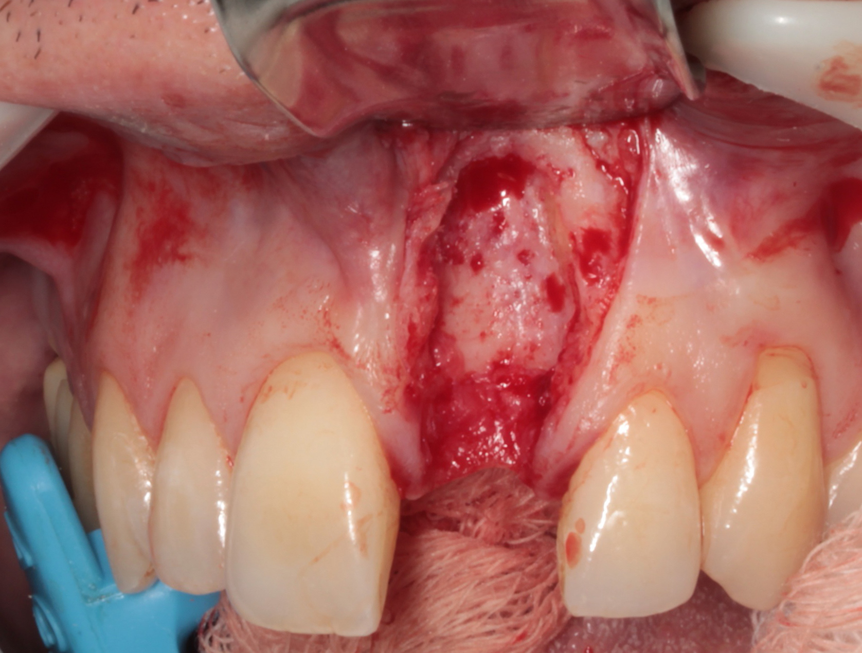
![]First, a full thickness flap was elevated.](https://osseonews.nyc3.cdn.digitaloceanspaces.com/wp-content/uploads/2018/11/07_A-FA-A_S_L-3379.jpg)First, a full thickness flap was elevated. The massive bucal defect was extending up to the apical third of the extracted tooth.
![]CERASORB M Granules were mixed with plated rich fibrin (PRF).](https://osseonews.nyc3.cdn.digitaloceanspaces.com/wp-content/uploads/2018/11/07_C-FA-A_S_L-3379.jpg)CERASORB® M Granules were mixed with plated rich fibrin (PRF). Additionally, two PRF membranes were created to cover the defect.
![]Bucal view of the defect after cover with a PRF membrane.](https://osseonews.nyc3.cdn.digitaloceanspaces.com/wp-content/uploads/2018/11/08-FA-A_S_L-3381.jpg)Bucal view of the defect after cover with a PRF membrane.
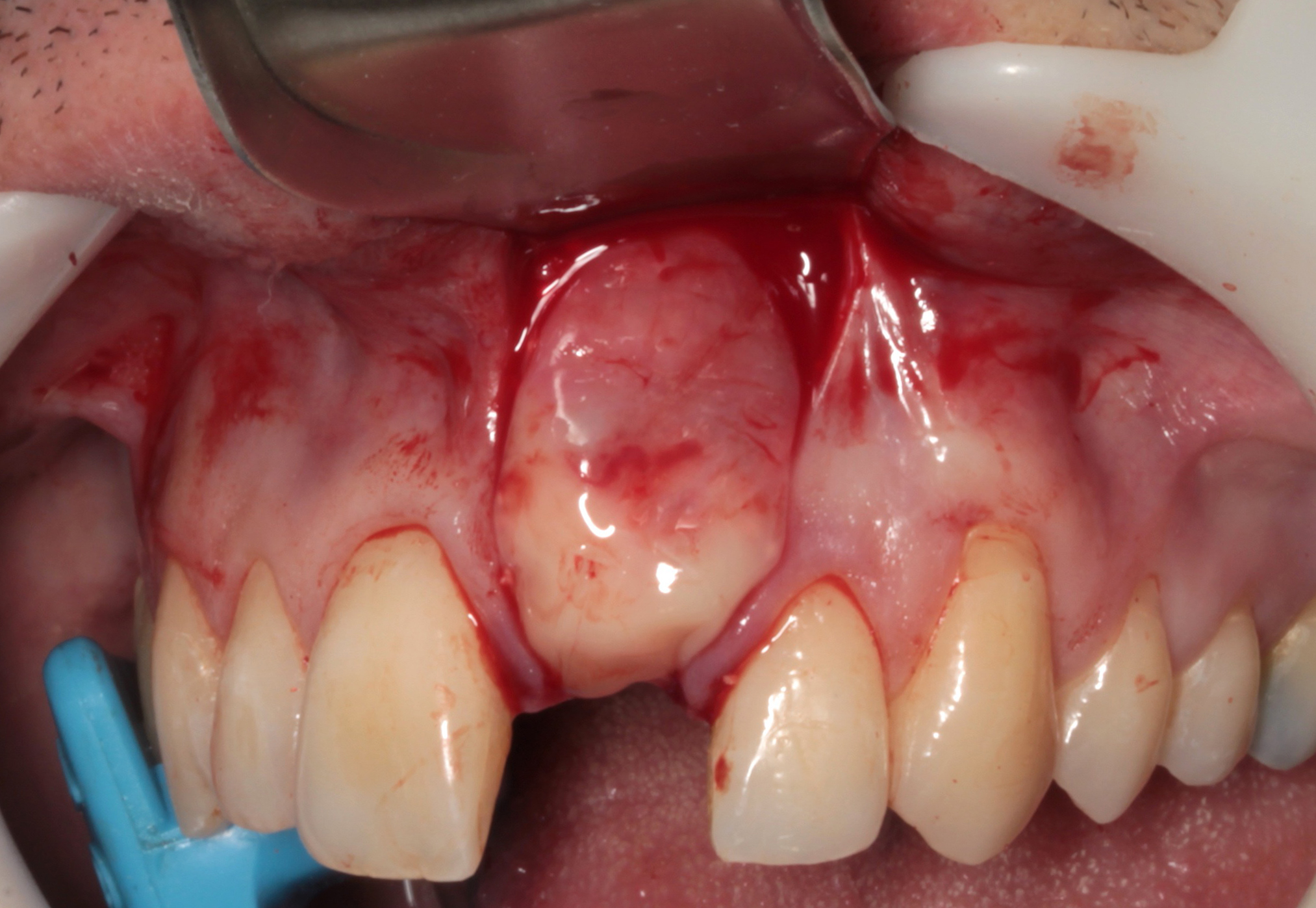
![]The flap was advanced coronally and the margins of the defect were adapted by interrupted sutures.](https://osseonews.nyc3.cdn.digitaloceanspaces.com/wp-content/uploads/2018/11/09-FA-A_S_L-3384.jpg)The flap was advanced coronally and the margins of the defect were adapted by interrupted sutures.
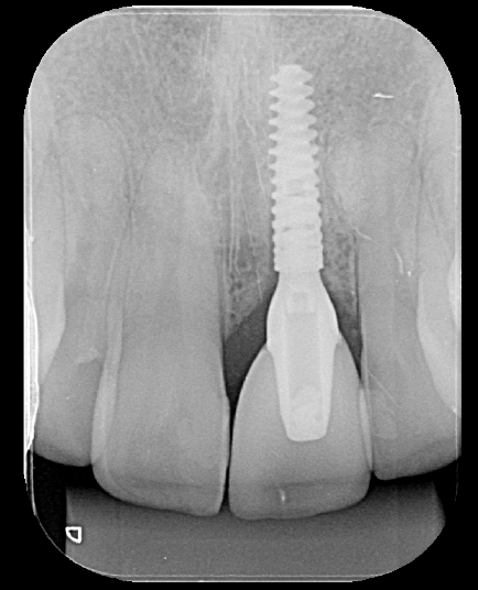
![]After 4 months (120 days) an implant with temporary crown was placed.](https://osseonews.nyc3.cdn.digitaloceanspaces.com/wp-content/uploads/2018/11/13-FA-A_S_L-3603.jpg)After 4 months (120 days) an implant with temporary crown was placed.
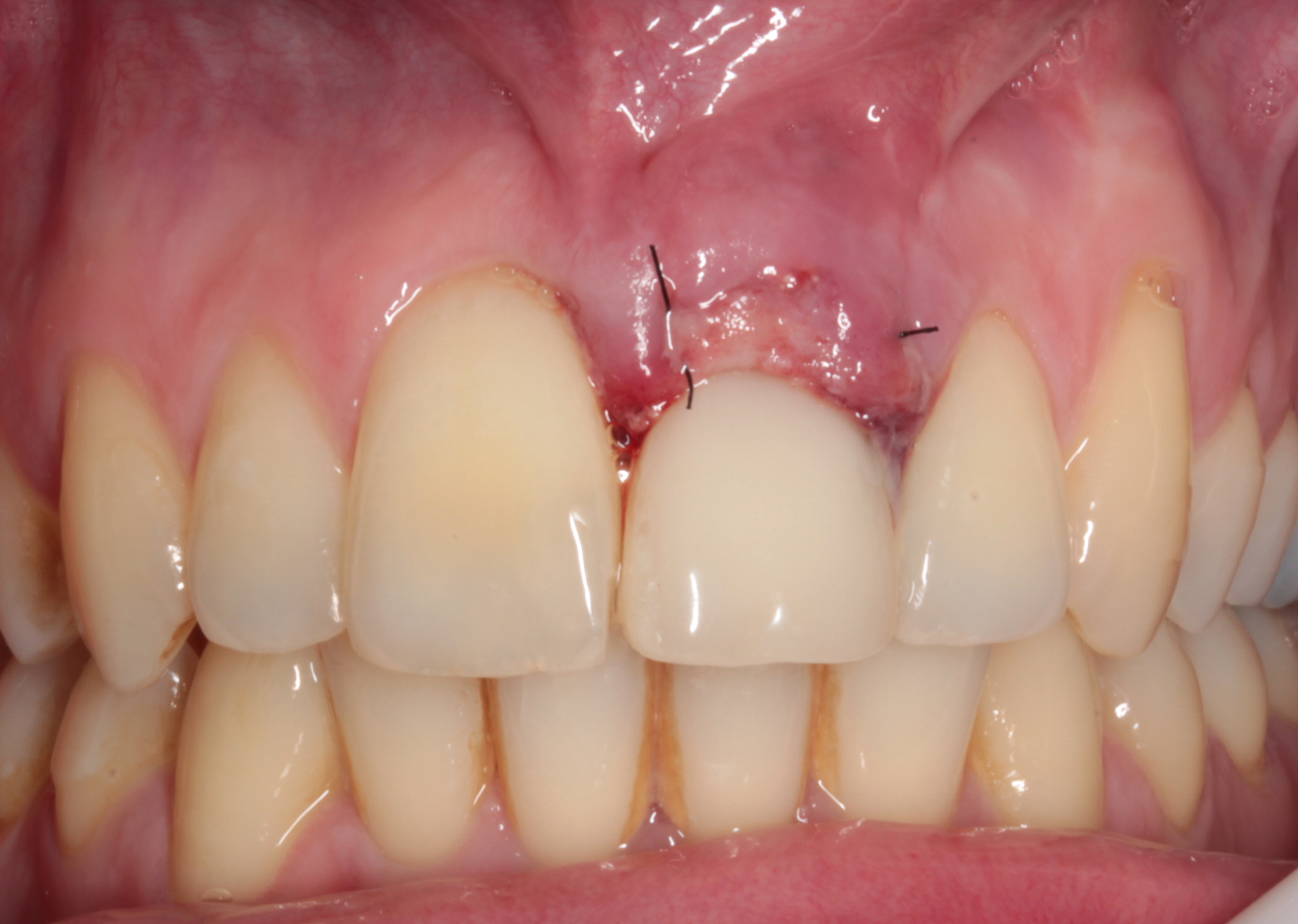
![]Vestibular view of the emergenece profile, 3 months after implantation.](https://osseonews.nyc3.cdn.digitaloceanspaces.com/wp-content/uploads/2018/11/15-FA-A_S_L-8204.jpg)Vestibular view of the emergenece profile, 3 months after implantation.
![]Final restoration / vestibular view.](https://osseonews.nyc3.cdn.digitaloceanspaces.com/wp-content/uploads/2018/11/16-FA-A_S_L-8211.jpg)Final restoration / vestibular view.
![]Comparative x-ray scan between the intitial situation and 120 days after the augmentation procedure.](https://osseonews.nyc3.cdn.digitaloceanspaces.com/wp-content/uploads/2018/11/11_AFA-A_S_L-3253.jpg)Comparative x-ray scan between the intitial situation and 120 days after the augmentation procedure.
![]One year postoperative periapical x-ray scan.](https://osseonews.nyc3.cdn.digitaloceanspaces.com/wp-content/uploads/2018/11/18-FA-A_S_L-8213-3_YEARS.jpg)One year postoperative periapical x-ray scan.Key Takeways from this Case:
Clinical)
Pre-operative)
First)
CERASORB%C2%AE)
Bucal)
The)
After)
Vestibular)
Final)
Comparative)
One)
- Fully resorbable, biomimetic pure-phase bone graft materials, such as CERASORB® M, can offer appropriate solutions in the dental esthetic area.
- Osseointegration of dental implants takes place in bone-tissue, not in bone substitute material. Therefore bone regeneration materials such as CERASORB® M are a promising treatment for long-term results in the esthetic region.
- The quality of the regenerated bone tissue is the same as natural bone, and the results are always reproducible.
5 Comments on Augmentation in the Esthetic Zone with Cerasorb and PRF
Dennis Flanagan DDS MSc
11/20/2018
Ed Dergosits
11/21/2018
Paul
11/28/2018
DrK
12/30/2018
Featured Products
Classic 50/50 Mix
Promotes osteoconduction
Provides structural integrity
Convenient Syringe!
50/50 Cortical/Cancellous
Available in 3 sizes.
Eliminate hassle of mixing particulate grafts
Sold in packs of 5 or packs of 10.
Proven safe, and clinically effective
Resorbable collagen membrane derived from purified porcine pericardium
Fast hydration and excellent tensile strength
Good adaptation to various defects
Excellent tear function and duration
100% allograft
Eliminates mixing hassle
Moldable after hydration
Gregori M Kurtzman DDS
11/20/2018