Coronal radiolucency around implant: is there a problem?

I had installed an immediate implant in #12 site [maxillary left first premolar; 24] in a young patient. The tooth had a periapical granuloma which I thoroughly curretted out after the extraction. The extraction was atraumatic and the buccal cortical plate was left intact. The buccal cortical plate showed perforation in the apical area due to the granuloma. The implant placment was done towards the palatal side without any contact with the buccal bone. Novabone putty graft was placed in the buccal side and sutured. Primary closure was achieved. The patient was supposed to come after 6 months for second stage but for some reason got delayed and the patient finally turned after 15months. Until then there were no complications. The implant was submerged and healed well. I have attached the IOPA at implant placement and the IOPA after 15 months. I see a coronal radilolucent lesion around the implant. On uncovering the implant and laying a uccal flap, it revealed good buccal bone, but I did not reflect the palatal flap, just to be more conservative in my surgical approach. I am not sure how significant the coronal radiolucent lesion is when I can see a good amount of bone all around the implant clinically except the palatal area which I did not reflect. Is there a problem with the palatal bone? Your opinion will be highly appreciates.


At Implant PlacementAt Implant Placement
After 15 MonthsAfter 15 Months

24 Comments on Coronal radiolucency around implant: is there a problem?

New comments are currently closed for this post.
CRS
6/9/2013
Retrograde peri-implantitis bacteria from the original infection.
s
6/9/2013
but clinically there is no sign of infection. And the bone has only formed around the implant in the perapical area. Can this not be a thin buccal and palatal plate?
m
6/10/2013
Clinically the site is asymptomatic, no pus discharges, no pain, no redness, no mobility, the implant is firm. If it a retrograde infection wont we see some radiolucency in the apical area as well?
CRS
6/10/2013
The appearance can be like this the original bacteria is still there in the bone. This will be difficult to fix. Never place an immediate implant when there is pathology.
m
6/10/2013
CRS sir, with due respect, I dont understand why the bacteria is still there and has prevented the bone formation crestally. If you look at the radiograph carefully after 15 month there is some bone over the cover screw and this I had to remove during the second stage. Bone over the cover screw formation is a good sign that bone is forming, In that case I dont understand why bacteria will be present in the bone. I totally agree that it is more predictable to install implant in a healed site but there are several studies which say that if the periapical pathology is removed thoroughly one can achieve similar results compared to a completely healed site. As a student, I would humbly ask you why should I not consider this as a thin buccal or palatal plate. I saw a good bone on buccal side during the second stage. Your opinion will be highly appreciated.
CRS
6/10/2013
It is very difficult to sterilize with curretage or site prep. The best way is a laser which will kill the pigmented anaerobes which hide in the bone. Grafting and allowing resolution, even the graft can be affected.When you cut the implant threads you expose these pathogens and can drive them into the implant site. Retrograde refers to backwards not a periapical infection like a natural tooth. You stated the bone covered the cover screw and both plates were intact so where do you think the bacteria came from? This is hard to treat and the implant may fail over the next few years. I have experienced this and and am developing a management protocol for infected teeth. I have had to redo grafts The bacteria have had a head start..
m
6/10/2013
CRS sir. As I was reading more literature on retrogade implantitis.The article mention that most of the time the retrograde implantitis would develop immediately after implant insertion. the definition of the retrograde implantitis as in a article says the following It is defined as a clinically symptomatic periapical lesion (diagnosed as a radiolucency) that develops shortly after implant insertion while the coronal portion of the implant achieves a normal bone to implant interface (for a review, see Quirynen et al. 2003). A retrograde peri-implantitis is often accompanied by symptoms of pain, tenderness, swelling, and/or the presence of a fistulous tract (Fig. 1). It should be distinguished from a clinically asymptomatic, peri-apical radiolucency, which is usually caused by placing implants that are shorter than the drilled cavity or by a heat-induced aseptic bone necrosis (McAllister et al. 1992; Reiser & Nevins 1995; Ayangco & Sheridan 2001). source : Predisposing conditions for retrograde peri-implantitis, and treatment suggestions Clin. Oral Impl. Res. 16, 2005 / 599–608 ....what should be the treatment line for this case then?
m
6/10/2013
It has been 15 months now and there is no sign of infection clinically, that's what i do not understand.
CRS
6/11/2013
Thank you for the eight year old information. Not quite sure what this has to do with this case. This is what a failing implant looks like radiographically in the early stages when placed in an infected site. Usually bacteria, sometime aseptic necrosis due to overheating or poor osteointegration either a patient/t host issue or who knows what. Perhaps you should go ahead and restore it and see what happens. I don't have a crystal ball and perhaps it will be just fine. We don't live in a perfect world. I am not going to debate the diagnosis since it is based on the history you presented.Not sure why you would not accept the differential diagnosis but so be it. Thanks for reading.
Dr. Hossam Barghash
6/10/2013
on periapical x ray film you can not judge on either on buccal or palatal bone , what you see is in interproximal area. which may be because you were more concerning about the buccal bone and you neglect to graft the interproximal area
Zaki Kanaan
6/11/2013
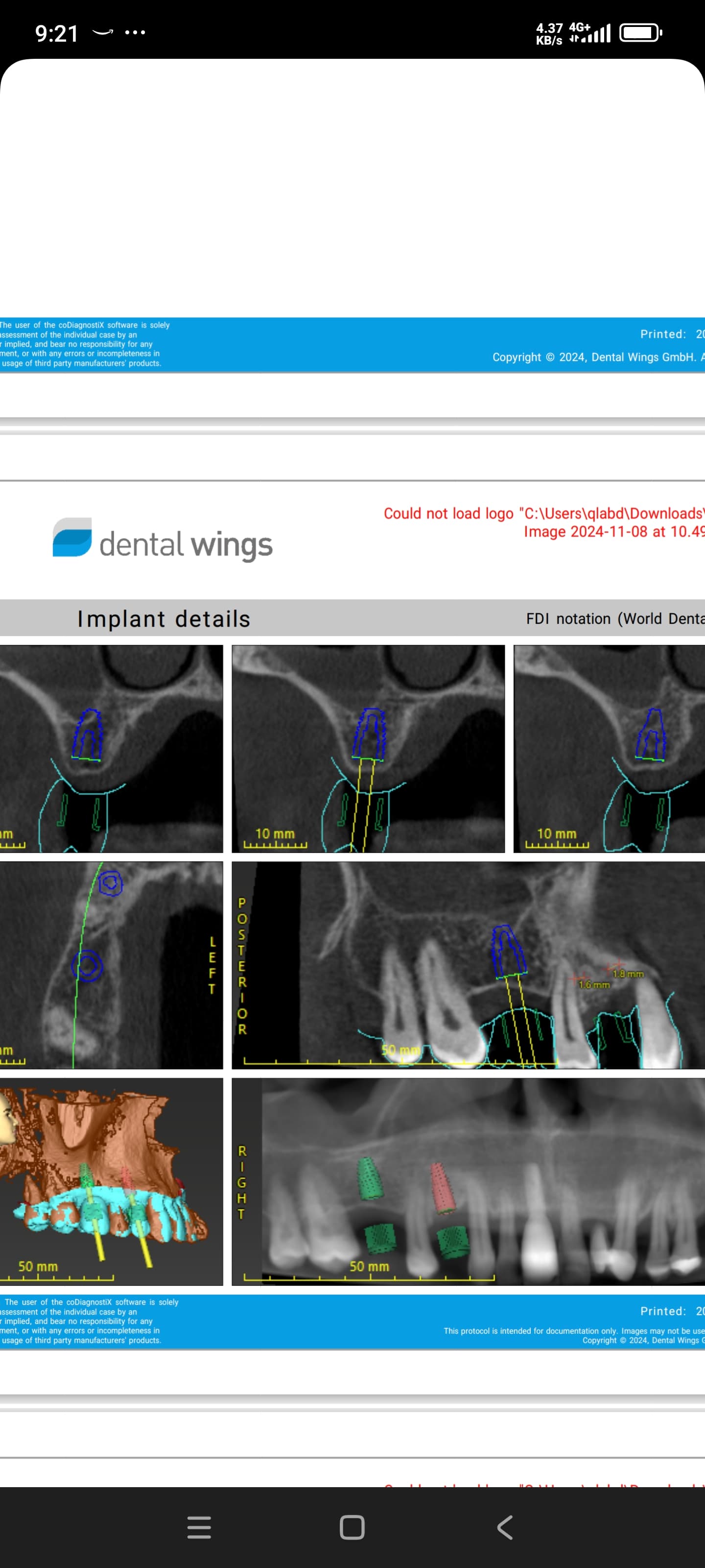
Why not take a CBCT?
Raquel Gonzalez
6/11/2013
When placing immediate implants there could be a risk of a gap between implant and bone, and also if not loaded at the proper bone healing stage could lead to immature bone or granular tissue... I personally don't think there's infection, or damage to the bone should be greater at 15 months! Tell me what you think
Dr John Beckwith
6/12/2013
It appears to be granulation tissue between the plant and bony wall as part of healing from immediate placement, if the space was 2mm or more during placement and was not grafted at that time you run that risk. The implant appears integrated. Go back in remove the tissue, place bone with a membrane, wait 3-4 months and restore
CRS
6/12/2013
That actually is what I would do also for treatment. Next time I hope the poster realizes that whatever he gained on an immediate placement on the front end of treatment is lost on the cost of treating this problem on the backend. Now the patient should get a cone beam to determine if the palate is perforated that is nearly impossible to fix. Implant sites need to be optimally prepared prior to placement. These immediate cases that are placed in perfect bone or get lucky with grafting are not a sure thing when you have to treat the poor healing It increases chair time and profitability when try to do too much at once. It is not like treating a tooth. And the idea that an inert titanium implant will magically cause bone to grow around it is foolish. The blood supply is what makes this happen. Implants can serve as pace maintainers but I don't place immediately unless the extraction site is perfect and there is no pathology. If there is good bone and blood supply yes I have bone growing over the implant.
Peter Fairbairn
6/12/2013
Hi CRS , agree that this is granulation tissue fronm the surface not a retrograde peri-Implantitis issue , there is a fair bit of more current research ( than m showed which is all 95 , 02 etc ) wwwhich is mainly from China . Retrograde peri-implantitis is more an infection at the apical area of an Implant from residual site infected tissue which sometimes is hard to currette out ( close to IAN or sinus ) it there can become acute when the patients defence mechanism is low and result in an acute episode . This can lead to a spontaneous loss of the Implant even after many years and no apparent cervical bone loss or issues . It has happened to me a few times over the years . I have seen acase where the Implant had the apical section removed a la apicectomy but did not see the result long term In this case the bundle bone issue will come into play when immediate placement is used and here grafting using a bio-absorbed material which will then allow for turnover in the future to maintain living bone is critical . regards Peter
CRS
6/13/2013
For lack of a better term I use retrograde meaning backward to describe infection coming from within he implant site vs from the oral cavity. Residual bacteria in the bone from infected teeth, sometimes in the periapical area or in the walls of the osteotomy while sealed off from the oral cavity as this is can cause a perimplantitis. It could also be avascular necrosis. If you do a search you will also find radiographs of radiolucencies mid implant vs just periapical it is just a description it is he same process remember an implant is not a tooth so it cannot get a periapical infection from a diseased root but it is the residual infection within the bone. That is my understanding of the process, but thank you for educating me. Bacteria especially p. gingivitis can remain in privileged sites and is difficult to eradicate just using curettage or chemical means that's a good reason not to place immediate s in chronically infected sites.
Baker Vinci
6/14/2013
There is a lot of cell level speculation. You are probably correct in suggesting that the infection is not from "walled off" bone, that was trapped at the initial procedure. I am an advocate of the immediate implant, with the exception of a questionable infection. You need to scan these cases. Exploratory surgery will become a thing " of the past ". Bv
CRS
6/15/2013
I feel that laser sterilization with a Nd-yag is showing some promise in killing the bacteria in the privileged sites in these cases.
E
6/23/2013
How predictable or successful is waterlase in decontaminating an implant before bone grafting? Could better results be achieved if the implant site is sterilised with waterlase before placing implant? For cases with pathology, would waiting 4 weeks after ext be sufficient or should it be longer?
CRS
6/26/2013
The problem with the Waterlase (erbium) laser is that it will "see" water and hydroxyapatite not pigmented anaerobic bacteria which are the bacteria which hide in the privileged sites of the bone (p. gingivitis) it is great for detoxifying the implant surface since it is dark and the laser won't hurt the surface. However the nd-yag laser at 1064 wavelength can melt the implant surface so one has to be extremely careful. I am new to laser dentistry so please consider that with my comments as I am exploring this newer technology for implants. As always thanks for reading
Sam Jain DMD
6/25/2013
I think primary closure caused this problem. Most probably there was a pin point seeping hole to the coverscrew female hex.... That led to this fibrotic granulation tissue. Why did u primary closure over the immediate implant. You need to place a screw retained temp crown on the implant and not a prim closure. The purpose of immediate implant is defeated if u don't place a SRT.
CRS
6/26/2013
I disagree since the poster stated that there was bone over the cover screw so it is doubtful that the primary closure caused this. I don 't see the logic in that comment.
Seth rosen
6/29/2013
Ummm could be scc. Biopsy.
alessandro aversa
7/23/2013
you need a 3D xray examination to understand more

Featured Products

OsteoGen Bone Grafting Plug
Combines bone graft with a collagen plug to yield the easiest and most affordable way to clinically deliver bone graft for socket preservation.
CevOss Bovine Bone Graft
Make the switch to a better xenograft! High volume of interconnected pores promotes new bone. Substantially equivalent to BioOss and NuOss.