Delayed Bone Resorption around Uncovered Implants?

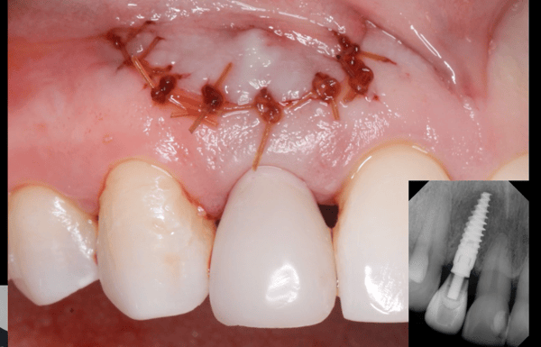
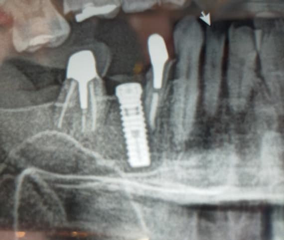
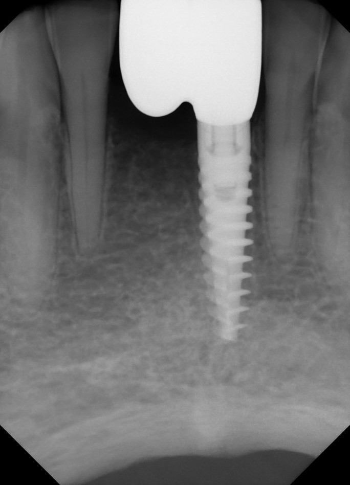
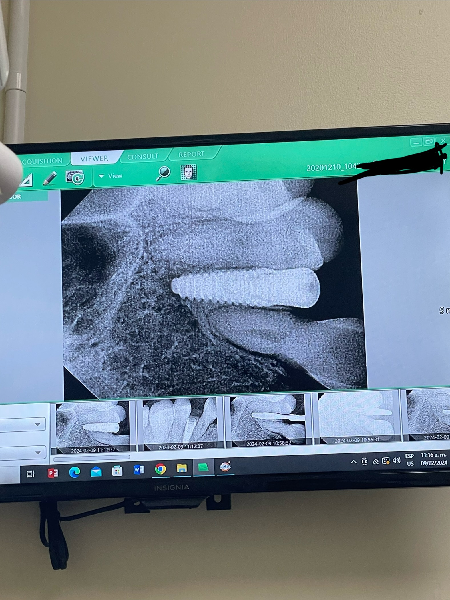
I had placed 3 single dental implants in three different patients with no contributing medical history. All implants are placed in the maxilla, and all implants are Alpha Bio Neo implants. The implants where placed minimum 2 months after the extraction no graft was placed and the torque was not exceeding 50N/cm. Normal healing was observed and a control X-ray was taken 2 months post op with no alarming findings. By the time of the second stage surgery a fistula was detected and no pain was reported by any patient. Xrays revealed bone lost in one case 3mm in the two other less than that . The covers screws where in place and the implants where stable. Do you have any idea why there was such a delayed response? If it was an infection, shouldn’t have it happened immediately after implant placement? Any thoughts here?


27 Comments on Delayed Bone Resorption around Uncovered Implants?

New comments are currently closed for this post.
dr.foukas
9/12/2019
I apologies for my spelling mistakes by :non exiting i mean not exceeding .
Alfonso
9/13/2019
Hi! Did you wash, clean and dry the interior side of the implant after places? Could be hapend, rests of blood stay inside the implant and this descompouse by time. Sorry for my writing.
Alfonso
9/13/2019
Hi! Did you wash, clean and dry the interior side of the implant after places? Could be hapend, rests of blood stay inside the implant and this descompouse by time. Sorry for my writing.
Dr. Moe
9/12/2019
Hi, So in order to figure it out, the next set of questions are: How much bucco-lingual bone was there after the implants were placed (Width) ? How wide are the implants? Were they sub-crestal or not? Was the patient wearing any prosthesis? Was this prosthesis Tissue borne? How good is Patient's home care? This should get us going down some reasons why the bone loss. Unless you have taken all of those things into account, I guess what is Patient's medical status? Among other things. My $0.02
Dr. Moe
9/12/2019
Sorry, Also, I would like to add, were the cover screws exposed when you went ahead for second stage surgery? For more clarity.
Roadkingdoc
9/12/2019
Possibility of heated bone during osteotomy ?
Dennis Flanagan DDSMSc
9/12/2019
Healing caps generally prevent the gingival overgrowth and subsequent plaque collection and infections
Leonidas
9/12/2019
All three patients are healthy the x-ray is from a 34 year old male with bad oral hygiene. Prior to the surgery all dental problems had been addressed. Antibiotic Prophylaxis was used prior to the surgery and primary closure was achieved The cover screws were not exposed in any of these cases this implant it’s 4.2 mm x 11.5 mm alpha bio neo . All Three implants had at least 1.5 mm of bone in all directions no fracturing of the buccal plate was noticed.In one of the cases the soft tissues covering the implant was thinner Than usual But it was not visibly perforated.During the healing process which was normal the Patients Didn’t report any swelling or pain. I personally removed the sutures and the healing was satisfactory. The implant was placed at the level of the crest worst case scenario 0.5mm sub crestal. No prosthesis used as a temporary during the healing phase in all cases .
Proka
9/12/2019
It can be electric activities, and this can accelerate osteoclastic activities
Dorian
9/12/2019
Please add more detail.
Wiiiiam R Dapper DDS, FAA
2/13/2020
Docs “usual suspects “ are always fundamentally “In play”. And should always be respected . Smokers, Diabetics and HTN PTS as well due to compromised healing propensities And they should be identified pre- op. I completely concur with Dr Flanagan ‘a rational for utilizing the appropriate trans mucosal healing abutments-and would also be my choice. However. in this case the cover screws were placed and the flaps sutured , I’m assuming primary closure was attained. If in fact the tension with which the wound was closed was taught and not passive then there is always a risk for avascular flap necrosis. The incision type itself in conjunction with a overly taught closure can result in suture line dehiscence , a very small opening at times , seen only under magnification . Nevertheless this can definitely develop into a problem with the potential to propagate and ultimately compromise the integrity of the Osseous crest and associated keratinized gingiva . As aforementioned above, this small dehiscence can be difficult to visualize at times, however, it can easily act as a portal of entry for Bacteria migration and ultimately result in an infection and subsequent subtle fistula development . The path through the decision making process should address the depth of the fistula , the origin and length of the fistula and the classification of the bony defect surrounding the fixture . Most infections are mixed. Metronidazole and Amoxicillin are a good regimen especially if purulence is Present and the defect is minimal. Obviously the time in which the issue is discovered and the physical nature and extent of the bony defect will weigh heavily on the decision whether to remove it or attempt to maintain it.
canbayrak
9/12/2019
How long did you wait for the second stage surgery. I don't think it's delayed bone resorption but tipical failing Implant radiographs. Can't figure out what went wrong sometimes it happens. Many factors to consider
DrT
9/12/2019
It seems that the bone loss is occurring the entire length of the collar of the implant, which was placed subcrestal. Please describe the collar. Thank you.
Mazin
9/12/2019
I think mostly the cause is over heating for the cortical bone only ( not cancellous )may be due to over pressure during osteotomy , or less commonly over torquing , again for cortical collar only and not the hole osteotomy site.
Dok
9/12/2019
Usual suspects to be explored: Pressure necrosis ( over torque )....... avascular necrosis ( not enough circumferential bone )....... temperature necrosis ( overheating the bone )........medically compromised patient......allergic reactions.......periodontally susceptible ( prone to gum disease)......primary stability issues. Anyone want to add to the list ?
Dr Dale Gerke, BDS, BScDe
9/12/2019
I suspect two main issues. Firstly some delayed bone loss post extraction. My thoughts are that post extraction, the crestal bone levels may seem stable, but the bone may still be subject to vertical loss. Also you do not say if you used a flapless technique, but if you did, there may have been some loss of buccal plate vertical height which you did not notice when you initially placed the implants. Secondly, you mention there was a fistula when you went back for the second stage. I think this is the most likely problem. I would suspect significant (albeit minor) submucosal infection leading to inflammation and consequent crestal bone loss. I note that it has been mentioned by a number of operators that they usually do a one stage technique in preference to a two stage because of issues they have with the two stage.
Suresh
9/12/2019
Could you please share the post extraction cbct or xray images prior to implant placement. .. Was the extraction atraumatic
Leonidas
9/12/2019
Thank you all for your comments . I didn’t use a flap less technique what is unusual for me in this case is the delayed response of the bone to all the factors that you mentioned above . The last X-ray is 4 months after the implant placement . I will post the initial X-ray within the day . I think that over pressure is the main reason for this complication . In this system there is no profile drill so you usually under prepare the osteotomy and you rely on the cutting implant design. But the bone was soft ! Do you think that I should keep the implant or removed it? I think that keeping it and monitoring is the best solution in this case since that is a relatively long implant and significant bone damage is anticipated upon removal .
Dariush Radman
9/12/2019
My best guess is there was an undetected connection between the oral cavity and the crestal bone from the day one which led to infection and caused peri-implantitis . bearing in mind a fistula takes time to show up as well as the bone loss to be visible on X-ray.that is why you found that by the time of 2nd stage surgery. to avoid that, I always examine the ST 2-3 weeks after the surgery to find any pus or exudate leaking from ST, and if I notice that, I will immediately place a healing abutment and the problem get resolved quickly. To treat that, peri implantitis, you may follow the peri implantitis treatment protocols ( one of them set by Dr.John Suzuki ). Good Luck
Vipul Shukla
9/13/2019
I think I agree with Dr. Alfonso. After the implant was surgically placed, you may have trapped blood and other biomass inside the implant chamber and since your cover screw does not appear to be watertight, this decomposing detritus found a way to escape around the cover screw creating superficial pus and fistula, typically this takes 6-10 weeks after placement. I doubt this implant will fail. My suggestion: Open via Stage 2, clean the inside of the implant with a bonding agent applicator brush dipped in Chlorhexidene mouthwash, then stimulate some bleeding around the implant crest with an explorer tip, remove obvious granulation, and add a sterile and dry anatomic healing abutment, suture tight around it, and leave alone for 6 weeks. Things should get better. Good Luck!
Dr Dale Gerke, BDS, BScDe
9/13/2019
In regards to your question about whether to remove the implant/s. It is difficult to decide this in the forum. If the implants can be reverse screwed out then it might be worthwhile. Simply replace with a new implant positioned to the crestal bone height. However it maybe that after placing a healing cap, the soft tissue will be stable and cover the implants. It seems to me this will be possible in at least 2 of 3 patients (where the bone loss appears to be minimal). If you can obtain this result then it may be worth leaving the implants but of course you would have to give the patients a guarded prognosis of long term survival. However if the implant protrudes through the soft tissue then I think it would be wise to remove the implant since you will not likely have a long term result and that is (after all) what the patient expects. The reality is that once the threads of the implant are supragingival, it is notoriously difficult to maintain the implant and thus the implant will almost certainly fail long term. If you have to remove the implant, you should be able to minimise bone loss by reverse screwing the implant (opposite to placing the implant) and if this is not possible (say around 50N) then use a trephine which is just a little wider than the implant and drill down about half the implant length and then try reverse screwing the implant again. Continue this method until you eventually succeed. If you can reverse the implant out and have some bone remaining then you should be able to place another implant immediately – possibly using some allograft. You mentioned the bone was soft, so I would guess that reverse screwing should work relatively easily.
Dorian
9/14/2019
Hi Dr. Gerke, Once the implant has been removed by a trephine the osteotomy site is considerably wider. Please expand on your technique of placing an immediate implant in this situation. Perhaps with Osseodensification? If a wider diameter implant is installed it sets up other issues like amount of surrounding bone. I am curious to learn how you do this.
Yossi Kowalsky
9/14/2019
I've seen similar problems . wish we knew . its quite frustrating. no one really answered you because we don't know.
Leonidas
9/15/2019
Thank you all for your input it is very good To hear different opinions share our knowledge and experience .Apparently as most things in dentistry this complication is the result of many factors most likely tissue residuals and over pressure in my opinion . I will monitor my patients better and i will change few things during implant placement . I decided to keep the implant inform the patient And be more conservative.
Dr Uzman Haq
9/16/2019
check vitamin d and cholesterol. I've seen late bone loss which has been resolved with vitamin d supplementation in individuals with deficiencies
george
9/17/2019
what kind of vitamin d levels?
lisa
9/17/2019
Yes, please share more details on Vit D levels that could cause this delayed bone loss and how much Vit D supplementation is suggested. Thank you.

Featured Products

OsteoGen Bone Grafting Plug
Combines bone graft with a collagen plug to yield the easiest and most affordable way to clinically deliver bone graft for socket preservation.
CevOss Bovine Bone Graft
Make the switch to a better xenograft! High volume of interconnected pores promotes new bone. Substantially equivalent to BioOss and NuOss.