Dr. Scott Ganz on CT Scanning
Dr. Scott Ganz is a maxillofacial prosthodontist who has pioneered the development of CT scan use in implant dentistry. He maintains a private practice in Fort Lee, NJ and is an Associate Clinical Professor of Prosthodontics and Implant Dentistry at the College of Medicine and Dentistry of New Jersey.
Osseonews (ON): Many of the world-class clinicians in implant dentistry that I have interviewed in the past have criticized the undergraduate dental school curriculum for failing to provide basic training in placing and restoring implants.
Dr. Ganz: I agree. The most significant problem that I see is that undergraduate dental students do not receive adequate training in placing implants and restoring implants. When they graduate from dental school they need to have this skill set. Dental students are given the tools to diagnose decay, periodontal disease, endodontic therapy, crown and bridgework, etc. They should know how to diagnose for an implant. Dental implants are mainstream dentistry today.
ON: What about the argument that dental schools do not have room in their timeline for teaching about implants.
Dr. Ganz: Implant therapy is just like any other area of dentistry. In fact, in my practice, it is always the first treatment option for a missing tooth. Treatment planning for any edentulous area must, in my opinion, include consideration of implant therapy. This is a medical-legal issue as well. Patients must be given the opportunity to choose the appropriate treatment option. Ideally the dental student should be exposed to dental implant training while an undergraduate, not after leaving dental school. That’s my political statement for this interview.
ON: Is there a particular area of implant training that you feel the undergraduate dental student should master before leaving dental school?
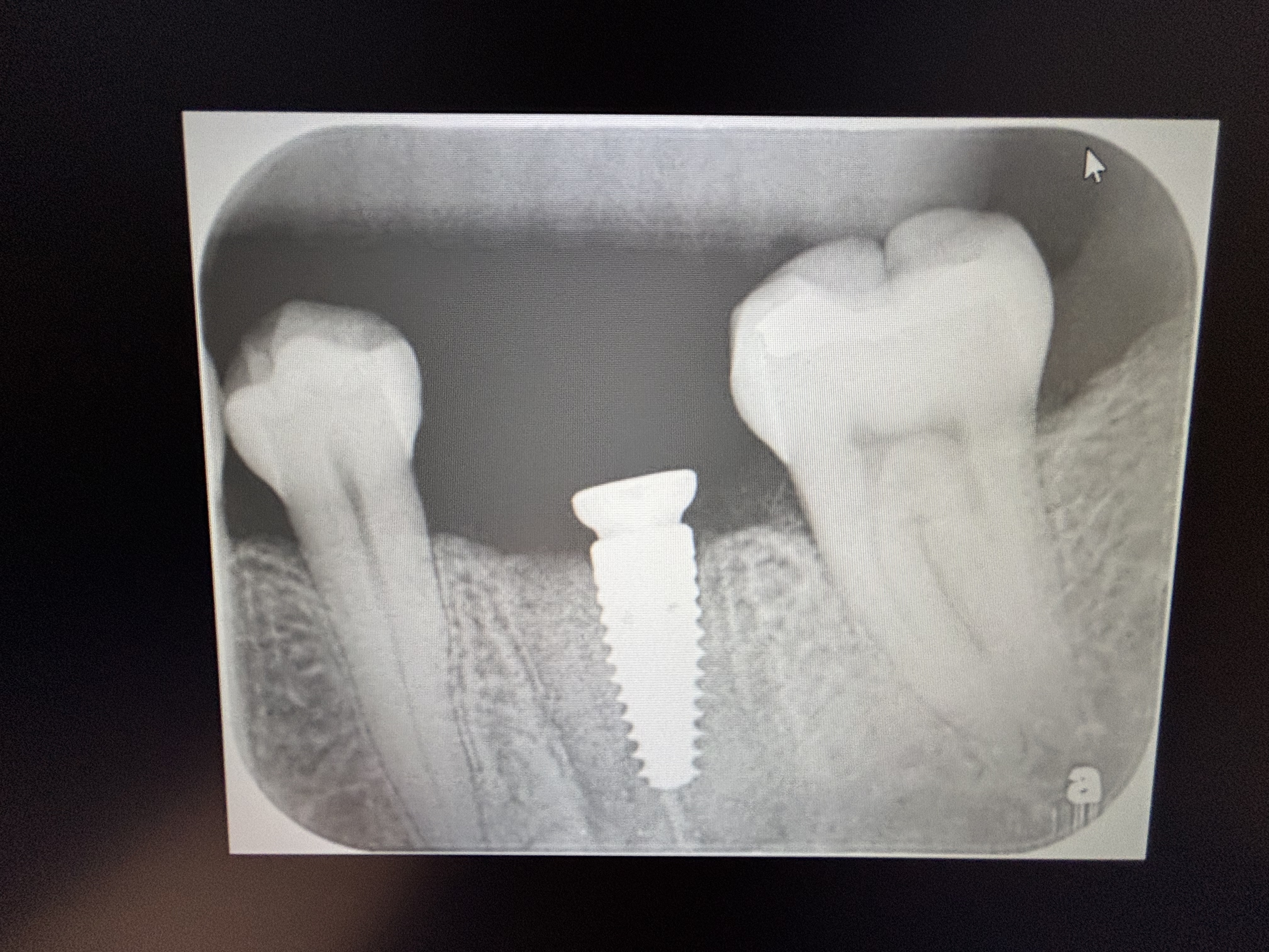
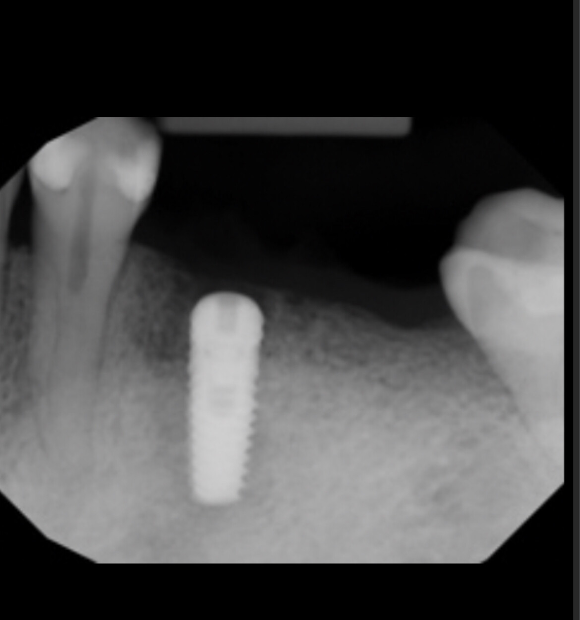
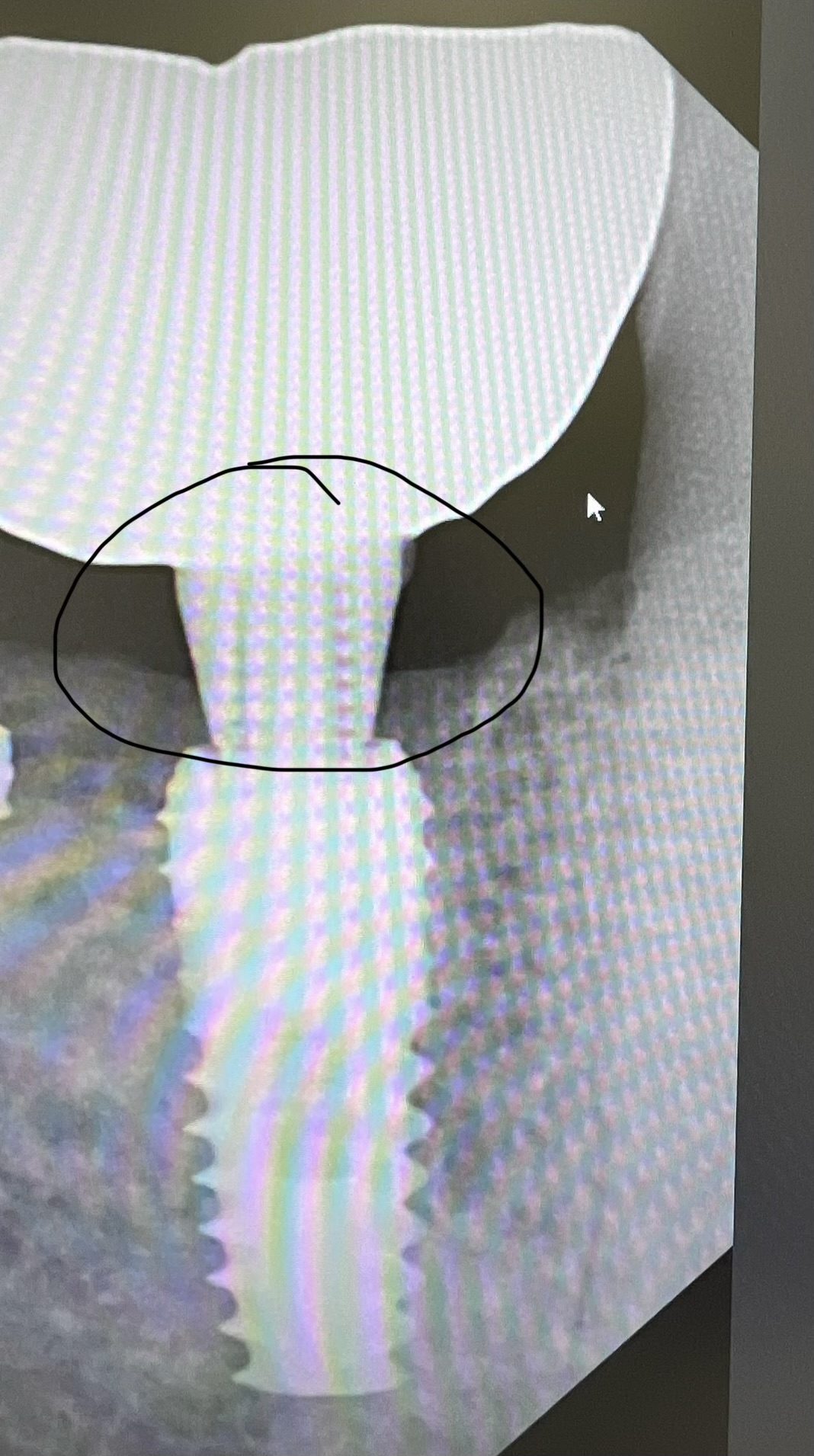
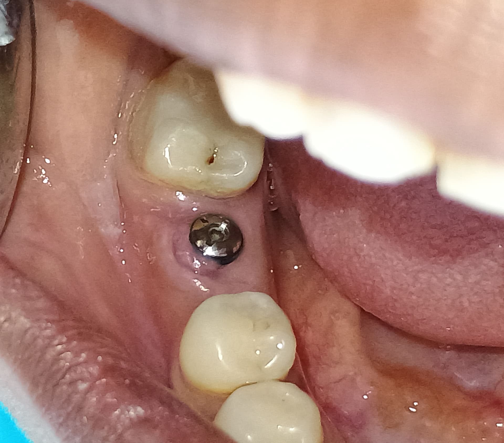
Dr. Ganz: How to read a CT scan. The dental student – whether undergraduate or post-graduate – should possess the skills to read a CT scan just like he reads a panoramic or periapical radiograph. A CT scan can be used to accurately analyze the bone quality and morphology and is far more accurate in assessing important anatomical structures than conventional imaging techniques. CT, with the 3-dimensional reconstruction available today, gives us incredible insights into the patient’s anatomy, not just for implants, but for other procedures as well. Of course you can use the CT scan to reproduce the anatomy of the bone to determine the most advantageous receptor for placing the implants.
ON: So this makes treatment planning as accurate a procedure as possible.
Dr. Ganz: Exactly. With the CT scan there is no guess work. You do not need to estimate. All of the vital anatomy is available in multiple views, prior to surgery. Everything is about as exact as we can have it today. A technique I pioneered involves taking the scan data and producing a stereolithographic model. The model can be examined in your hand allowing all aspects of the jaw to be evaluated. Taking this to the next level, you can place replica implants into the model, guided by the CT derived template, giving the laboratory the basis to create provisional or permanent restorations prior to the surgical intervention. At the time of surgery, the surgical template is used to place implants exactly where you want them, nice and parallel. For immediate load cases, the prefabricated prosthesis can be delivered with greater efficiency, in less time with less patient morbidity.
ON: Do you foresee a major paradigm shift in implant dentistry where a CT scan will routinely be used in the manner described?
Dr. Ganz: When the panoramic radiograph first appeared you would generally find it first in the oral surgeon’s office. Now most dental practices have one. For those practices where a relatively high volume of implants are placed I think you will soon witness the appearance of CT machines, especially now that lower priced Cone Beam CT machines are available .
ON: Could you summarize what you consider the crux of the issue is for the dentists now engaged in placing or restoring implants?
Dr. Ganz: Just remember, “It is not the scan, but the planâ€. The dentist must be able to fully understand and utilize the information contained in the CT scans to generate accurate treatment plans from software applications like SIM/Plant (Materialise, Glen Burnie, MD). Assessing the desired tooth position in relation to the underlying bone can be accomplished when the patient wears a radiopaque scanning appliance during the scan. Remember the “goal of implant dentistry is the tooth that we replaceâ€. It is then the CT generated plan that permits the optimal placement and restoration of implants via surgical guides, virtually eliminating the problem of malposed implants, making implant dentistry far more predictable, which will eventually result in higher volumes of implants placed, and potentially reducing the cost for the patient.