Evaluating the Vertical Dimension, RP5 Design..
Dr. Jameson is a board certified Prosthodontist who has done considerable work in disseminating information concerning the concept of linear non-interceptive occlusion. He was a consultant in Prosthodontics to the Surgeon General, USAF prior to his retirement from active duty and has been a consultant to the Department of Veterans Affairs. In this third interview, Dr. Jameson discusses some important topics in implant prosthodontics.
Osseonews (ON): For simplicity sake, let us assume we have a patient with an implant supported mandibular overdenture who now wants a more stable maxillary complete denture who is wiling to accept implants and the modifications necessary to produce a nonlinear interceptive occlusion. Let us also assume that the patient currently presents with 30-degree teeth arranged on a Curve of Spee and Curve of Wilson and has bilateral balance. What steps would you take to evaluate or establish the vertical dimension, centric relation and the plane of occlusion?
Dr. Jameson: My approach is predicated on the evaluation of the condition and estimated longevity of the existing prostheses. We will assume the fit is adequate and occlusal wear to be minimal with a life expectancy sufficient to warrant modification and use after implant fixture placement. The occlusion can best be evaluated on an articulator. To this end, I would duplicate the prostheses and reproduce in improved stone. I would then attach a striking plate parallel to the occlusal plane in the palate of the maxillary prosthesis and the bar/scribing pen in the central bearing area of the mandibular prosthesis. Vertical dimension of rest is determined and the pen opened or closed to contact the plate at this position. An intra-oral needlepoint tracing is then accomplished at vertical dimension of rest and buccal stone indices made. The stone replicas are then attached to an articulator using the stone indices and the occlusion analyzed. This permits a more leisurely and hopefully thorough evaluation. Discrepancies can then be eliminated intra-orally by closing the pen down gradually and adjusting premature contacts until centric occlusion and centric relation are synonymous.
To evaluate the existing plane of occlusion, I would observe the position of the dorsal aspect of the relaxed tongue relative to the mandibular occlusal surfaces. It should be level with, not below or above said plane. For an anatomic arrangement such as mentioned, the occlusal plane should be from half way up the retromolar pad in the posterior to the cusp tip of the mandibular canine in the anterior. I would also verify contact of the musculature at the corner of the mouth to be in contact with the buccal surface of the first premolar when the mouth is opened slightly.
In determining the acceptability of the existing maxillary prosthesis, it should also be evaluated as to its esthetic attributes. Does it provide the desired lip support from a frontal as well as profile viewing? Are the anterior teeth properly positioned and visible appropriate for age and gender? Do the incisal edges contact the lower lip to perform properly during speech? Also, the position of the maxillary anterior teeth labial to the residual ridge will influence the optimal number of implants necessary to provide the stability desired by the patient. With an anatomic posterior tooth form, vertical dimension from rest position is customarily closed from 2-3mm to provide interocclusal rest space, an anterior vertical overlap routinely occurs. If the maxillary central incisors have been placed 8mm labial to the middle of incisive papilla, to offset rotational movement around the maxillary residual ridge with protrusive contact, posterior implant retention is advantageous.
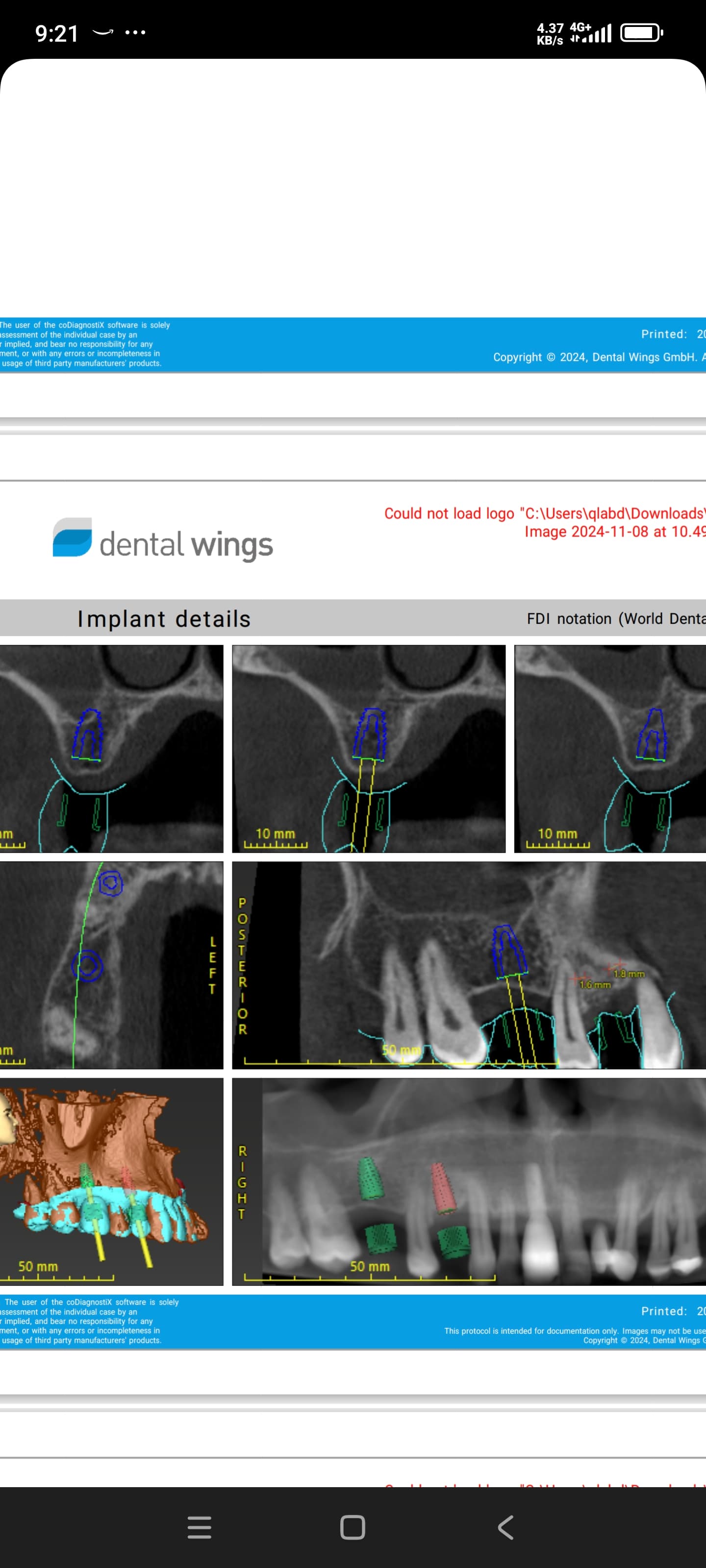
If all the afore mention criteria are positive, I then would place ball bearings on the denture in those locations of potential implant placement and obtain a tomograph radiograph with cuts in these locations. This allows a more accurate evaluation of the bony undercuts, sinus locations and quality of bone available than with a panographic radiograph.
ON: One common way to establish the orientation of the plane of occlusion in an edentulous patient is to use the inferior aspect of the alar of the nose and the most superior point on the tragus of the ear. Some use the upper third of the tragus. What is your protocol?
Dr. Jameson: With the patient standing erect, I make certain the wax rim from canine to canine is parallel to the horizon and even with the upper lip. The maxillary anterior teeth are arranged on this wax rim as dictated by age, gender and personality with the central incisor incisal edges parallel to this wax rim. Vertical dimension of rest is determined and an intra-oral needlepoint tracing developed and registered on separate recording bases. This recording is used to mount the casts in the articulator. Once this is accomplished, the recording bases are removed and the wax rim with its anterior arrangement is placed on the maxillary cast. A flat plate in then positioned from the top of the retromolar papilla on either side to the incisal edges of the maxillary central incisors. These three points constitute the horizontal occlusal plane when linear occlusion is employed. If an occlusal scheme other than linear occlusion is to be used, the vertical is closed the customary 2-3 mm, the lower anterior teeth arranged with the desired vertical overlap and then the lower posterior teeth arranged using the tip of the canines and the retromolar papillae to establish the occlusal plane.
ON: If the patient has a square arch form, optimal bone quality and bone volume in the maxilla and premaxilla, would you recommend the placement of two free-standing implants in the canine areas for retention?
Dr. Jameson: This may sound simplistic, but I never recommend placement of implants unless they are needed. But if retention is lacking or the patient needs that added security that can be provided through implant retention, then two freestanding implants will usually suffice. If there had been considerable loss of the anterior residual ridge, placement of the central incisors back where they were before loss, usually on average 8mm labial to the middle of the incisive papilla, lack of anterior support results in functional rotational problems and could necessitate additional implants. But in this scenario, additional anterior implants would do little to prevent posterior downward rotation. Unfortunately, placement of molar implants are frequently complicated and compromised by the maxillary sinuses especially with sinus enlargement frequently seen with advanced combination syndrome.
ON: In general, would you recommend an RP5 design for a maxillary overdenture where the prosthesis is supported by the tissue and retained by the implants?
Dr. Jameson: As I have said, if the maxillary denture is adequate in fit, function and aesthetic quality, then two implants would be sufficient for retention with a tissue supported overdenture. Implants should never be considered as anything other than a supplemental tool in our restorative armamentarium and definitely not as a means of compensating for poorly designed or improperly fabricated prosthesis. What frequently happens is that a patient has overcome a troublesome mandibular complete denture with two or more implants for retention and now is more conscious of any movement in the heretofore trouble free maxillary prosthesis.
Gary J. Kaplowitz, DDS, MA, MEd, ABGD
Editor-in-Chief, Osseonews.com


