Fractured Upper Molar Case: Bone Regeneration and Sinus Lift

Case submitted by: Dr. Gerald Rudick with the assistance of Arta Prenga & Shlomit Dayan

Forty year old female in excellent health presented having pain in the upper right quadrant. Xray shows tooth #16 ( 3) had previous endodontics, post and core and a crown several years before and there is evidence of a root fracture….. the prognosis of keeping this tooth is hopeless (Fig. 1).

Following local anaesthesia , the tooth was extracted flaplessly with a conventional forceps. However, the apical portions of the two buccal roots separated. With a periosteal elevator, one of the broken roots was easily dislodged and removed, and the remaining root tip left and will be removed at the time of the bone grafting surgery. The patient was given antibiotics and analgesics and told to return in a month’s time so that some healing and detoxification of the socket can take place, as well as soft tissue closure. A full thickness flap was opened showing the amount of destruction that had occurred because of the apical pathology, and a clear view of the remaining root tip (Fig 2). It is important to note that unless successful bone regeneration can be accomplished to correct the defect, there is a strong possibility that the adjacent teeth will be lost.

Prior to opening the flap, 4 vials of blood were drawn in empty red capped tubes and spun in the centrifuge to obtain fibrin clots . The clots were then pressed to make membranes (Fig. 3).The fluid that was extruded from pressing the fibrin clots is Fibronectin and Vitreonectin, which is used as the wetting agents for the particulate graft mixture of Osteodemin, Allosorb and Osteogen, with a sprinkling of Metronidazol powder. The graft mixture is compacted into the defect and covered with the sticky fibrin membranes which are rich in growth factors. They also prevent the graft from being washed out (Fig .4 ).

A piece of titanium mesh is cut and folded to a saddle shape to fit over the graft site (Fig. 5). The titanium mesh was not screwed down , and it is covered with a piece of PTFE membrane to block the holes of the mesh while bone formation is taking place.The gingival mucosa was approximated as much as possible but because the area is bulked out it was not possible to get complete closure of the soft tissue. Gut sutures were used to secure the soft tissue (Fig.6).The disturbance to the soft tissue sets up RAP ( Regional Acceleratory Phenomenon), as described by Carl Misch some years ago, and this disturbance can speed up healing 2-3 times. The soft tissues will mend and close to a greater extent. As time goes by in the four month healing period, however, the titanium tends to become exposed and the PTFE that was covering the pores in the metal gets sloughed off….but new bone is growing under the titanium mesh, even though it is exposed. If the titanium becomes exposed, it could be annoying to the patient, however the irritating area of the mesh may be folded or cut off.

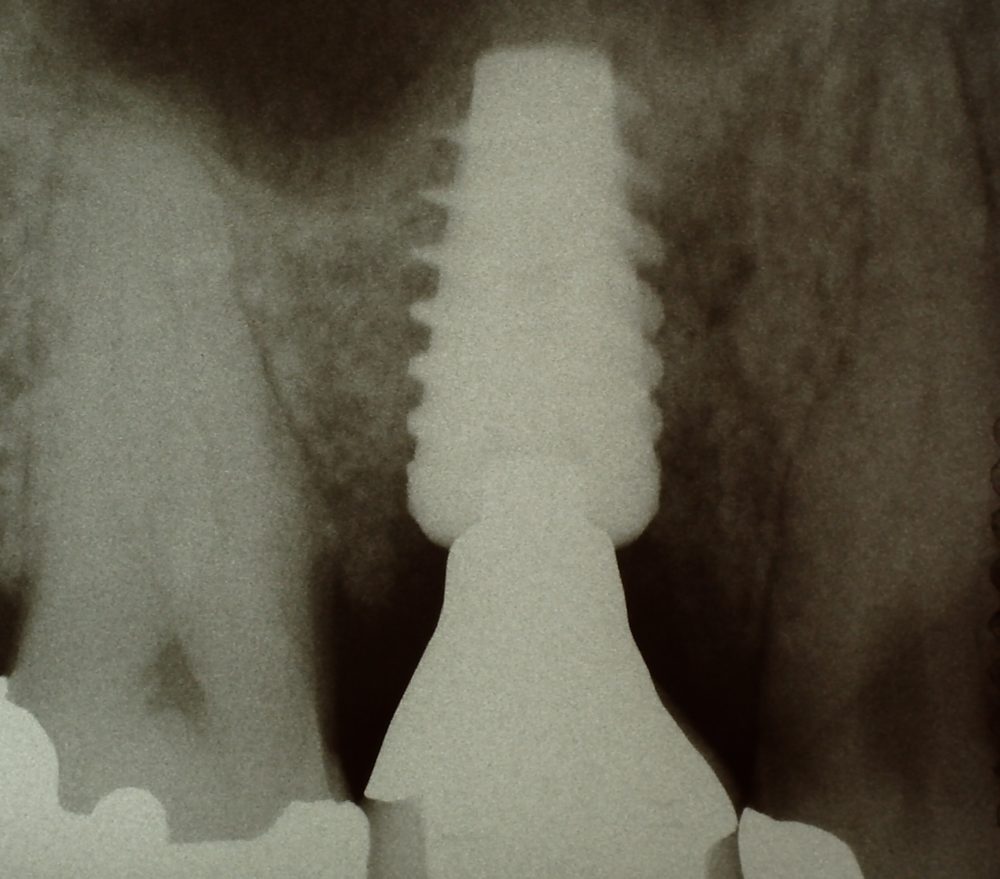
When the the four month healing period has expired (fig 7 ), the mesh which is completely immobile, is ready to be peeled off, (fig.8) revealing new bone that has developed, and the soft tissues are resutured giving complete closure. After another month or two allowing for further mineralization of the newly developed bone, an osteotomy is prepared in a closed technique, using conventional drills to the limited of the floor of the sinus, and with Osteotomes, the sinus membrane is lifted slightly,and a fibrin plug was placed in the hole with some grafting material, before the implant is screwed into place. ( fig. 9), which is evident in the xray. Waiting 3-4 months after implant placement, an impression is taken and a porcelain to metal screw retained crown is fabricated ( Figs. 10, 11).

Fig. 12 demonstrates the different steps that were taken. This case is now five years old, and is working out very well. It is important to note, that when the titanium is folded to a saddle shape, it is not necessary to screw it down, and is easier to remove when the healing period is completed.


My beautiful picture


![]prognosis of keeping this tooth is hopeless](https://osseonews.nyc3.cdn.digitaloceanspaces.com/wp-content/uploads/2019/01/19579-24-10-c461962c473b-e1548108377916.jpg)prognosis of keeping this tooth is hopeless

17 Comments on Fractured Upper Molar Case: Bone Regeneration and Sinus Lift

New comments are currently closed for this post.
Dr Dale Gerke, BDS, BScDe
1/22/2019
An interesting presentation thank you. Obviously relatively messy, but the end result seems very acceptable. Can you clarify; the photo with all sutures in place shows a bit of white material exposed. I presume this was the PTFE membrane but could you confirm?
Dr. Gerald Rudick
1/22/2019
Exposure of the titanium mesh in these type of cases, is not uncommon, and does occur even if the mesh is tacked down or screwed down. …….patients need to be reminded of conventional orthodontic cases, where a lot more hardware is exposed during the treatment; and that oral hygiene is the key to a good success, along with using Peridex daily to keep the mesh relatively bacteria free.
Andy
1/22/2019
Turned out OK. I have some not so pleasant experiences with Titanium Mesh. I would have placed the Collagen barrier membrane UNDER the titanium mesh and overlaid the mesh with PRF membranes. If the PRF becomes exposed.....no worries and it facilitates faster healing of flap. Also I'm not familiar with your bone allograft but, if I have a Schneiderian membrane perf at final diameter that I am not going to then access and repair with Tatum procedure, I will place a wad of PRF and bone graft the osteotomy well short of sinus floor or I will select shorter implant.
Dr. Gerald Rudick
1/22/2019
Many dental implantologists are not particularly thrilled with Titanium Mesh…. it is sort of a love/hate relationship.....but the end result usually turns out well. In this case PRF was used under the titanium mesh as it was protecting the graft material, and PTFE is stronger than collagen, and it was used to cover the mesh and block the holes in the mesh....while bone was forming under the mesh. The Schneiderian membrane did not appear to be damaged, and the PRF plug is used to protect the membrane from the graft mixture that was placed into that portion of the osteotomy that was in the sinus cavity to protect it as the implant was screwed into place.
Doc
1/22/2019
Although you received a good result I do need to question your approach. Not tacking down the titanium mesh, and leaving it exposed is a bit risky wouldn't you say? Although a good outcome this personally would not be my approach. I would definitely stabilize the membrane and get primary closure. I think you got lucky here.
Dr. Gerald Rudick
1/22/2019
When possible, I would tack the membrane down, but if you look at the first intreatment photo, it is clear that there is no place to screw to without damaging the adjacent teeth....the bone was blown apart by the pathological situation. The operation was done one month after the tooth was extracted, and the soft tissue allowed to close the extraction socket.....but remember, when the graft material and the mesh were placed, the area was bulked out, and the soft tissue was not placid enough to connect the edges of the cut together completely....Placing a piece of a free gingival graft to completely cover the titanium mesh and connect the two edges of the cut would not work at that stage of the process. ….perhaps if a CT scan was done, and a virtual model was made in advance of the surgery, a balloon device could be placed over the palatal tissue, and inflated a couple of weeks before the surgery, to stretch out the soft tissue so it would be pliable enough to stretch it over the site....but this was not the case.
Yossi
1/23/2019
Gerry , Your the MAN. Thanks for showing not perfect work . That's how we learn . Not from Photoshop of the one perfect case that's not routine. I almost always section upper molars to avoid root tip fractures. I do not use mesh. i have been having very good result s with Osteogen plugs . I find 4 months is minimal time to wait . i go towards 6
Dr. Gerald Rudick
1/23/2019
Hello Yossi, your observations are always spot on...… I did not section the tooth in this case at the time of extraction, because a root fracture was suspected..... so I don't think it would have made a difference...because those roots were probably already separated. I think Osteogen plugs are very good and I use them routinely, but in this case the damage to the bone was so extensive, I wanted to overfill the site......all the best Gerry
roadkingdoc
1/23/2019
Dr Rudick, thanks for posting a difficult case with a very nice result. We can all learn something from it. Keep up the good work!
Dr. Gerald Rudick
1/23/2019
Dear Roadkingdoc, I appreciate your comments, and from what I read about you, I take this as a compliment...so thank you.
DrK
1/25/2019
Thankyou for showing us all this case. I have one doubt, was this case pre planned for a particulate bone graft and membrane or was that decision made after the fracture of the buccal roots? If it was preplanned would it be ok to use luxators/elevators to loosen the tooth a bit before applying forceps so that maybe the root tips even if fractured during extraction are easy to remove in the same appointment rather than scheduling it with the graft placement appointment. Regards
Dr. Gerald Rudick
1/25/2019
Dr. K....here is a response to your questions....nothing was preplanned.....from the patient's symptoms, the xray and a physical exam, I knew the tooth had to be extracted....and when it came out, very easily, it was then that I saw the missing portions of the roots. One of the roots was easily removed with an elevator, and the other, purposely left in place during the month that was left for some healing to take place, the soft tissue to close, and the bacteria and pathology to be sloughed off. It was on the second appointment, when a full thickness flap was opened, did I see the extent of the damage to the bone, as seen in the first photograph. I knew the bone had to be rebuilt, and I chose to place a titanium mesh molded into a saddle shape to contain and guide the bone graft......perhaps if there was a CT Scan taken prior to the extraction I would have seen the problem sooner.... but it made no difference, as what was done on the second appointment had to be done. Thank you for your enquiry, and I hope I answered your question.
DrK
1/27/2019
Thank You. Yes it does answer my question. Regards.
roadkingdoc
1/27/2019
For what its worth I section and remove about all first molars. Careful to conserve as much surrounding bone as possible. If the patient declines a graph,I still feel I have done them a good service.
Dr. Gerald Rudick
1/28/2019
In this particular case, it would not have made a difference....the photos show you the damage which was there long before the extraction......
roadkingdoc
1/28/2019
Dr R,I think you handled the case excellently. I am not doubting your clinical judgement. I said about all first molars I section. this would not have been one. I practice in rural Indiana and remove many teeth unfortunately.I think the sectional and removal is a skill all dentists doing removals should have in their arsenal. Please keep posting cases. I have practiced for 41 yrs and am still learning!
Dr. Gerald Rudick
1/28/2019
Dear Roadkingdoc….I agree with you...… better to cut up the tooth to be extracted rather than accidentally fracturing the alveolar ridge.

Featured Products

OsteoGen Bone Grafting Plug
Combines bone graft with a collagen plug to yield the easiest and most affordable way to clinically deliver bone graft for socket preservation.
CevOss Bovine Bone Graft
Make the switch to a better xenograft! High volume of interconnected pores promotes new bone. Substantially equivalent to BioOss and NuOss.