Gaining attached tissue while uncovering implants: best technique?
I have a patient who had 2 implants installed. In order to achieve primary closure the flap was pulled up to cover the alvelar ridge increasing the zone of unattached tissue. Now the implants have osseointegrated and are scheduled for uncovery. What is the best technique to increase the zone of attached gingiva around the implants? Pedicle flap?
6 Comments on Gaining attached tissue while uncovering implants: best technique?
New comments are currently closed for this post.
CRS
9/4/2013
I would do a connective tissue graft prior to implant exposure or a free gingival graft. It is really hard to advise without a photo. Where would you pedicle the flap from? Is this mandible maxilla esthetic or posterior? Post a photo!
Peter Fairbairn
9/5/2013
This is one of the most important aspects for long term stability of peri-implant bone especially in the lower 6 area .
It is so case dependant , hwether upper or lower , What adjacent tissue i like , Tissue biotype ? etc so hard to give you a definative answer without more information .
The upper generally best to bring tissue from the palatal aspect using split and then full thickness incision to move the attached tissue buccally .
The lower is more difficult and may in extreme cases necessitate the use of a palatal tissue graft and the use of soft tissue screws to resist muscle pull .
But as I said very case dependant so more info please.
Peter
mike
9/9/2013
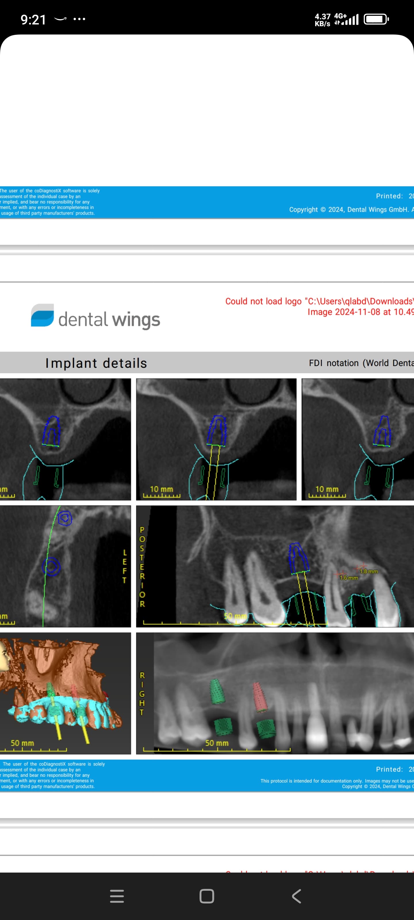
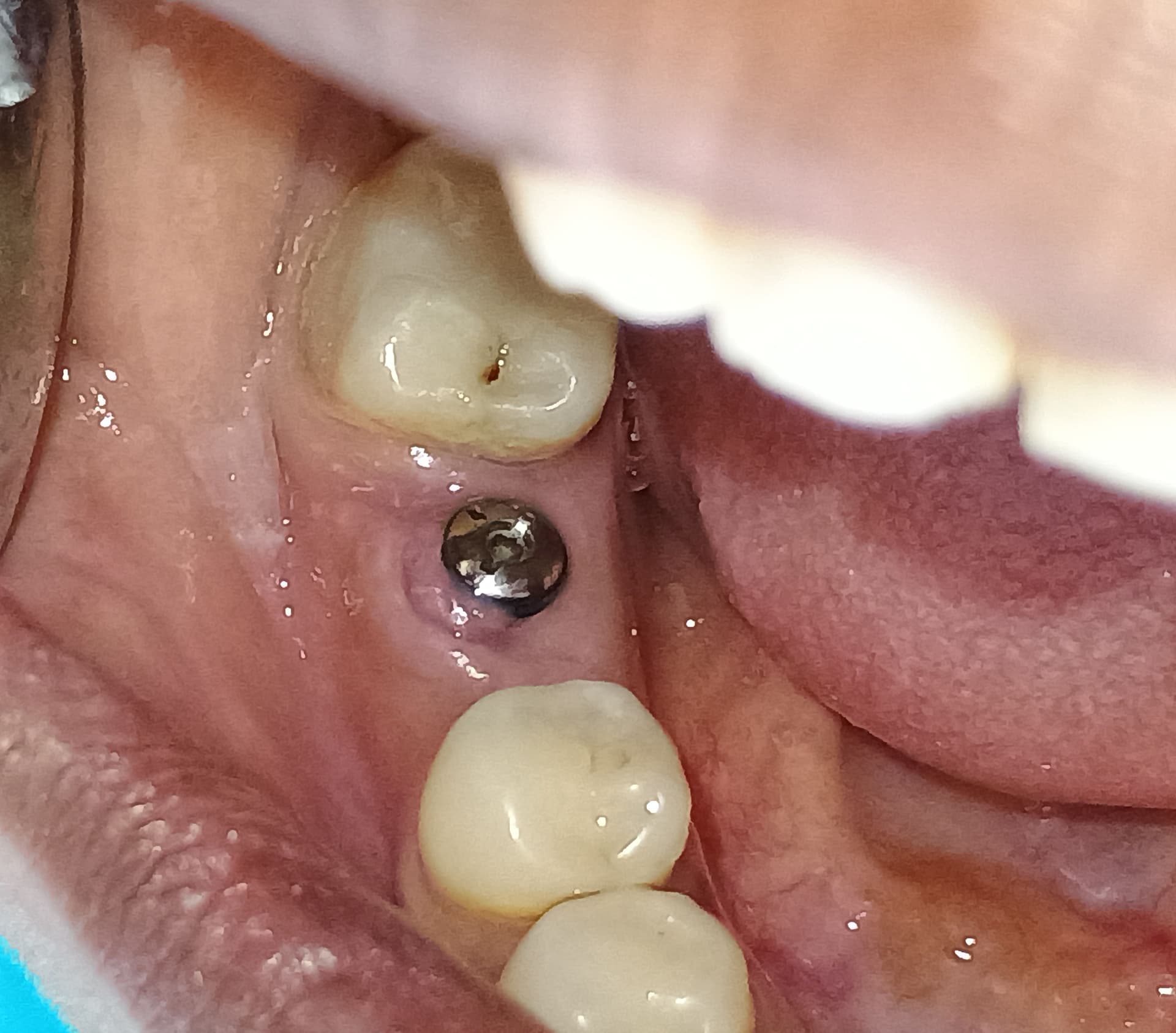
Thanks everybody for your reply. Sorry no photos, I guess you can't read my mind....Anyway, the site is on the mandible replacing #18-19. The mucogingival line is on the top of the ridge. I was going to do as you suggested by taking a split thick flap from the lingual, uncover the implants then go full thickness and reposition the flap. My concern is what happens to the denuded bone interproximal and potentially lingual to the implants. Also, I can harvest some connective tissue and place it under the labial flap, but the flap there is full thickness. what happens to the CT blood supply with no periosteum underneath? As always, thanks for the advice..
Peter Fairbairn
9/10/2013
Hi Mike , it is generally this area where the most difficult issues are to correct and as you know correcting them is important for a better long term result.
It is hard to imagine and as all cases are different hard to advise .
The area lingulal to the implants should be split thickness so the periosteum is retained and it will regenerate .
If you have experience with palatl harvesting , that can work well when used with soft tissue screws as I said , but be wary as never to far from failure here
The last option is use a Collagen type fleece or even a Matribone type product which is collagen with HA .
Peter
mike
9/10/2013
thanks for the reply but after making this slpit thick flap, what do we do with the exposed bone interproximally. I can do a free gingival graft and suture into place in between the implants but what of the blood supply to it the graft??
John Manuel, DDS
9/10/2013
The best solution to lack of attached tissue is to address this potential before placing the implants.
For example, if you are seeing an edentulous, thin mandibular ridge with attached tissue margin at the ridge crest, a good plan is to use a "2 Stage, Windowed, Ridge Split at implant placement (possible with Bicon 4X5 or 4.5X6 short implants) or before placing standard implants.
After measuring and planning - 1st Surgery is to lift a Buccal flap from a crestal incision within the attached zone, cut the "window" through the cortical plate only, along crest then down front and back and connect the cuts above the external oblique ridge, leaving a long, slightly rectangular "trap door" . Replace the flap and have the patient return in about 3 weeks. (I know some prefer 4 weeks, but the callous can get pretty firm by that time).
At the 2nd appointment, the crestal cut alone is "re-incised" making certain to leave 1-2 mm of attached tissue along the edge of the Buccal flap edge. NO REFLECTION FROM THE SIDE OF THE RIDGE IS DONE! THIS WOULD ELIMINATE CIRCULATION TO THE NOW MOVABLE "TRAP DOOR". A fine chisel is used to gently tap and cut the medullary bone enough to allow the previously outlined window (trap door) to be "bumped out to the Buccal - similar to an old Ford Model A Rumble seat.
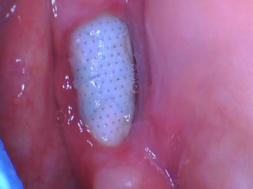
The tiny, short implants are then placed into the open slot with about half or more of the implant stabilized in preps in the open groove base, with the implant tops about 3 mm below the crestal bone edge. Granular and autogenous graft particles are filled over the sunken implants, and halved or quartered Collagen plug strips are gently sutured over the tops to allow clotting before epithelial invasion.
IF the patient has adequate width attached ridge crest tissue, the crestal slit is gently brought to full or almost full closure, this allowed by the flexible callous outlining the movable window after you've cut ONLY the crestal slit.
IF the patient has inadequate width of crestal ridge crest tissue, the crestal slit is not full closed, but "a lot" of 5-0 Chromic gut sutures are placed to hold the collagen strips over the graft atop the implants while the crestal slit heals by secondary intention. This will leave you with 4-7 mm wide brand new attached crestal tissue width, right where you need it!
There are some videos on Bicon.com. This is not a new procedure.


