Immediate temporization failure to integrate: any thoughts?

Virgin tooth #4 in this 60 y.o. male fractured vertically and was extracted atraumatically. Immediate implant placed to the crest of buccal bone or about 3mm below the gumline. Socket was thoroughly curetted out, but no osteotomy drills were ever used because the socket had almost the perfect dimensions to place the 5.0 X 11.5mm implant. Handpiece insertion torque was 45 NCM with a ratchet driving the implant the final 2mm. Gap between implant and buccal bone was less than 2mm, so no bone graft used. Immediate temporary placed and kept out of occlusion. A week of antibiotics was given followed by another week of a different antibiotic because the area never stopped hurting. Ultimately the implant became loose and I removed it with my fingers. Does anyone know why this failed to integrate?


extracted tooth #4extracted tooth #4
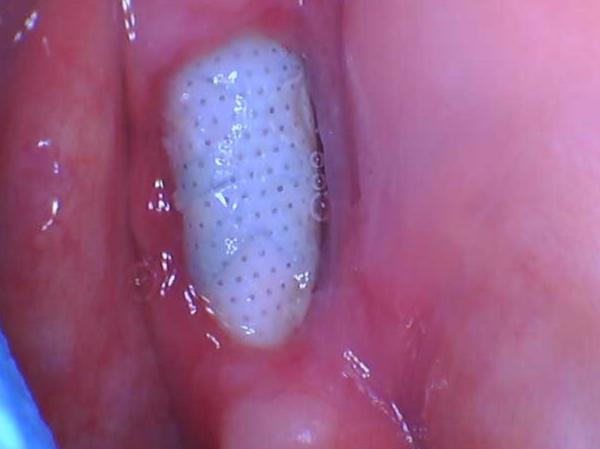
immediate temporaray - note not in occlusion. Also note excellent tissue adaptation giving high hopes for aesthetic final result.immediate temporary – note not in occlusion. Also note excellent tissue adaptation giving high hopes for aesthetic final result.
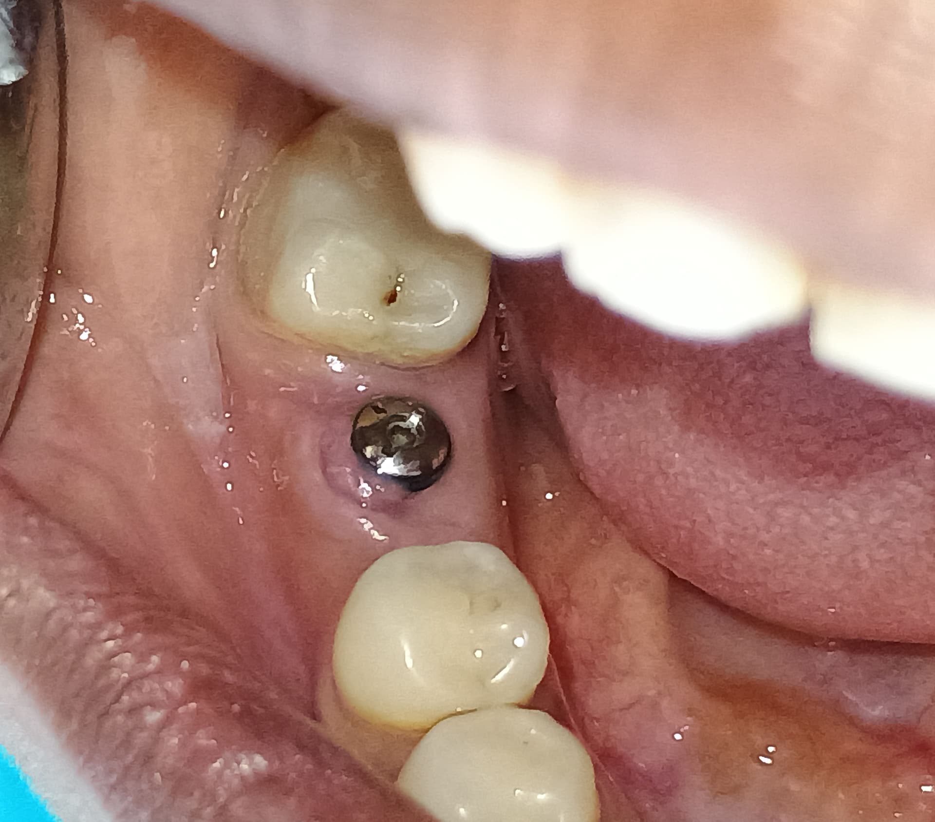
 implant with temporary removed three weeks later using fingers onlyimplant with temporary removed three weeks later using fingers only
pre op x-ray tooth #4 with vertical root fracture.pre op x-ray tooth #4 with vertical root fracture.
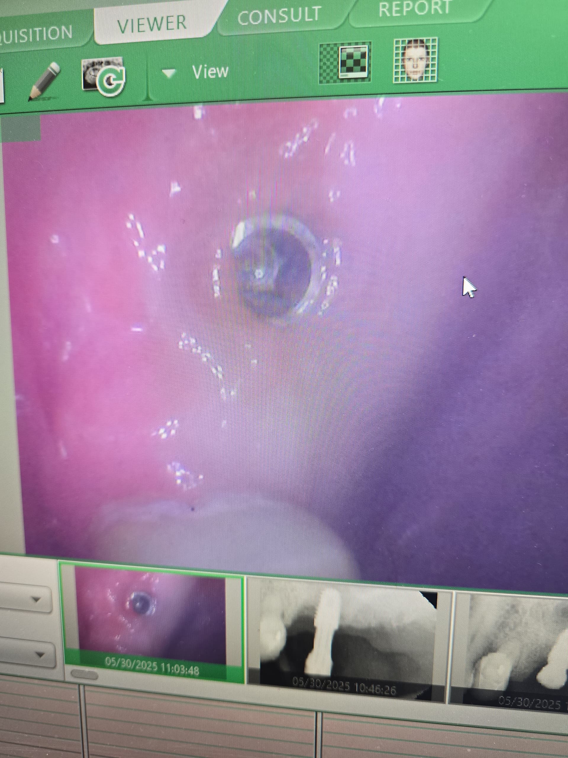
final x-ray with temporary same day as surgeryfinal x-ray with temporary same day as surgery

34 Comments on Immediate temporization failure to integrate: any thoughts?

New comments are currently closed for this post.
CRS
8/27/2013
Traumatic occlusion. How did the original tooth fracture? There was micro movement of the implant , I would have buried it not restored it with a temporary and probably treated the parafunctional habit or malocclusion. Just start over bury it and determine what is going on with this patient's occlusion. The red flag is the " virgin" fractured premolar just because it is out of occlusion a food bolus or a habit could have caused the trauma. What is key is that the "area never stopped hurting" that's not an infection but trauma. That's the diagnosis.
kevin W.
8/28/2013
Traumatic occlusion.100%!!!
Richard Hughes, DDS, FAAI
8/28/2013
Traumatic occlusion is a large factor. You should of prepared the socket by decorticating and osteotomy preparation. I still would placed some particulate graft material in the site (OsteoGen) to increase the BIC. With the tell tail sign of the fractured tooth........immediate temporization is out of the question. The occlusal table looks to large B-L. Hard lesson.
CRS
8/28/2013
Dr Hughes I have a question have you had experience with maxillary bicuspids being sort of a "home base" for patients when they guide their occlusion. I heard this at the AAID meeting and that sometimes these patients need to have their occlusion deprogrammed with a neutral splint. I have a case I 'm treating now, the patients actually fractured bilateral bicuspids one month apart and is undergoing myofunctional therapy. Any thoughts?
Peter Fairbairn
8/28/2013
Hi CRS and poster, I feel the key here is the staement that "it never stopped hurting and changed anti biotics " which would infer an infected site issue . With root fracture cases ( especially here as no osteotomy was done ) there will be a bacterial situation . the best solution is very agressive curretage and then leave for soft tissue closure . Then open at 3 weeks and clean again ( can use CHX especially if you use xenograft material ) then place and graft yes even if 2 mm as soft tissue will always get there first and this can have have an effect on the viabilty of the buccal plate ( Bundle Bone ). But it was the extended pain that was the tell tale to failure . No problem wait a few months and follow traditional prtocol . Regatrds Peter
CRS
8/28/2013
I like it! How would you handle the occlusion?
Mingfu Ye
9/5/2013
I had a case exactly the same as this one, so I think the infection is the primary factor.
Dr. Alex Zavyalov
8/28/2013
Good educational case. Thanks for sharing. Torque 45 N is a sufficient level to hope for a positive outcome. Why “the area never stop hurting”? I think the showed occlusion was not an initial cause of failure.
Dr. Alex Zavyalov
8/28/2013
Sorry, correction: " the shown occlusion"
JS
8/28/2013
Thank you all for your feedback on this which is my case. I have had four of these upper bicuspid vertical root fractures with immediate implant placement and temporization this year - which is a lot for a general dentist like me. This is the only failure and fortunately my overall percentage is still excellent like most of you. I just really like the way the immediate temporary preserves the gingival contour that the natural tooth had. Now if you all were to say to me that the only way to consistently do that would be a gum level custom healing abutment that would be an acceptable compromise. It also seems to hold the graft particles in place nicely if you do pack some into the buccal. Peter, I will take your recommendation to heart and work some graft particles into the buccal gap in cases like these.
sb oms
8/29/2013
I don't think you can blame this 100% on occlusion. First, any one can fracture a virgin premolar with a single bite on a pit, seed, shell, etc... And I don't see wide PDL spaces or any other type sign of traumatic occlusion. There are wear facets on the adjacent teeth, but what 60 year old patient doesn't have those. Second, the poster said in his own words, "No preparation was done. This means he screwed a round implant into an oval whole. His 45 NCM of insertion torque may have come from one thread of this fixture. Most likely very little BIC, and not ideal. In these cases the x-rays can make the situation look much better then it really is. And that does not look like a five mm diameter implant, looks more like 4mm. Third, the immediate temporary looks way out of occlusion. It's functioning like a fancy healing abutment. Unless the patient is purposely chewing on it, I'd like to assume it's out of occlusion. What I have learned from immediately placing and temporizing hundreds of implants: 1. Pain is a bad sign. Especially pain that does not go away over the first few days. These patients always eventually get some swelling and the implant gets loose. 2. The pain in these cases comes from infection. While you can clean the heck out of any socket and sprinkle holy water and antibiotics galore, there will be still be some bugs. And now you have locked them in place with your implant. These are the cases where you do everything right, and you still have a failure now and then. Immediate temporization, when done correctly, should not lower success rates. If the temporaries are made correctly (screw retained, no contact in centric or lateral - excursive movements- then they are just fancy healing abutments. In my opinion, our presenter had an infection (not an obvious one with swelling and pus) and poor bone - implant contact. These cases do not respond well to antibiotics, as the drugs just don't get to the bugs. Cellulitis, abscess, whatever you want to call it. My pearl for this presenter, prepare the socket. Open up the bleeders, get cells to the seen. Go beyond the apex and get a good bleeding socket if you are going to do immediate placement. I give an intravenous dose of antibiotics (clindamycin) to every immediate or graft that I do. Just a single dose at the time of surgery, and this works well in my hands. I routinely place immediate implants into infected sites (that have been cleaned beyond thorough by the way). I know the drugs are there as the implant is going in, and the patient is bleeding antibiotic into the site.
Peter Fairbairn
8/29/2013
HI sboms , nicely put I agree the best instruments are vital . As to Clindomycin we ahve been keen on its use in implantology for years and recently I had a lively debate with a MFOS friend of mine who was adamant that it should never be used and when asked why he stated that he heard it in a lecture ( Wallace ) . So good to see some more support for its ability. Peter
JS
8/29/2013
OK several of you have made the point about bone to implant contact (bic). Here is where I may be confused. I thought the rule for immediate placement was all about having great initial stability more than bic. I see immediate placement in molar sites after all. The other rule was that any gap greater than 2mm between the implant and bone needs to be grafted. Those two rules have suggested to me that as long as you have initial stability the bone will ultimately grow into the implant surface. sb oms - Thank you for your well reasoned comment. If you look at the shape of the extracted tooth compared with the extracted implant, the tooth is much more severely tapered. I would say it takes a good 5-7mm from the apex before the root has flared beyond the dimensions of the 5mm implant. When I was inserting this one (yes after thoroughly curetting) I considered the implant to be acting like a threaded osteotome into D3 or D4 bone. That is why I thought I had decent bic. Although that bone would have been quite compressed. Bacteria likely played the biggest role in this case as the socket was infected at the time of extraction. Patient had waited a couple of weeks after the root fracture diagnosis was made and infection ensued.
CRS
8/30/2013
Basically with an immediate you are wedging the implant into the set vs cutting precise threads which allows for intimate contact an logically easier osteointegration. When an implant is exposed to the oral environment it is exposed to movement, food and the oral flora. Pain can indicate trauma vs infection. Often infections can be without symptoms but in this case it really doesn't matter, the implant failed within 2-3 weeks. Now you being a restorative dentist what do you think is better, an implant placed in an exact osteotomy site after a healed bone infection with a graft preserving the space or an implant wedged into an infected space after a tooth was fractured and exposing it to the oral cavity? The gingival contour is simple to regain after exposure when the implant is allowed to osteointegrate. If you had one of four immediate s fail that's a 25% failure rate, too high for me. Implant placement is a clinical decision based on guidelines not rules and marketing a tooth in a day. Biology doesn't change. I personally like to minimize risk the magic antibiotic will not fix the clinical issues described. Now you have to redo the implant oh and by the way an immediate implant is usually placed in nascent bone in other words you prepare the osteotomy site based on this X-ray 2-3 mm deeper to fit the base of your implant since it is narrower and you'll have an exact fit in healthy bone, graft the gap bigger than 2mm cutting the osteotomy into the dense bone of the sinus floor gives stability vs compressing it which can lead to necrosis. I am starting to add the NG-yag laser to sterilize these sites on contact. I would advise brushing up on the immediate implant technique vs wondering why the implant failed. Thanks to the posters on management some great comments! Thanks for reading!
sb oms
8/29/2013
Dr. Fairbaum- I know the guy who made the clindamycin comment (Wallace), and I know his lectures well. He was talking about it's use for sinus grafts/sinus infections, not routine dento-alvaolar surgery. And I aggree, clindamycin has no place in the treatment of sinus infections. Why do I know his lectures well? He is my father!!
Peter Fairbairn
8/30/2013
Hi sboms , I knew and a great inspiration to have as a father , I have seen his lectures many times and he is a true Implant legend . My mentor began sinus aumentation in the early days ( late 70s ) and has always relied on Clindamycin ( as have a number Sinus research papers ). My only question was why , as the normal respiratory ( Aerobic bacteria ) are generally not the issue , and the Oral ( anaerobes ) introduced during the procedure may be of greater importance and thus in Pen allergic patients we feel Clindamycin to be a suitable option . In the 16 years I have augmented sinuses I have been lucky enough to have never had an infection , but that may have been luck ( or only using synthetic materials for the last 10 of them ) . Anyway that is neither here nor there . Back to the topic yes JS the bic is an important issue and hence I feel peri-implant grafting is important and always use a fully bio-absorbable material to leave only host bone in this site long term ( HL Chan HL Wang etal , JOMI May ) BTcp appears to have host upregulatory effects ( Zhao , Watanabe et al BONE 2012 ) leading to imroved bone quality in the BIC . Another useful tool; to get is an Osstell meter to able to fully check initial primary stability and integration. Regards Peter
Peter Fairbairn
8/30/2013
Then again , Tarnow feels there is no need for this grafting ( Tarnow et al , a new classification for immediate implants , JOMI may 2013 ). This is the joy of implantology many things work for many diffferent surgeons . Peter
greg steiner
8/31/2013
There are proponents of using implants to spread ridges in the buccal and lingual dimension and so you might think that wedging an implant in this case would be acceptable. However while I don't advise spreading ridges with implants at least in that instance the bone has space to move into when you are widening a ridge. That is not the case here. Interproximal bone is often very dense and it will not crush easily. In this case the implant placed tremendous force on the interproximal bone that was crushed against the adjacent teeth and that is the source of your pain. If orthodontics can be painful this is ortho forces times 100. The antibiotics were ineffective here because there was no infection as the poster noted. Placing graft material such as allografts, xenografts and growth factors will not result in implant integration and Tarnow is correct because grafting around immediate implants with the standard materials only makes the radiograph look better and do not provide any support for the implant. Next time prepare your osteotomy for the implant with the appropriate burr and you will never have this problem again. Greg Steiner Steiner Laboratories
CRS
8/31/2013
Peter I liked the fact that Tarnow centered the molar implants instead of placing in one of the root sockets. My rule of thumb is that the socket has to be perfect with good quality bone and good primary stability, and yes we don't really know what makes these things work! It is a miracle that it does sometimes , so I tell my patients one miracle at a time! I think that a Tarnow article is better than a lot of these one hit wonders showing a perfect result with immediate placement and temporization. I think that is why this technique is popular although all the clinical parameters need to be weighed. Good to hear from you as usual!
nailesh gandhi
9/1/2013
This is traumatic only. When tooth was vertically broken it was indicative that the implant has to be submerged !
Peter Fairbairn
9/1/2013
Hi CRS , looking again more closely at the final x-ray above I feel a little concerned about the area at the apex and distal to it . But the time scales suggest infective site ( difficult for poster to asses that "there was no infection ") . As for the Tarnow paper placement into the furcation bone is always the aim and I have done hundreds over the years with practice it gets easier . Always possible on 6s but can be an issue on 7s due to root morphology. I always place 3 weeks post extraction to get soft tissue closure and as I said graft with BTcP and get great consistent results where not only do you retain better residual ridge dimensions but you retain more attached gingiva which is critical for long term success. The body wants to heal and get many things done at one go is in fact natural and best for the host. As for the Tarnow "type C " sockets I generally socket graft ( if cannot get PS due to Vital tissue ) and place at 3 months at 3 months . Peter
JS
9/2/2013
( difficult for poster to asses that “there was no infection “) . Poster never made that claim.
CRS
9/4/2013
Looking at the apex, perhaps it is necrotic bone from pressure, torquing it to 45 without preparing the apical bone, the implant end is blunt. I have seen this happen at about three weeks post op. Or there could very likely be infection with a fractured root we'll never really know. I would redo and bury the implant until integrated.
Richard Hughes, DDS, FAAI
9/2/2013
CRS in reference to your question to me on 18 August 2013. I have not heard this about the maxillary bicuspids. I have seen this many times in clinical practice. I have also seen the ML cusp of the maxillary second and sometimes first molar lock the occlusion posterior. When I reduce the ml cusp the patients occlusion has a tendency to slide anterior. I use permissive splints or anterior deprogrammes from time to time. I apologies for not responding sooner. I do not look at this blog every day. Will you be at the AAID this year?
CRS
9/4/2013
I just attended the western AAID conference on implant complications in Chicago this May it was excellent.
Larry J. Meyer DDS
9/2/2013
Peter, nice comments. Can you explain your usual procedure on a molar tooth upon re-entry at the 3 week visit? I am interested particularly in how much debridement should be done in order to graft. Also, I assume a flap is needed, maybe you could give an idea of that also. Do you usually do one stage or two stage? As always I look forward to your comments. Larry J. Meyer DDS
Peter Fairbairn
9/3/2013
Hi Larry we always split the roots on extraction as is shown in the Tarnow article but prefer to then leave for 3 weeks for two reasons ,mainly to get a form of soft tissue closure ( albeit sometimes frail ) but also as these teeth are often very infected ( acutely ) we like that period to allow things to resolve a bit . placement is then into the inter-radicular bone but as you stated curretage with sharp currettes is vital to debride the site of the adjacemt root sockets prior to grafting suing a full absorpable material and then after ensuring the material has set ( I use materials that have a set ability hence become stable and are their own "membrane " thus never use a traditional membrane to allow for improve blood supply to the healing site and allow the Stromal sell derived factor 1 ( induced in periosteal tissue at bone damge site ) to help with regeneration . Always 2 stage but another benefit especially in the 1 st lower molar area with preservation of the site prior to modelling is that we retain more attached gingiva in an area where its loss is easy and can have issues in the long term survival of the Implant . Regards Peter
Baker Vinci
9/3/2013
I did not look at the other responses, but will, because I am under house lockdown, secondary to a posterior "gutting". If you are not willing to stick to the basic principles, then you are going to get these kinds of results. This is routine procedure, but if you didn't use the osteotomy drills provided, then the torque value received was very likely in a very insignificant portion of the implant. I feel as If you must engage about 5mm of apical bone at the first stage of your drill sequence. Bv I understand the desire to get immediate temporization, but look where we are now, with this case. A 25-50$ Essex temp. and 3 months of integration, saves you and the patient time and money. To suggest 2mm of exposed space next to an implant does not need to be managed, makes little sense to me. The tooth failed and to be honest with you, I have already forgotten why. Any reactive, inflammatory or infective process, brings with it, granulation tissue, neo-angiogenesis and the likelihood of soft tissue integration.
Gregori Kurtzman, DDS, MA
9/3/2013
Two factors may have caused this.................... 1. temp had proximal contact with adjacent teeth, which when the natural teeth are loaded due tot he PDL they can shift mesial and distal slightly causing pressure on the implant temp. Immediate temps should no proximal contact, should be able to easily pull a piece of shim stock thru. 2. pt didnt stick to a soft diet and was chewing on the provisional
Dr. Gerald Rudick
9/3/2013
The comments posted by the caring and capable dentists who have offered their opinions in this situation are excellent and of high scientific value ......but we are not Gods, and do not have all of the information. When a virgin tooth fractures,it indicates an abnormal amount of chewing force and bacteria does invade the nano crevice, and could bring the infection through the entire length of the root. Judging by the initial radiograph, the infection my have gone into the sinus as well.......and stayed there while the implant was torqued into place. No scan was offered, so we do not know how thick the cortical plate was, or if it had been perforated, as no attempt to prepare a drilled osteotomy directed in the palatal direction was done. Maxillary bone in the bicuspid area may be the density of a piece of pine wood at best,and not the maple or oak hardness that is found in the mandible........so the immediate loading could have played a role in the failure. More attention must be devoted to growth factors, which are readily and inexpensively obtainable from drawing and centrifusing the patient's blood to obtain Plasma Rich in Growth Factors (PRGF) specifically Platelet Rich Fibrin, which is compressed to form a membrane and the exudate liquid referred to as Fibronectin and Vitronectin. In the gap that was left because of the difference of shape between the ovoid root and the round implant,a piece of the crushed PRF sprinkled with Metronidazil powder could have been inserted into the socket, and the implant dipped into the liquid exudate (Fibronectin and Vitronectin sprinkled with Metronidazol powder) before being screwed to place. The PRF forms an ECM ( extra cellular matrix) which is an excellent scaffold for cell growth. "Bioactive modifiers, or growth factors, are naturally occurring substances capable of stimulating cellular differentiation and growth, in other words, these factors can stimulate non differentiated mesenchymal cells to become specific functioning cells - i.e.osteoblasts or bone forming cells." as printed in Implant News & Views Sept/Oct 2013 Regeneration and Repairing Bone and Soft Tissue - An inexpensive Device for Compressing the Fibrin Clot (PRF) to Release Growth Factors.( written by Gerald Rudick) By taking advantage of the information and technology readily available in our chosen field, we can alter the playing field, so that our procedures have a better chance of succeeding than in the past. Gerald Rudick dds Montreal
Baker Vinci
9/7/2013
You must respect the premise of guided tissue regeneration. If there is a 2mm gap, what tissue will be more likely to fill that space, first? B Vinci
DrSSG
9/21/2013
Immediate placement requires preparation to minimum 3mm beyond apex. One of the basic tenants of implant dentistry was omitted...in that there would likly have been poor adaptation to bone....or 45Ncm on one or 2 threads. This has already been said in several ways by the comments above...sb oms is right on target. BTW ...the photograph and x rays do not match???
JS
9/21/2013
The photo does match the x rays. What looks like lateral and central incisors in the photo are in fact the worn down first bicuspid and canine.
Scott Noren
12/18/2013
I think micromovement is the enemy here..the comment about no interproximal contact or occlusal contact seems key here and may be the cause for failure..the pain issue also may be from pressure on the adjacent teeth. Call me 'old school', but I will graft and come back in about 95% of my implant cases...

Featured Products

OsteoGen Bone Grafting Plug
Combines bone graft with a collagen plug to yield the easiest and most affordable way to clinically deliver bone graft for socket preservation.
CevOss Bovine Bone Graft
Make the switch to a better xenograft! High volume of interconnected pores promotes new bone. Substantially equivalent to BioOss and NuOss.