Initial Lingual Die-Back Around the Gingival Margin Follow Up: Did the BioHorizon Laser-Lok Surface Help?

Dr. B comments,

This case is a follow up to an original case: Initial Tissue Die Back after Flapless Implant Placement Case: Suggestions?.

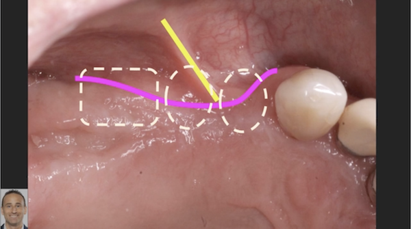
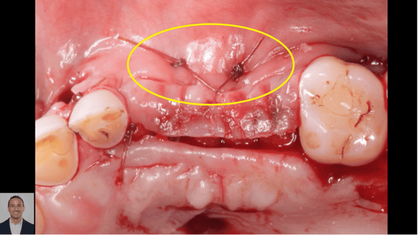
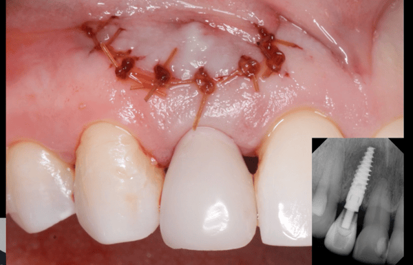
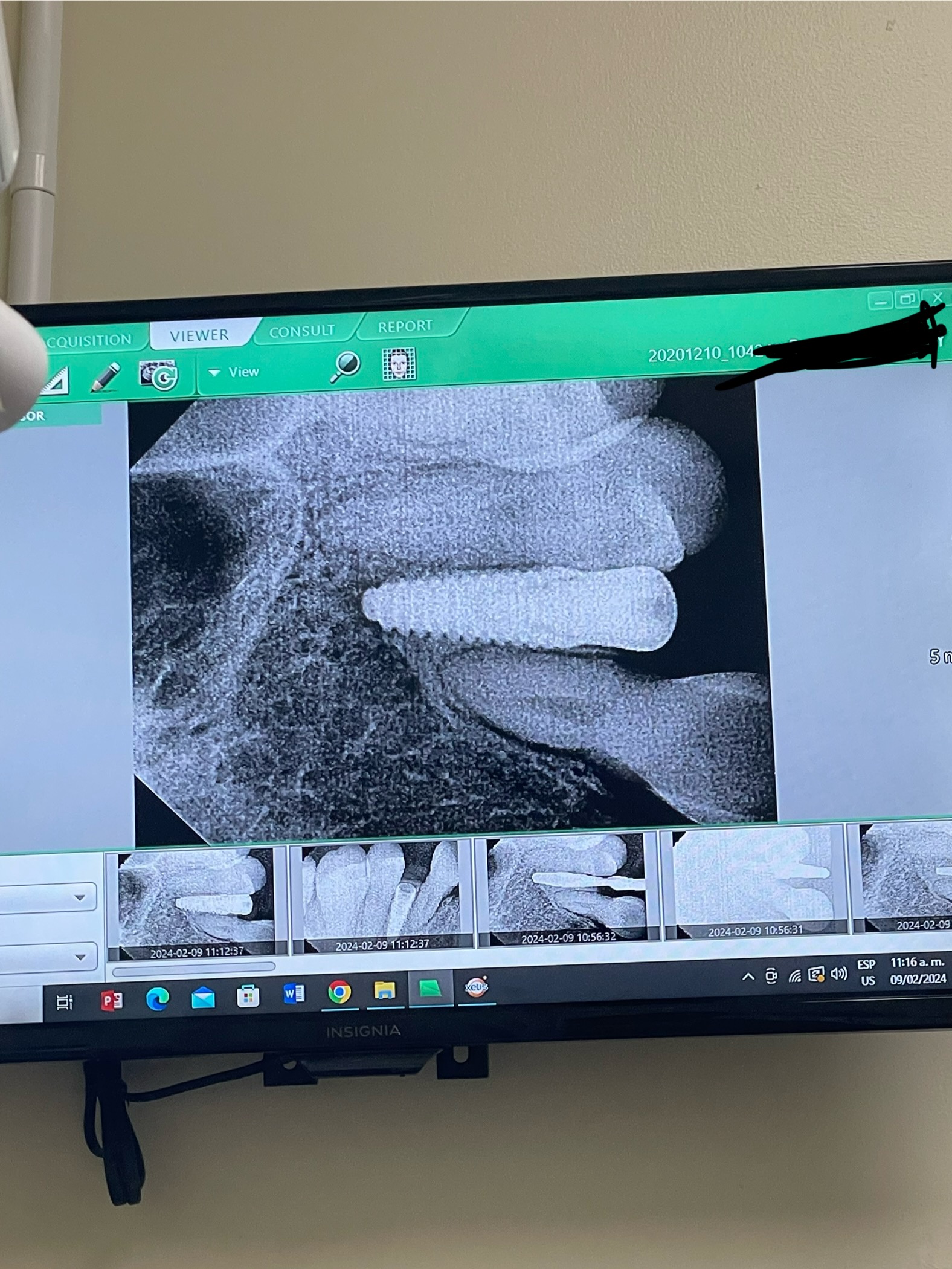
I recently restored the Biohorizons implant from the above posted case that had initial lingual die- back around the gingival margin immediately after placement. We treated conservatively with Periogard swabs of area 3 times a day for 4 weeks. The tissue came back nicely. I wonder to what degree the Laser-Lok [BioHorizon] surface played into this? How much of the success of this case can we attribute to the unique physical characteristics of the Laser-Lok surface? I have attached photographs of the area at 12 days, 4 weeks, 6 weeks, and then 6 months where we had nice healthy tissue grown up around the healing abutment.

12 Days Post Op

4 Weeks Post Op

6 Weeks Post Op

6 Months Post Op

9 Comments on Initial Lingual Die-Back Around the Gingival Margin Follow Up: Did the BioHorizon Laser-Lok Surface Help?

New comments are currently closed for this post.
dr.keyhan
3/29/2011
laser lok technology is recommended for better results in osseointegration ,decreas suserisition around the bone margin according to micromovement of implants. it's the hypothesis for this company. i dont think any relationship exists between die back and this design.
King of Implants
3/29/2011
Seems like the die back is showing bone. So the laser-lok has nothing to do with gingiva growing over bone.
Dantis.eu
3/29/2011
I use the biohorizons laserlock implants last 3 yers, hawe placed over 135 . All internal tapered model working wery fine,hawe a great initial stability, are fine for crowing. In this case you must hawe a 2 implants in lower jaw for better displacing of biting forces.laser lock is not laser blast gun from star wars movie,realy :))
Iyad
3/30/2011
Well, first I would like to comment on Dr.Dantis for the decision of placing to implants I think we cannot judge that till we know the exact extent of the upper dentition, on the other hand, I have been using the Laser lok for more than 2 years and placed around of 250 of them, and in the case of yours, yes I believe that the laser lok helped in triggering the tissues towards the implant body and that because all the new researches that they were published shows that the Laser Lok enhances the attachment of true soft tissue attachment to the body of the implant (like to a natural tooth)and I had a similar case but in the maxilla which I had an overgrowth of the gingiva over the polished collar!!!
Dr Sanjay Jamdade
3/30/2011
From the what I undestand from Dr Jack Ricci whom I heard first hand at NYU the upper few rings of the Laser Lok are designed towards attracting fibroblasts from the connective tissue and providing attachment substrate for them. So certaily Laser Lok has a role to play in developing a soft tissue collar around the neck of the implant. This is based on the phenomenon of contact guidance. Contact guidance has been recognized as a cell behavior phenomenon since the early fifties. It's been researched by various cell biologists since atleast 2-3 decades. It's a phenomenon appearing in all cell biology literature. So it's an established phenomenon. There is no reason to doubt it's ability to enhance hard and soft tissue adhesion. In your particular case I don't know. Multitude of possibilities. You probably had a soft tissue dehiscence with alveolar bone showing through. There could be shortage of attached gingiva, I can't tell much from a picture. If you incise gingiva in to two halves you can send one half lingually and the other half buccally and then suture. In cookie cutting, if you have less attached gingiva to begin with you are left with nothing at the end. Connective tissue below the gingiva is the saving grace for all dentistry! Dr Sajjad Khan's advice is bang on target and you seem to have produced results and that's good for you! You want to know what if it was any other implant system? Right? My hunch is the soft tissue would have grown back. Because live exposed bone granulates and eventually you would have had a tough attached tissue there no matter which brand of implant you use! The soft tissue is growing on TOP OF THE BONE NOT THE IMPLANT surface! (that's what it looks like to me in the picture) Incidentally I am not a Biohorizons user. But I am sure it is a good system.
BAYNON
4/5/2011
Of 3 implants placed on this patient on that day, this was the only one that had me concerned at the initial follow up visit. I realize that the tissue was to grow over the bone to resolve this, and not the implant, however my concern was that the bone would not persist in this area to allow this. With hindsight I believe that my use of the tissue punch was to blame for this denuded area in the 1st place. Does anyone have any more tissue sparing "flapless" implant placement recommended techniques to share? Perhaps using a downsized tissue punch (say a 3.8 for a 4.6 implant) may be helpful?
BAYNON
4/5/2011
(I must add that the Flapless placements are all being done with a scan and prefabricated surgical guide)
Pieter Linssen
4/6/2011
looks great at 6 months that's all I care about. As long as probe is wnl enjoy
Andres Paraud
4/29/2011
I think the problem here was the lack of gingiva atachment, i dont suggest tissue punch in this cases with to much free gingiva around the site, mucosa + implant dont get along well. Sometimes i suggest gingiva graft with connective tissue improve the site before placement of the implant or at least before connect the implant if you did a submerge 2 stages technique. greetings from chile.

Featured Products

OsteoGen Bone Grafting Plug
Combines bone graft with a collagen plug to yield the easiest and most affordable way to clinically deliver bone graft for socket preservation.
CevOss Bovine Bone Graft
Make the switch to a better xenograft! High volume of interconnected pores promotes new bone. Substantially equivalent to BioOss and NuOss.