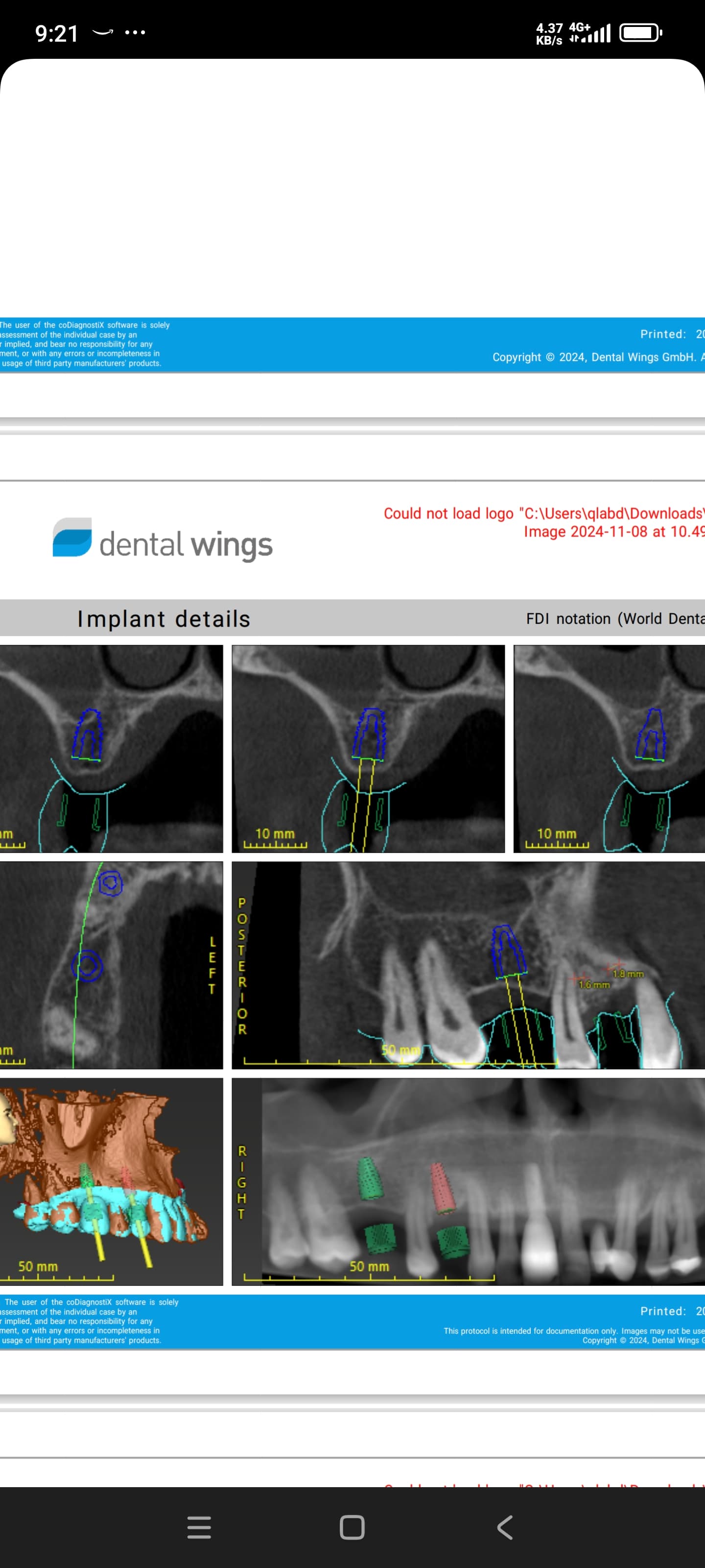
Internal sinus lift immediately thru the palatal root socket?

I have a 60 year old patient with no contributing medical history. There is a 30 year old root canal with a distal root completely fractured with some possible trifurcation involvement. I have not taken a CBCT yet, since I do not think this is an ideal case for immediate implant placement.

I have done many successful internal sinus lifts through the osteotomy at time of implant placement mostly using “Steiner Sinus Lift” protocol. My question is: if there is no large perforation in the sinus membrane, at the time of extraction, can I just do my internal sinus lift via membrane elevation thru the palatal socket? Has anyone done this? Just seems with several millimeters of the palatal root already above the sinus floor that this would be a good case to attempt. Or do I just socket graft, CBCT in 3 months and then do lift? This will most likely be a future Conelog implant.

Any input is greatly appreciated.


17 Comments on Internal sinus lift immediately thru the palatal root socket?

New comments are currently closed for this post.
Carlos Boudet, DDS DICOI
1/16/2019
I have not done it, but I have had to restore them. You are more likely to perforate the membrane trying to elevate it when it is the highest point than when it is level with the adjacent bone at the floor of the sinus. You can restore it adequately with a custom abutment and crown (can show you pictures), but you would be doing a surgically driven instead of a prosthetically driven restoration. In your mouth, which would you prefer?
smile
1/16/2019
Thanks for the input. Did not consider higher likelihood of perforation with the membrane at it's highest point. My thought process was if I did it at time of extraction I am still not placing an implant immediately so in 3 months when I take the CBCT scan I can evaluate the lift. If I am not entirely satisfied with the lift I can then do an additional lift through my implant osteotomy. I also work from a prosthetically driven restoration perspective as you. Place the tooth virtually on the CBCT scan with merged dental model and place implant in relation so future screw access hole is in correct place for screw retained crown and usually custom abutments.
Carlos Boudet, DDS DICOI
1/16/2019
Elevating membrane at apex of palatal root may not place the graft in the necessary position for implant placement in the ideal position. CBCT would show how far apart the roots are. I think you'll do better grafting the extraction socket and then doing your sinus lift through the osteotomy or in the ideal position if there is not enough vertical bone to place the implant at the same time. The consensus is 5 mm, but some surgeons do it in as little as 3 mm. Good luck!
Carlos Boudet, DDS DICOI
1/16/2019
It is not done very frequently these days, but have you offered a distal root amputation to allow the patient to keep his crown if the rest is in good shape?
smile
1/16/2019
I did discuss distal root amputation when reviewing treatment options and success rates of those options but pt did not find it a desirable solution.
Peter Hunt
1/16/2019
We have been doing a lot of these over the last three years, so much so it is now relatively routine, and I am pleased to say, successful. The one thing I would not attempt to do is to place the implant down the palatal root space, it's often quite angled and outer bone plate can be quite thin. The problem with maxillary molars is that the labial and palatal bone about the residual roots is thin, often fenestrated and receded as well. It’s no wonder there is so much collapse of the alveolus following the extraction. It’s possible to fill the root spaces with bone graft following the extraction and this is the key to an effective Socket Regeneration procedure. The question then comes to be one of whether it is possible to place an implant at the same time. The key to success lies in whether the implant can be stabilized adequately in the residual bone. When the tooth is removed, which we do by removing each root individually, a triangular core of bone can generally be seen in the central part of the socket, this is supported on webs of bone from the adjacent sockets. Often it’s possible to generate a starter channel down into this central core with an ultrasonic instrument or a trephine. Care has to be taken because the central core can be very shallow. Generally we get through the cortical bone to some softer cancellous bone. This can be expanded outwards and upwards with osteotomes. Usually, this will be the start of a Sinus Lift as well. Once we know we are into the sinus, we add bone graft into the region and then osteotome this up by hand. It’s possible to get good stability in a relatively small amount of bone because the outer rim of the region is cortical bone. Then it is a matter of placing the implant. We use a tapered system which does not have an aggressive thread pattern. The platform of the implant is placed down below the surrounding bone line. We place a 4.0mm gingivaformer in the implant and then bring additional bone graft up and around that gingivaformer. We cover the graft, implant and region with a thicker and tougher membrane than usual. Instead of two or three surgical procedures (extraction + socket regeneration, Implant placement 3 - 6months later, second stage exposure finally) this is all done in a single procedure. You can get Primary Stability for the implant in the web of bone and in the sinus floor. Secondary Stability comes with healing in the Socket Regeneration region. We never place a provisional restoration at the outset as this would break the Primary Stability. Usually these can be restored in 4-6 months.
OsseoNews
1/16/2019
Very informative. Thanks. Can you post a case, by any chance? https://www.osseonews.com/post-case-photos-and-get-feedback/
Dr. GELFAND
1/16/2019
Why not just place the implant into the palatal socket immediately following extraction and be done with it?
Peter Hunt
1/16/2019
Because: 1. The implant will be considerably off axis. 2. The augmentation will be all to the labial: greater bulk leads to greater shrinkage in the region of greatest bulk. 3. Placement of the implant may mean the thin walls of the palatal socket become fractured.
Miguel Martinez
1/16/2019
hello. all roots look real far apart. why not go right thru the middle; at its best position. just my thought
Miguel Martinez
1/16/2019
i agree with peter hunt. im doing them now with the versah burs. i could share some really cool sinus cases demonstrating it if interested.
Yaron Miller
1/16/2019
Yes I second this and have started doing all my immediate upper first molars( and in fact all my immediate placements) like this. Just did one yesterday. Very predictable and less banging with osteotomes in the patients skull. If you can get a good purchase in the bone between the roots the versah burs really help to form a nice densified osteotomy, around which you can then graft as per your protocol. I really think that placing an implant in the palatal root is a poor treatment choice and I would never do this.
Miguel Martinez
1/16/2019
in regards to your original concern. if there is a root tip fracture of buccal roots, you can find them thru buccal and not effect placement of central, ideally placed implant in great healthy most solid bone. also, i often use mallet and periotome (assistant pressing in forehead) to sever pdl all around tooth. to facilitate extraction. i dont have physic forceps and some extractions wouldn’t allow them anyways. what do you do yaron miller before taking tooth out? did a cool lift yesterday also along with digging out root tips left behind years ago on a #13. molar #14 taken out and a 5x11.5 placed. versah in transport to there symposium as we speak, lol cool stuff
Yaron Miller
1/16/2019
I always section the roots and take them out individually. To date I have never had an issue with fracture of the buccal plate. I guess I will see you at the symposium then- safe travels.
Dennis Flanagan DDS MSc
1/16/2019
There is the Fugazzotto technique. Using an appropriate trephine, cut through the inter-radicular pyramid of bone and carefully elevate the cut pyramid superiorly so as not to tear the sinus lining. Introduce particulate mixed with CaSO4. If there is enough bone the implant can be placed. If there is inadequate bone then graft and cover with a collagen barrier and suture.
mark
1/17/2019
the adjacent teeth are already crowned. Just do a 3 unit bridge
Greg Kammeyer, DDS, MS, D
1/17/2019
Peter Hunt is right on. I've been doing immediate maxillary molar implants for 7-8 years. About 95% of cases are great candidates for this AND you get a prosthetically driven implant position. The sinus floor provides nice primary stability.

Featured Products

OsteoGen Bone Grafting Plug
Combines bone graft with a collagen plug to yield the easiest and most affordable way to clinically deliver bone graft for socket preservation.
CevOss Bovine Bone Graft
Make the switch to a better xenograft! High volume of interconnected pores promotes new bone. Substantially equivalent to BioOss and NuOss.