Issues with healing after placement of cancellous bone and membrane: recommendations?

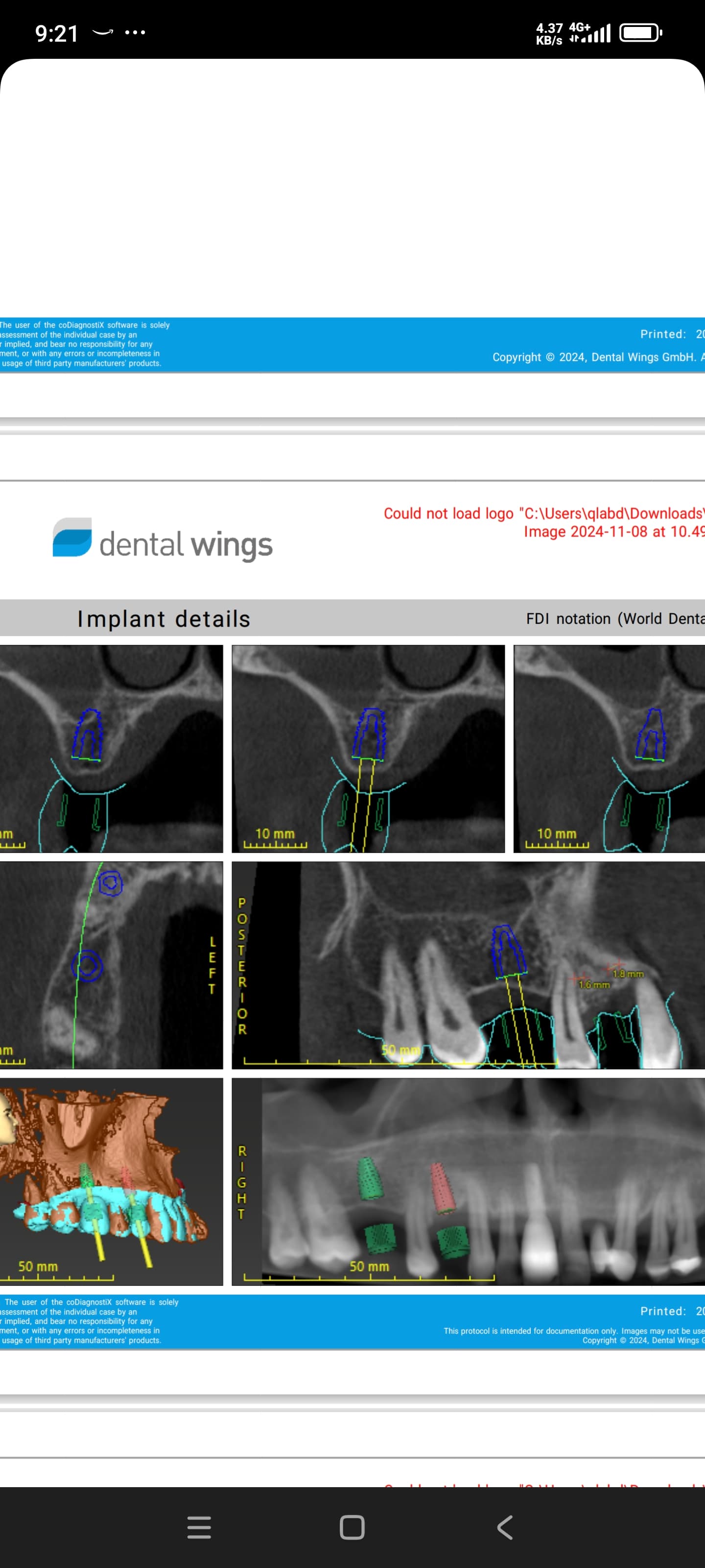
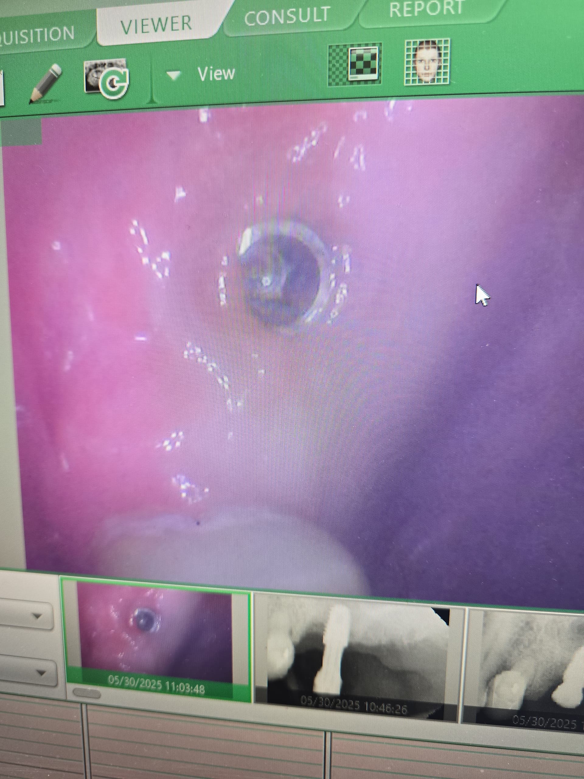
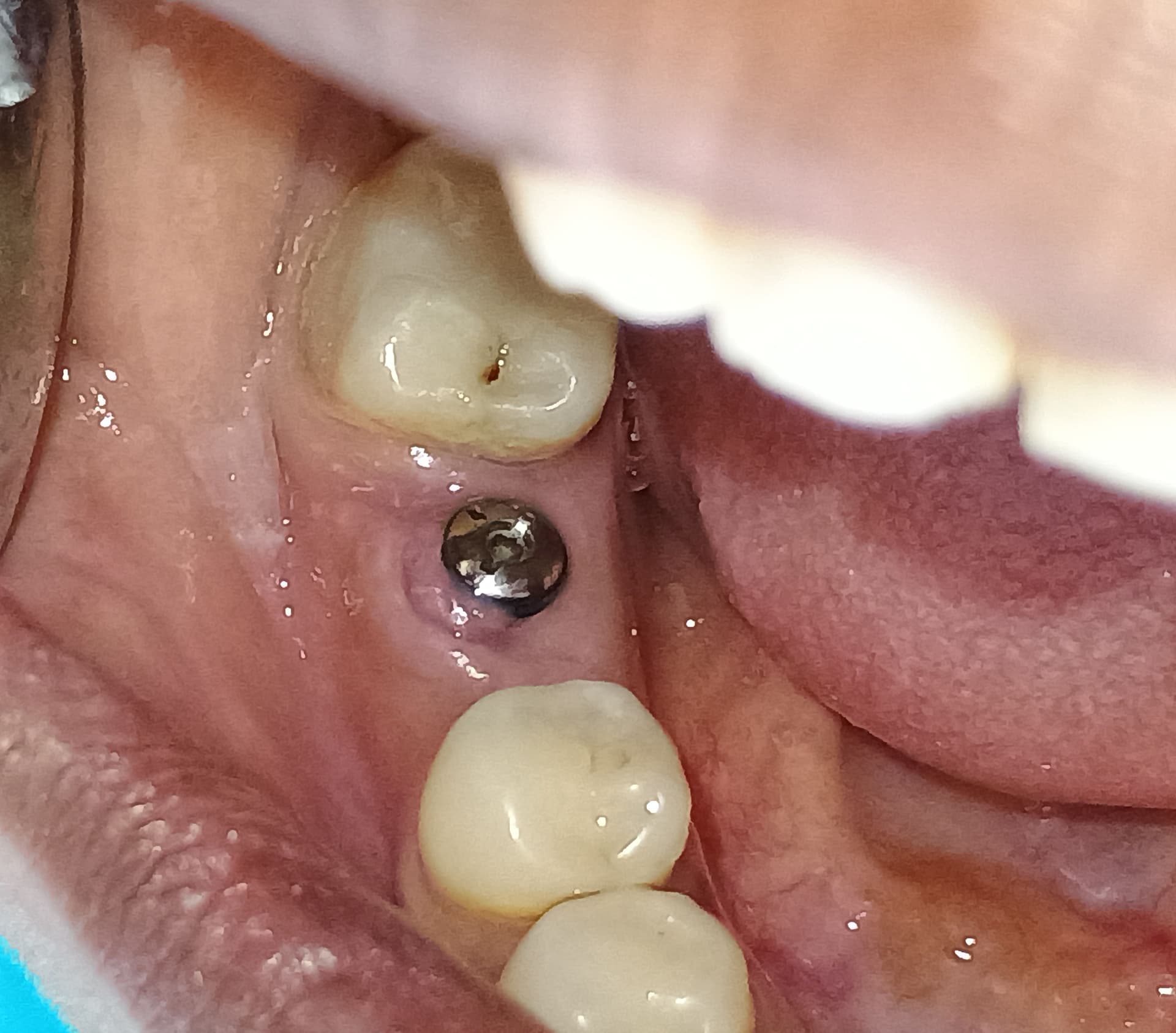
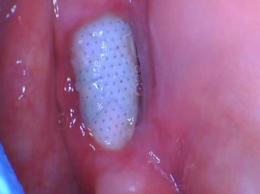
In March 2018 I removed #8,9 and placed NuOss cancellous bone and resorbable collagen membrane and sutured the area. Patient returned in 2-wks for suture removal and post-operative check and all was WNL. January 2019 returned to site for osteotomy. 1st x-ray is pre-op, 2nd x-ray is pre-tx, 3rd x-ray is post-tx. Upon laying a flap to place implants, the bone consistency was similar to the granules that were placed in March 2018. I checked its density by removing this material. Enough of the material was removed to decide to abandon osteotomy. I placed same material back into socket area, sutured area, and instructed patient to return for suture removal in 2-wks. I have some questions:
Q1: I have used this protocol of extraction to implant placement, using same materials, and had similar results, yet not to this extent. Are the materials in question?
Q2: Is my technique in question?
Q3: I placed the same material into socket and sutured. Did I just create an issue in the future?
Q4: Any suggestions?


16 Comments on Issues with healing after placement of cancellous bone and membrane: recommendations?

New comments are currently closed for this post.
Dr. Moe
1/8/2019
Hi, I think you are doing the right thing, I do the same. Some times, it is what it is. Bone grafts Don't always take. Actually, at a course I took just one month ago, the instructor is vehemently against doing allografts and membranes. She was explaining the same thing, the material stays as material at the site with not bone remodeling. She is a big believer in Autografts, which happen to be the gold standard. I think this type of stuff happens and we can all be Monday morning quaterbacks when we go in again, but there is no way to predict this. As a checklist, did Pt had a lot of bleeding in the site? Is there such as thing as condensing the allograft too much? Did you create some bleeding points, if not enough bleeding or if the Cortical bone looked too dense? I am well aware that I am talking about cortical bone in the Anterior maxilla, not the area where it's like in posterior mandible. Again, trying to find reasons for the bone not turning over. I guess my last point would be, Since you didn't get any bone formation with the bone graft, would you have been better at removing the bone graft, debriding the area, and allowing the body to heal the area on it's own?, that is a possibility. But overall, I am not sure it's necessarily your technique. Let's see what other pearls we can gleam from more experienced Docs on the site. My $0.02
Jah
1/9/2019
I will be more observant, yet these are the questions I pose to myself in looking at my protocol. I have done a lot of these- extr, bone, membrane, suture, PO and suture removal. To your last point, I would be fearful of leaving the patient with insufficient bone leading to extra surgical procedures in the future if this patient were to proceed w/ Implants.
R Gangji DDS, AFAAID, FIC
1/8/2019
NuOss, is deproteinized, bovine origin, cancellous bone ( (Xenograft). And just like allografts ( from Human) they have the disadvantage that their preparation during freeze drying and irradiating lowers the materials Osteogenic powers,And the body’s response to it ( Immunological) may lesson in terms of incorporating it to bone. So if you’re not using an autograft, (Osteogenic,osteinductive, osteoconductive) ,all other grafts have some , rare , but failure potential. Especially if protocols like the ones Dr Moe stated Are not performed. We know the convenience of the bone in the bottle but sometimes they can fail. Xenograft especially are frowned upon here in the States( Because of their poor turnover ) and but widely used with great success in Europe. From what I was told there are some ethical issues of using allograft in Europe as well so Xenografts like Bioss are used) However, the Best European teachers like Danny Buser will scrape some autologous bone and mix it with their xenograft In order to stimulate precursor cells for osteogenesis . So poor turnover of xenograft, along with maybe as Dr Moe stated over compaction, less bleeding of socket, maybe not hydrating graft particles, maybe less than ideal primary closure w/ smoker, systemic issues w pt health , diabetics?. So many factors, When I place the graft I look at my socket and scrape it until I can see the bone and if it does not bleed I make it bleed ( your x-rays show they are endodontically treated 8;9 therefore you will get little bleeding in that socket w Endodontically treated teeth as PDL fibers are not as vital)Take a round (2-6)bur or a sharp hand instrument (curettes) and scrape ,decorticating (RAP phenomena) so blood is there , Also I prefer Allograft and recently have had great success with a quick alloplastic osteogen plug material by Implantdent . So I think you did everything correct maybe pay attention attention to bleeding , clean socket, Good closure and some good luck After I tell you all this , on rare occasions ,I go back and I get the same result, but I think the results get more successful w experience . You did the right thing though try allograft instead of xenograft, as white xenograft particles do not remain in socket when you open , allograft turns over fast 50/50 cortico canc 250-1micron ,I use) Hope this helps there are other clinicians who are much more learned here ,would be interested in their opinions.
Jah
1/9/2019
Excellent
Dennis Flanagan DDS MSc
1/8/2019
Since no endo treatment produces a sterile outcome, if the extraction sites were not thoroughly debrided then there will be slow to no bone growth. The primary purpose of graft material, no matter the source, is to maintain space for angiogenesis and subsequent osteogenesis. I don't believe that cytokines (which are proteins) survive processing to be biologically active.
Doc
1/8/2019
There was no mention of patients medical history, and social history. NuOss and collagen membrane is a decent protocol and this should have turned over in the amount of time you provided. There are many other factors including medical, smoking, denture irritation, inadequate degranulation of socket before biomaterial placement, underlying infection at time of bone placement.
Jah
1/9/2019
This gentleman is a healthy 26 year old male, w/o any significant med Hx nor social Hx. He may smoke pot, I did not ck extent of usage. A Good Athlete.
Dr AG
1/8/2019
Xenograft in a socket is the least efficient way to get bone. It take lots of time to be transform is any at all into bone. Lack of bleeding, over compress the particule, poor healing potential from patient are all possibilities. To use the same material again and expecting better healing is something I don't understand. Better remove all and either let it heal or place other material. Buser (ITI) protocols are the best in this situation. Try to read the ITI book concensus on delay implant placement. Early type 2 or type 3 protocole should work better.
Jah
1/9/2019
I hear you!
Joseph Kim, DDS, JD
1/9/2019
>>>Q1: I have used this protocol of extraction to implant placement, using same materials, and had similar results, yet not to this extent. Are the materials in question? I have used a bovine xenograft / collagen membrane combination for many years with very predictable results. However, having vital bone form between the particles requires a bony vascular supply, not overcompressing the particulate graft, and a barrier to epithelial invasion, preferably resistant to collagenase, in my practice, dense PTFE. In your background, you don't describe the type of closure, if any, which was achieved, or if you left the collagen membrane exposed. Q2: Is my technique in question? See above. Imagine that doing nothing after extracting the teeth would have most likely resulted in bone formation. If your technique impeded the natural regrowth of bone, you should try to identify the pattern of patients or procedures you are doing that may be contributing to this. Q3: I placed the same material into socket and sutured. Did I just create an issue in the future? Maybe, maybe not. Q4: Any suggestions? Next time, place your implants at time of extraction, into a bleeding socket (not just from osteotomy). Consider a collagenase-resistant barrier such as dense PTFE or PRF instead of leaving collagen membranes exposed to the oral cavity. Ensure graft particles are not overcondensed at placement, and remove any prosthetic load against the graft. Look for a pattern of failure and you will likely narrow down the offending variable.
Jah
1/9/2019
Q1/A1: Interrupted sutures at 7 and 10. Continuous sutures from 7 to 10. I will be more observant toward compression of graft material. I will explore the use of PTFE membranes as a protective barrier. Q2/A2: I am knocking my brain to find a pattern, will continue since this has been brought to the forefront of concern. Q3/A3: I will be adding Allograft to my armiamentarium. Q4/A4: I fear placing an implant at the time of extr leave a gap at the most coronal aspect of socket, leading to susceptibilty toward loss of attachment of graft material onto the implant. If I learn to improve my closure w/ PTFE membrane, I may feel more confident w/ this side of Implantology.
R Gangji DDS , AFAAID
1/9/2019
PTFE comments ......If you are not drawing blood then agree I Agree w Dr Kim , Generic PTFE membrane is also cheap and I get predictable results , When you peel it off in one month you will get ittle “dog ears “ Keratinized tags around site but More importantly complete closure with Attached epithelium almost all the time Ptfe works , Not sure why people use collagen membrane as in the wrong hands it is not predictable And PTFE more resilient to patient abuse when they are eating in terms of Protecting the graft particles . I prefer resorbable collegen membrane when I have a good primary closure or I create primary closure when collagen membrane is used . I like the way Dr. Kim gave you steps in considering areas of concerns And prosthetic load ( An. ill fitting , pressure causing , flipper ) can definitely create issues Thank you Dr Kim
Jah
1/9/2019
Using this type of membrane allowed for ease of use during placement ( no memory). I will do research on PTFE membrane in the near future.
R Gangji DDS, AFAAID
1/9/2019
Ptfe very easy to manipulate, like a plastic sheet. I also use amnion chorion (BioExclude) placenta derived membrane, strongmembrane ,w growth factors , little expensive for cases that require good soft tissue outcome and when you are not drawing blood.prf gives me the best soft tissue outcome Remember, you want membrane that does not resorb when exposed to oral environment for at least 2 weeks. Or three. Unless u have primary closure of the mucosa.
Peter Fairbairn
1/12/2019
CaP materials have great osteo-inductive potential as shown in 200 high impact scientific journals on Pubmed . So my last 5,500 grafts take advantage of this and yes I never use a membrane , the Periosteum seems to be the miracle of nature . As Dentists we need to understand more about host healing and work with this . Only the host can regenerate bone , we simply cannot , we often merely integrate foreign material into the site reducing the space for host bone . My 2 cents
Jah
1/14/2019
Thank you Dr, may I have a site to explore this particular material more?

Featured Products

OsteoGen Bone Grafting Plug
Combines bone graft with a collagen plug to yield the easiest and most affordable way to clinically deliver bone graft for socket preservation.
CevOss Bovine Bone Graft
Make the switch to a better xenograft! High volume of interconnected pores promotes new bone. Substantially equivalent to BioOss and NuOss.