Placement of Mini Implant: best treatment plan for this case?
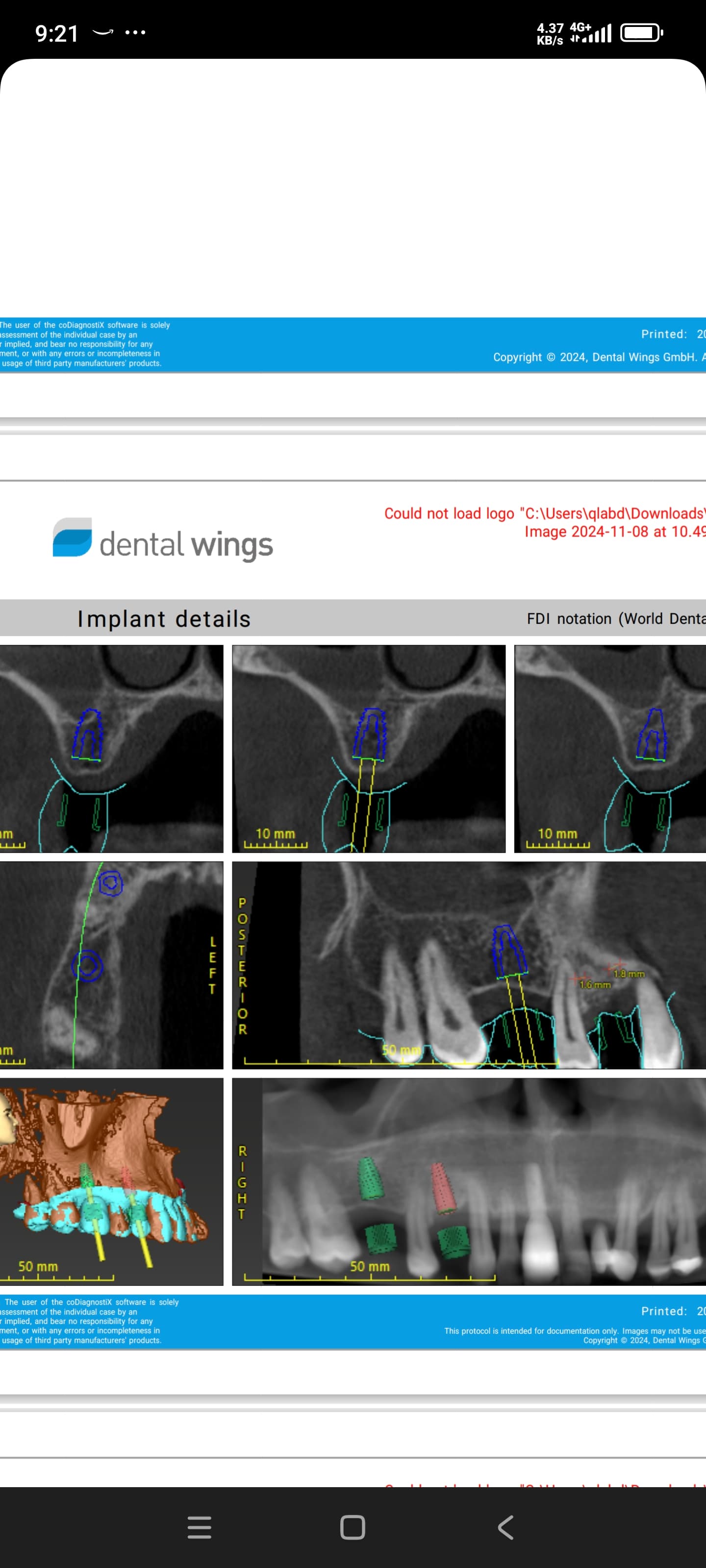
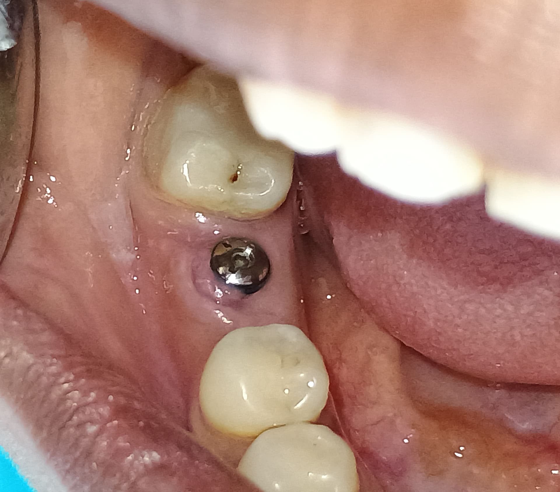
I have a 45 yr old male patient with no medical complications. He presents with a congenitally missing maxillary left lateral number 10 (2.2). The patient was referred to me by an orthodontist to restore the missing tooth. The patient terminated orthodontics when he achieved functional occlusion. He did not want prolong the orthodontic treatment to gain more space for the lateral.The space measured between the maxillary left central (9, 2.1) and canine (11, 2.3) is 4.0 mm. Is it possible to place a unibody mini implant into the site and restore with a crown or is a bridge the better option? Also what is the long term prognosis of a mini implant with a crown restoration?


25 Comments on Placement of Mini Implant: best treatment plan for this case?
New comments are currently closed for this post.
CRS
2/2/2014
Looks like with the undercuts on the natural teeth the space is even less than 4 mm, and a mini is your only option. I would suggest a resin bonded bridge.
Dr L
2/2/2014
Your patient made the decision to stop the ortho, now he can deal with the consequences. CRS is correct in saying the prosthetic space is probably less than 4mm. And yes, you can restore it with a bridge instead, but really, anything you put in will look like a tic tac.
Rand
2/4/2014
Amen!
peter Fairbairn
2/3/2014
Can work well with a mini long term on the laterals , have a case 10 years old and looks great .
Have an extreme case at 3 years , cannot tell not patients own teeth...
But agreed offer the options to the patient
Peter
John L Manuel, DDS
2/4/2014
Bicon makes a 3.0 W x 8.0 H mm that should fit as well as a 4.0 W x 5.0 H which is the better option. The 4.0 width is only at the incisal end where the taper into the abutment begins. The bottom of the 4.0 is only 3.0 mm wide and it can often be positioned slightly labial or slightly palatal where it resides not exactly between the existing central and cuspid.
Also, it looks like the Distal surface of the Central Incisor is rounded at center and a slight reshaping could give more room here, as could some room be made by a slight flattening of the Cuspid's Mesial surface. A slight overlap of the cuspid would allow even more Lateral Incisor width.
I often place a 4.0 x 5.0 mm just palatal to the Central and Cuspid roots with a slight division 3 flare so the crown would slide in place slightly labial to the Central and Lateral.
John L Manuel, DDS
2/4/2014
'Forgot to mention that, other than the pilot hole, the site preparation can be done with hand reamers which will not invade neighboring Lamina Dura areas, but slide along them due to the single cutting edge and solid guide planes laterally. I.e., they are "self-centering" in these tight areas and cannot invade cortical plate, nor lamina dura structures without a major effort.
John L Manuel, DDS
2/4/2014
Also, the tapered top of the Bicon Implant resides about 3 mm sub gingival with only a 2.0 mm abutment shaft protruding from the 3.0 x 8.0 implant and only a 2.5 mm abutment shaft protruding from the 4.0 x 5.0 implant. As such there is much room for circulation to support the gingival structures.
pisit
2/4/2014
Gain some space by proximal cut. Then ortho.you sill have new space more than 4 mm. May be 6 or7 mm.
DrSS
2/4/2014
This is the poster case for a mini implant.
They work fine! (selectively)
Yes it would be a one piece (implant and abutment)
It means you commit to angulation of abutment at placement stage...that is often a problem but this case appears to have ample bone.
Go for a mini (max)..11 or 13mm long...the maxillary type have a wider pitch thread for the softer bone.
Space is narrow for prosthetics....so you have some decisions to make.
a) Mesioangular rotation over the central?
b)Enameloplasty of the canine and the central to create a hint more space.
c)Accept the fact this lateral will be smaller.
Either way you will immediate load with a temp crown any way so play around with some composit and see what works for you and patient.
Sure the resin retained bridge will work...but why? You can have a single tooth with at least as good aesthetics...and not connection to neighboring tooth.
I don't see the downside of the mini.
ezgator
2/4/2014
well said!
Dr Bob
2/5/2014
space is very limited a mini implant would work but the tooth would look small.
Try bonding an acrylic denture tooth first the patient can then live with the look of what can be fit into the space. Afterward a decision can be made to do a two unit bridge off of the cuspid or a mini implant. If the bonded denture tooth looks OK and stays bonded it may even provide a long term solution.
Mike Heads
2/5/2014
I have been using Osteocare Mini and Midi implants for over ten years for exactly this type of case, they work brilliantly. They go down as narrow as 2.35mm but you need at least 13mm of length to get a reasonable amount of surface area for oseointegration. The hardest part of the treatment with this type of implant is preparing a crown prep onto such a small abutment. You need good loops and a very steady hand. I agree with using a temporary tooth to sort out the aesthetics first and I regularly find I need to steal a little space from both the distal of the central and mesial of the canine to improve the aesthetic result. In my opinion any one who even thinks about placing a bridge in this case is not giving their patient the best possible treatment available for them.
CRS
2/5/2014
My resin bonded bridge comment is based medico legally offering all options to this patient who did not follow thru with appropriate ortho to provide "the best possible option" for this non compliant patient who helped set up this difficult situation in not allowing the ortho to be completed. However I find your Osteocare comments very helpful and if everything goes well then a great treatment option. I tend to be leary of patients which may have different expectations just a precautionary comment based on a tricky clinical scenario. Thanks!
CRS
2/5/2014
I took another look at the films are the mid lines off and there is space on the distal of the other canine? Interesting, possible additional room to be created? Possible rapid ortho case? Sometime the early termination of ortho can prevent a good result for the implant. I would like to hear the orthodontic perspective since I can't even bend a Hawley!
John Manuel, DDS
2/5/2014
From a general dentist w/ 42 years of ortho tx experience:
The upper right buccal segments have migrated forward. The pre maxilla appears under developed. This is likely a division 2 case where the central incisors were more vertical at the outset and a small or missing lateral reduced the natural forces in development.
I prefer, in these cases, to first open the palatal sutures most heavily at the Mesial of the cuspid. These sutures open rapidly - a few months with fixed appliances. This movement alone would add several millimeters of lateral incisor space.
The last phase of fixed appliances torques the central incisor roots back Palatally at 1-3 degrees per month. This gives a more stable result and increases the inter cuspid width. More space for incisors.
Also, the posted photos show #8 to be thicker labio-Palatally and wider than #9. Could be a lens positioning thing...
In summary, while completion of comprehensive orthodontic treatment is ideal, one could open that right suture, Mesial to #6, several millimeters in just a couple of months.
John Manuel , DDS
2/5/2014
Correction on the side with missing lateral.
Also, CRS, incisive canal between 8&9 is large and pear shaped. We sometimes must tip the centrals mesially to close the midline contact and the maxillary midline often fails to match the facial midline. One cannot always take advantage of space on the other side of the arch.
As I mentioned, if you picture the bi's and molars as a "bus", the left side ( I previously got the sides switched ) the bus on the missing lateral side is parked forward relative to other side. You can get clear plastic graph lined arch form guides to lay atop your models which make it easier to see and measure Right-Left arch form discrepancies.
CRS
2/5/2014
Tough to make a diagnosis without a ceph but typically my orthodontists will strive to line up the dental mid lines with the skeletal base. Also in a 45 y o patient the palatal sutures are closed so a osteotomy is used to assist in a non growing patient. This could be a fascinating case for surgically sustenance rapid orthodontics. Do you have any experience with this? Please advise thanks.
John Manuel DDS
2/6/2014
If greater correction were needed slotting the cortical plate would help, but the necessary movements for this case should easily be accomplished with standard fixed palatal appliances, e.g., RMO 3D.
A clear template over a corrected size photo here shows upper left posteriors about 2+ mm forward and the upper left first bi to be about 3 mm closer to the palatal midline, so it's not too difficult to correct a deficiency using normal appliances.
Clearer records may show some forward tipping in these posterior teeth, and that's easily corrected.
Re: controversy over whether the pre max suture actually opens at this age, the fact is that it's very easy to get 5-7 mm inter first bi width increases in older adults. Some of this May be tipping. An AP ceph might allow measurement, but the apparent clinical result is more space.
The space Mesial to #6 on the other side is due to the undersized lateral incisor #7. We'd normally bring 6 back into contact w/5 and veneer or crown 7 to fill the space.
The crown size discrepancy between 8 & 9 could be explained by 9's having been more palatal and having suffered greater wear in a Division 2 case. When it's tipped labially the short clinical crown becomes more noticeable, and if the orthodontist puts the bracket on equally to 8, the you have this clinical result (which is what many patients prefer to avoid a crown).
Often, patients wit this Div 2 incisor tip back fruit ortho treatment as soon as it looks better. This often leaves an unstable and disproportional result.
Re: your question, I do not do much of the bone surgery/ortho combination, only in limited areas of extreme cases. That's what the "Big Boys", like you, are for!
CRS
2/9/2014
Thanks
Richard Hughes, DDS, FAAI
2/7/2014
CRS hit the nail on the head. There is more space distal to #6. The patient needs to return to fixed orthodontic treatment. I would like to see articulated casts and a ceph.
Do the case correctly or live with extremely compromised results. C&B from cuspid to cuspid is a possible and radical option.
I suggest a diagnostic wax up for the doctor and patient to evaluate prior to any irreversible action.
David Vaysleyb
3/21/2014
Do a bridge. Way easier and more predictable. <--- Trust me
If you decide to go for mini-implant, patient has to be ULTRA-cautious. Soft foods for a month, nothing sticky that could pull it out (caramel). ABS no immediate load.
Problem w/the mini-implant is that it is a 1-piece abutment+ implant. This means PT will be walking around w/a peg for a tooth for 6-months. Most won't like that.
Easier and simpler to do the bridge.
dr.m
5/24/2014
I will put a mini but defiantly keep the contacts light.
Richard Hughes, DDS, FAAI
5/26/2014
This is an interesting situation.
First I would measure the anterior teeth mesial distal. Then measure the B-L and M-D dimension of the site in question.
A diagnostic wax up is in order for several obvious reasons.
The shape of the papilla will be an issue.
Determine I'd any tooth structure can be sacrificed on the distal of the central and mesial of the cuspid. Either way use a MIS Uno or AB Dental I6 implant.
This has the potential to be difficult.
A Maryland bridge is an option.
sergio
5/27/2014
I thought after all these years, major doubts about mini implants for this kind of use were diminishing.
Clearly, I was wrong.
Use mini. It'll work well and the patient will appreciate that.
For some comment above where the patient will be walking around with a peg because it can't be loaded etc.., it just tells me that still there are lots of people who put their opinions without good amount of experience with minis.
Make a temp crown after placement. Put that out of a bite. Take an implaression before cementing the temp. Wait for about 5-6 weeks then cement the perm. crown.
It'll work well as long as it's done in hands of experienced dentist.
Bicon or any other 3.0 mm implants will work too except, to put 3.0mm implant in this tight space, you better be really good with your surgery. Otherwise, you cause problems make you deal with other teeth around the implant in the future.
sergio
5/27/2014
In my opinion, larger conventional implants tend to have gingival issue ( recession) over time every once in a while and force you to deal with them if they are in esthetic zone.
With minis, they DON"T look good initially but over time you have less issue with gingiva if a patient keeps it clean because gingiva was never manipulated in any way.
Both have pros and cons that way in terms of esthetics.


