Purulent discharge and radiolucent area around implant: Advice?
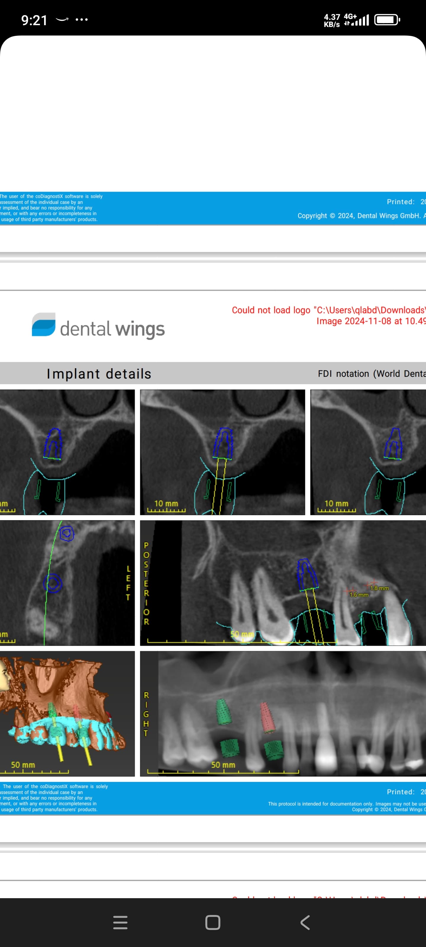
I placed an implant at area 32(lower left lateral) with bone graft and clindamycin 300mg tid for 5days. After 2 weeks the patient started to feel pain. I have seen him again after 18 days and there is purulent discharge and a radiolucent area around the implant. I then did a slight irrigation and prescribed Augment 625mg tid for 7 days. What do you advise?


13 Comments on Purulent discharge and radiolucent area around implant: Advice?
New comments are currently closed for this post.
roadkingdoc
2/19/2018
Pain,discharge and radiolucency add up to failing implant in my office. Placement pic looks good. Failure always disappointing for all parties. In my office watching and wishing adds up to more bone lose. Seems sometime everything goes well and the damn thing fails. Good luck with the case.
Doc
2/20/2018
Unfortunately implant failure sometimes happens, it's very disappointing I know.
I feel this implant is past the point of return and I would remove it. With a purulent discharge I wouldn't bone graft at the time of removal. I would remove, saline irrigation, allow to heal for about 6 weeks, and otherwise reenter and place another implant or GBR with a later stage implant.
I often like to revisit these cases and see where you may have gone wrong? Medical Hx and a systemic issue; drilling sequence; partial denture causing trauma; etc...
Matthew DMD
2/20/2018
Agree with roadkingdoc. Extract ASAP, curette area thoroughly, then bone graft. Wait 4 to 6 months and replace implant. Be sure to flap the tissue as to confirm buccal plate stays intact.
Good Luck!
Dok
2/20/2018
Take it out, curette well, graft and allow the site to heal fully. Don't want another failure in this site so next time be more vigilant with diagnosis, treatment planning and surgical protocol. Perhaps even have an oral surgeon/periodontist do the surgical work and you do the prosthetics. Don't want to make the same mistakes twice.
Fredrick Shaw
2/20/2018
I saw some multiple failures that looked very similar to this case. It involved several perio residence who went through a short period of time as a mystery. It was concluded in their program the surgical kit used by the department was not monitored regarding number of times surgical drills were used. It was investigated and turned out the heat created by the dull cutting instuments created pseudo periapical pathos is. Although the top of the drilling sequence could be adequately cooled with saline, the apical osteotomy incurred unfavorable high temperatures enough the bone “died backâ€.
Matt Helm DDS
2/20/2018
This implant is already failed, clearly! Flap it so you have good access, remove the implant, qurette thoroughly making sure that you clean up loose bone spicules and sharp bone margins, irrigate, and don't graft except perhaps for some collagen to encourage more bone volume, and revisit in 4-6 months.
And review your insertion protocol and drills. This looks like bone necrosis caused by dull drills overheating, or excessive rpms during the osteotomy, or excessive pressure on the handpiece (likely due to the dense bone), or maybe even a combination of all three.
Darwin
2/20/2018
I am a patient and this happened to me. All on 4 uppers. paid $40,000 total about. The OS described it as you did Purulent discharge that was like "jello consistency". Then because of that failure, I was required to have 4 more implants placed so it would not matter if I had another single failure and then purchase new porcelain uppers. Another 20,000. As the first time it was all done in a single surgery under general. The advice above is excellent. It is a clear failure. IDK about a bone graft in that site IF you can sustain the uppers with many other sites as I had done. I am really upset that my DDS did not accept any personal responsibility and had me pay so much more for the "fix". I have a degree in the "science" field and made over 17 trips to his office out of state at that time. I can tell you the OS/DDS I went to was one of the few who started the "all on 4" over 20 yrs ago and was a pioneer in this field who has since retired, last year.
dr abrams
2/20/2018
cat scan see if you perforated the bone. ck. site after removal.
Richard Hughes DDS, FAAID
2/20/2018
All of the above are correct. You need to find out what caused this to occur. These things do happen. Hopefully, this is and will be an infrequent event.
John Rodriquez DDS
2/20/2018
Explant first then currette necrotic hard and soft tissue, irrigate and clean with tetracycline/saline solution (assuming no allergies). Have the tetracycline capsules compounded to exclude fillers. Hydrate your bone with this solution and add calcium sulfate to the mix and use this for your grafting (old school) and cover with non resorable membrane. Remove membrane once site is almost closed completely. Check site in 4 months and it should be ready for new implant. The only failure is not learning from them. Remember it is the practice of dentistry and even the best have failures...
Raul R Mena
2/20/2018
Dok,
I fail to understand the magic of an OS or a Periodontist. Don't they also have failures?
John, that is very good advice including the Tetra. I don't hear many using it anymore, but it is part of my grafting technic in cases with infection.
Patric
3/6/2018
In the old days we used to use gelfoam soaked in an opthalmic solution called Tetracortrel which was tetracycline with a corticosteroid. Work like a charm, even on dry socket.
Dr Kamil KS
2/21/2018
Unfortunately this case is a failure from all the presenting sign & symptoms.
AB use is a loss battle.
Agree with removing the implant, cleaning & curettes & leave for 2months I suggest.
Most likely due to bone heating during placement & possible insufficient irrigation.
It is annoying & disappointment to have failure.
We all been through I think.


