Radiographic Stent using Nobel Guide Not Seated Correctly: Best Options?

Dr. S asks:
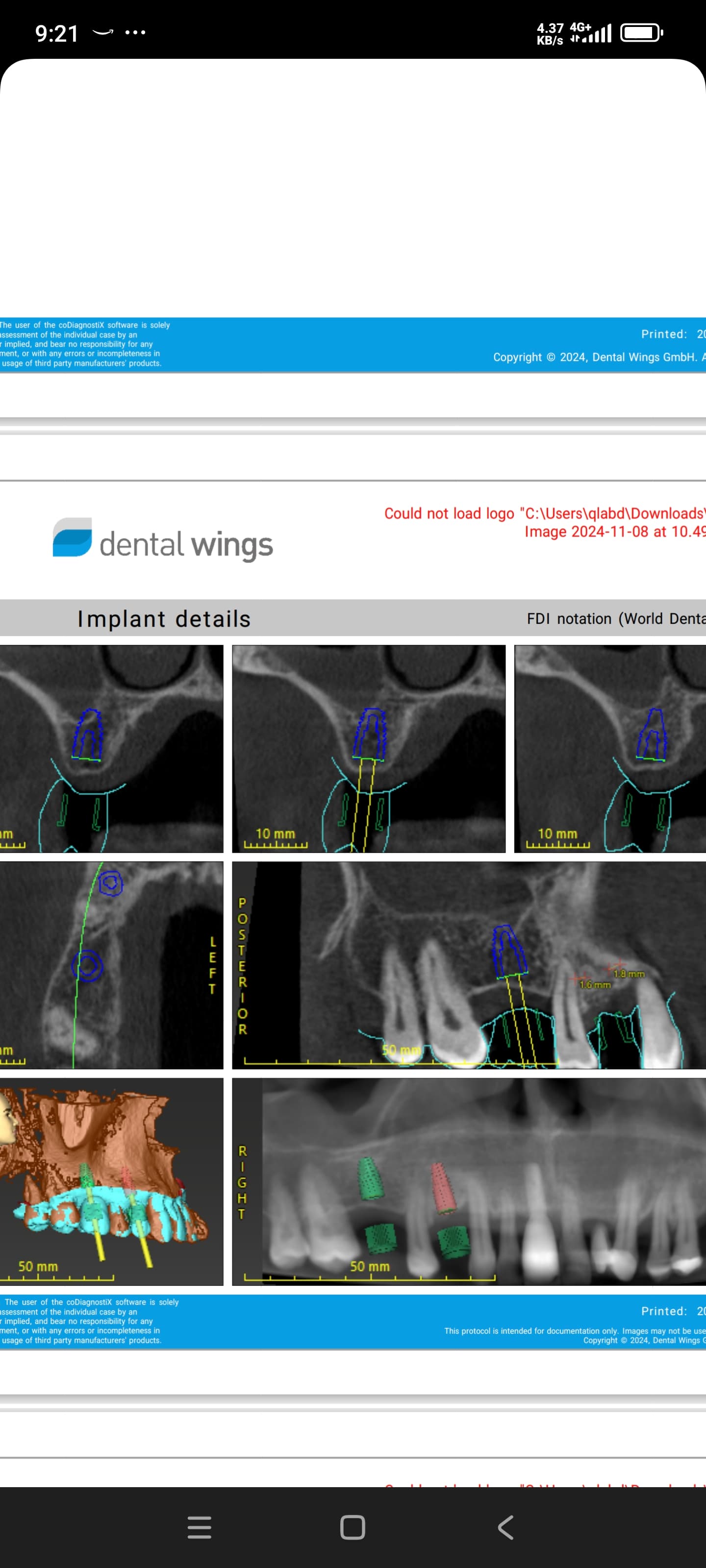
I have been treating a lady with dental implants for the past few years. One implant in particular in the lower left premolar region has been troublesome. The case was planned using the a Nobel Guide protocol. My assistant dentist did the work up and I did the surgery. Following the surgery it appears that the Nobel Guide scan did not go according to plan and I believe that my assistant did not seat the radiographic stent correctly, leaving it skewed which at the time of surgery had the left implant sitting much lower than planned and the right implants higher than planned.

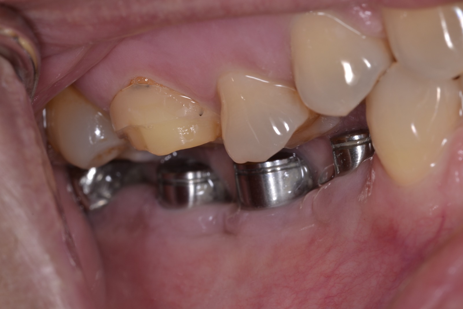
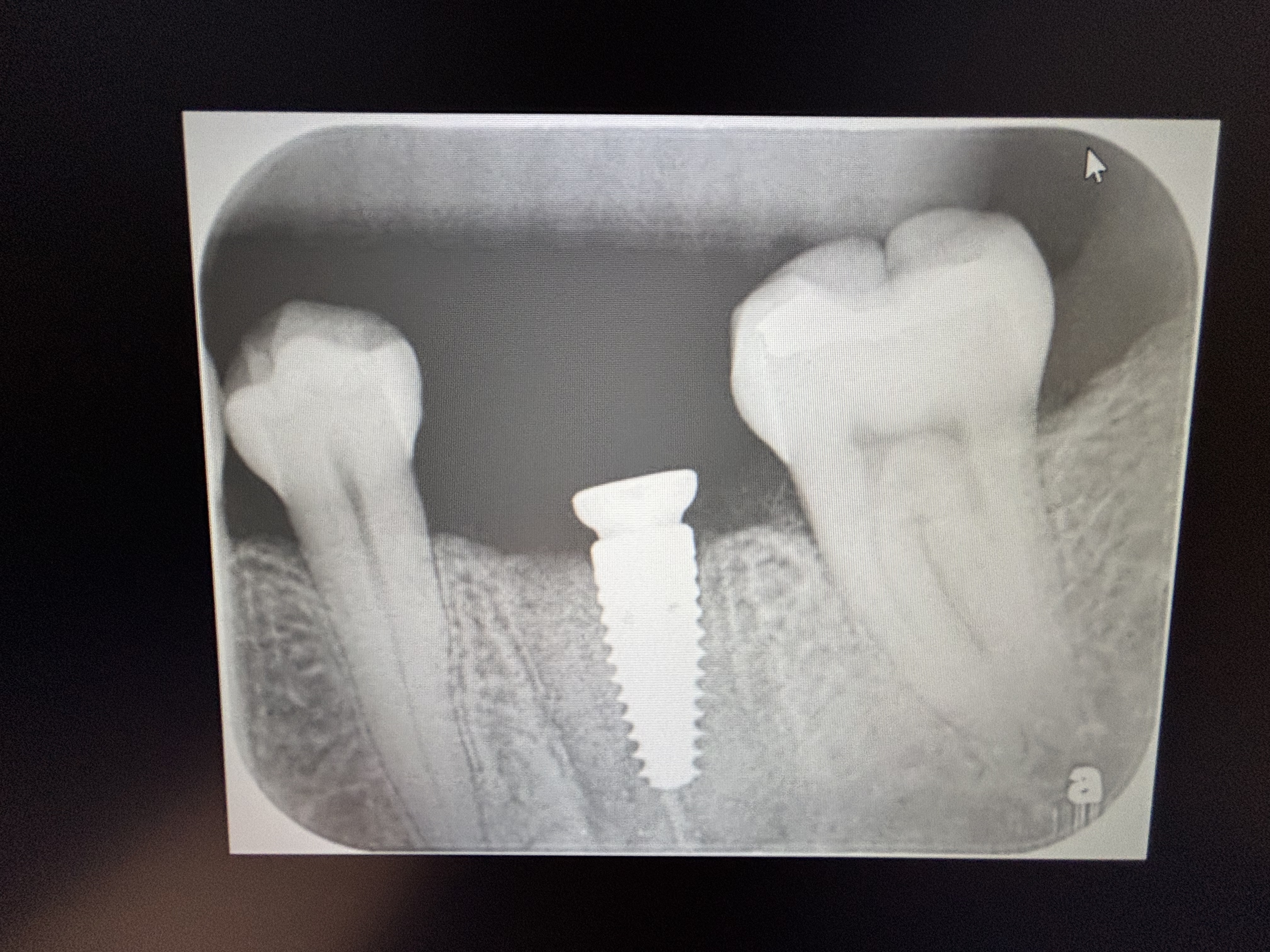
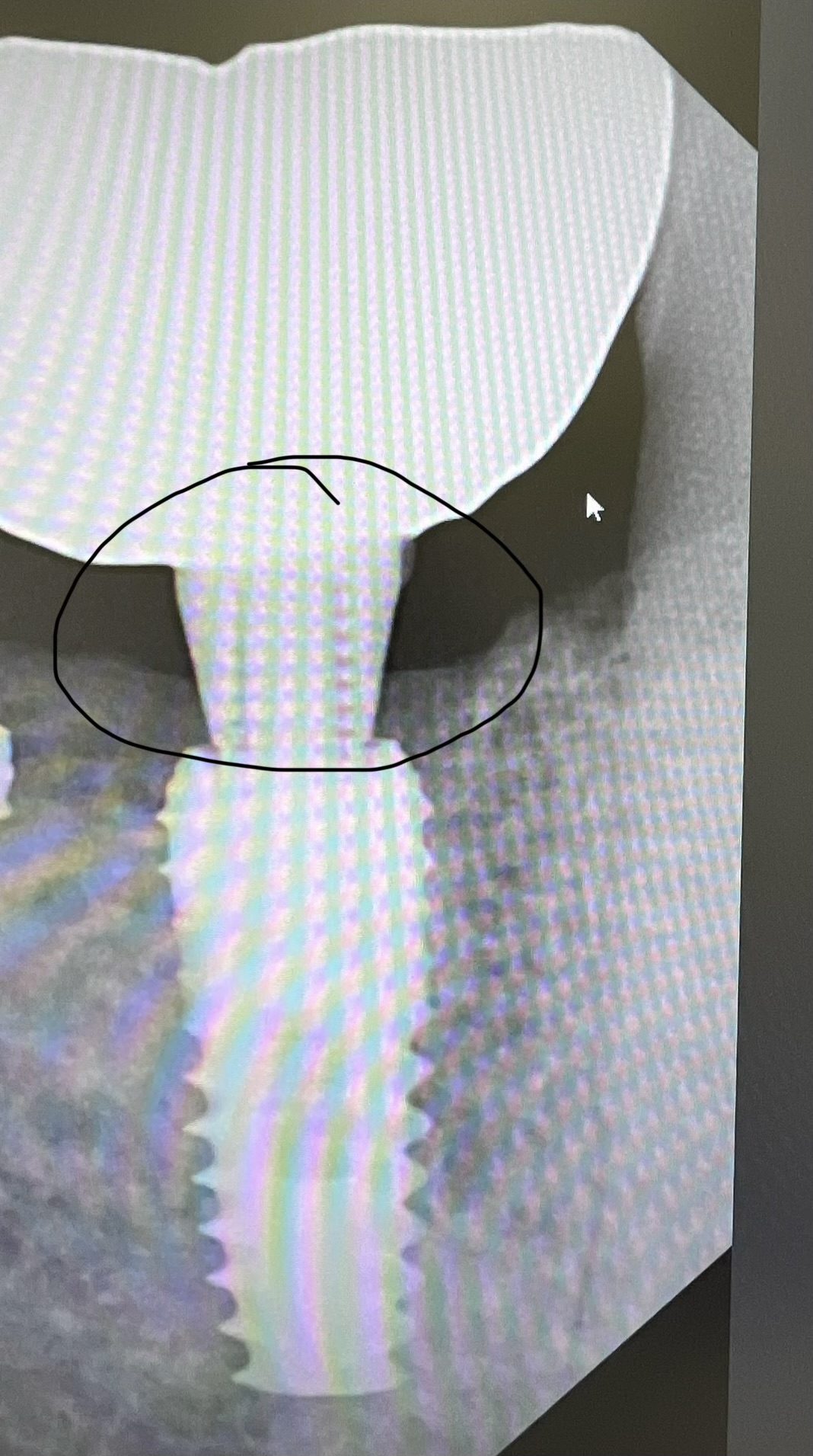
Anyway, I decided to leave the implants in situ and to assess with time. They have been in place for 3 years now. The left implant gives the patient a slight ache in the region and radiographically shows considerable bone loss around the coronal 2/3’s and good integration at it’s apical end. The radiographic image has not changed over the past 2 years as evidenced by the attached radiograph. The implant in question is a 10mm 4.3mm replace select implant and has been restored with an acrylic crown. I plan on removing it and augmenting the region but I’m unsure if this is the best thing to do. Could I have some of your thoughts please? Please note the molar has had the endodontic therapy redone.

January 2010

August 2011

37 Comments on Radiographic Stent using Nobel Guide Not Seated Correctly: Best Options?

New comments are currently closed for this post.
Italonet
11/14/2011
since you said that the Guide you used wasn't exactly in the right place and the crown you set in was made in resin there is a great chance that a malloclusion problem occured and overdose time the resin has soffered a natural wear and had its height decreased. That should has stop the bone load at some point. This considering that none pre operativa exam was shown in this time.
TOBooth
11/14/2011
take it out its a gonna!! vertical defect occlusion related most likely. Also why blame your auxilary?! You placed YOUR RESPONSIBILITY DONT PASS THE BOOK!
juan
11/14/2011
Hi! marginal bone loss is often related to marginal plaque acumulation and food impactation. also, povisional acrilic crown is prefered not leave in place too much time, especialy when the abutment is attached directly to the implant platform and too deeply. I do not would relate this with oclusal trauma, since it not fracture a simple acrilic crown.
naser
11/15/2011
radiographic stent has nothing to do with osseointegration bad oral hygien ,smoking. DM.and not least the acrylic crown might be the culprit best wishes for your assistant( redo)
Dr. Dan
11/15/2011
Bone augmentation won't do anything. The implant is partially integrated and will at some point fail...but I really don't know when to expect that. If the patient is comfortable with the temp she has in place, then stick with it. Inform your patient of the options and make sure she signs a piece of paper understanding the situation. FYI, the surgical guide had nothing to do with the implant placement or bone loss around the implant. Don't you take xrays to verify where you are going as you drill?
Dr Lee Nightingale
11/15/2011
My opinion ( for what it may be worth to you) is to remove the implant as a traumatically as possible, graft the site and re do the implant. The positioning and bone loss problems are too great to overcome, and I believe that grafting in this situation would be hopeless. Miscommunication between clinicians is a problem and errors do occur. Accept the mistake, put procedures in place to prevent it from happening again and move on. Nobel guide was designed to make implant procedures fool proof but the reality of the system is you really need to know what your doing to get it working to best effect. Good luck with the case. Lee
Vipul G Shukla
11/15/2011
Hello Dr. S, Your post seems to point the blame to your "Assistant Dentist" for not seating a stent correctly during scanning and none whatsoever for the dentist who placed these in the mouth! You also mention that the location is a lower second premolar and yet the X-rays are scanned wrong side up??? Did your "assistant" scan these too? I believe the NobelGuide technique makes you believe that an immediate prosthesis can be placed right after surgery and the immediate loading will not cause any problems, but it is not so every time. I think the problem here was loading too much too early and not enough time for osseointegration. Just a matter of time before it fails completely if not already. Want success? Remove the failing implant surgically, graft and close, implant again after about 3-4 months. And this time, wait for integration before the crown. 'Success comes to those who wait'
Pankaj Narkhede, DDS; MDS
11/15/2011
I would take it out, graft - redo implant after 6-9 months
Dr. David Goldberg
11/15/2011
Implant is failing. Remove, graft with GTR. And replace. A quick question, why did you use stent in this case. It looks like a single implant between 2 teeth.
dr. bob
11/15/2011
before you do anything retreat or remove the infected first molar, and remove the failing implant. graft and redo the implant in the bicuspid area also the molar area if you extract the infected tooth instead of retreating it. failure to take care of the first molar problem will bite you in the but given time.
Adrian
11/15/2011
It really doesn't matter who made a mistake here. I think the important thing is that the patient's wellbeing be put in front of everything else. I wouldn't try to retrieve this case as chances are you could make things worse. Any treatment plan should ignore cost and focus on what's best for this patient. I would suggest removing the molar and the failing implant, bone grafting and allowing a period of healing. Then replacing both teeth with implants 5-6 months post healing.
Leo Chen
11/15/2011
It's no business about X-ray stent. The implant should be higher than usual, if X-ray stent not seat in proper. There are lots of factors related to bone loss. Now I suggest you collect more information, like BoP, pocket depth, attached gingival. If these are all OK, no BoP, Pocket depth<6mm, enough attached gingival, patient is not uncomforted. I believe wait and see continue is a good choice. Otherwise, take it out or refer her to specialist. GOOD LUCK! Leo
Dr. Michael Gross
2/7/2012
Dear Dr. Leo Chen, you are mistaken. If the radiographic stent was not seated correctly while doing the CT or CBCT scan, the planning done in the wrong position to bone. Once the radiographic stent is designed to surgical guide, and this guide is placed correctly, what was too high in mouth now is deeper in bone. But not only there will be a vertical misplacement but also a rotational misplacement. Normally what was too high and then placed correctly will also result in a lateral misplacement. IMHO the lateral misplacement resulted in implant placement outside of bone in the coronal part and lead to bone resorption and failure. Michael
Baker vinci
11/15/2011
Here are my thoughts. Are you wearing a blind fold and flying VFR with these guides . You can't blame anyone for this ,other than the surgeon who placed this mess. This is my concern with these guides. They, by no means, were designed to replace an understanding of anatomy or implant science. Why are you using a ct fabricated guide in this situation. The reason most dental schools had/ have spatial relation assessments at interview time, is so the average dentist could have some basic skills sets ,with regards to depth perception, parallelism, symmetry and just plain simple engineering. I bet this patient is uncomfortable, her implant has been harboring a large pool of bacteria, acting as a breeding ground for bacteremias everytime she flosses or brushes. If you knew that the guide wasn't seated at time of placement, why didn't you toss if and eyeball the fixture or just come back. How much was the guide? Did the patient pay for it? I agree dr Dan he shouldn't pass the " book" or the "buck". Did this surgeon not expect a thorough " beat down". Let someone else take it out and tx appropriately. This maybe a bit tough! I personally think we are getting punked! Bv
jon
11/16/2011
I agree with Dr. Vinci. Way to try and pass the buck. It is always the "friend" that screwed up. Get a good lawyer. If you are a GP, you are held at the standard of a periodontist or OMFS. Blaming it on your associate (if there WAS really one as I can not imagine someone placing the implant not wanting to be involved in the process other than surgery) is a load of BS. Good luck in court on that defense if a lawsuit goes forth. Refer!! Get help as you need it. Pray you are not sued as this may cost you much more. Anything done from this point pay for it 100%. I think it is also BS that #30 was "retreated". Please show me the x-ray. If it was not you should do this N/C as well. Treat the patient, not the wallet. Good luck and sorry to be so harsh, I just call it as I see it.
jon
11/16/2011
Also, it looks by your x-ray the implant was more subgingival, not supragingival when placed (unless angulation is off). If it was guided surgery, can you post part of the cone beam slices at surgery time?
amgsl55
11/16/2011
Ask for a little advice and you get slammed. I'm not passing any buck,I was posting the sequence of events as they occurred and the intent is to remove the implant augment and then redo. At no stage has there been ever any mention of court or legal action, the patient is very understanding. The implant was not immediately loaded, the acrylic tooth was placed ( out of occlusion) to satisfy the patients short term requests. The placement being too subgingival was noted at the time of surgery however I decided at the time to monitor. Leo, you mention that the implant should be higher if it was not seated properly at the time of scanning, but consider that it was skewed at the time - which is what I believe is what happened. The right side implants are slightly higher than planned and well integrated but the left side sits too low. What I find interesting with this case is that there is no bleeding on probing, no exudate, placing rotational torque causes no pain and the radiographic appearance has not change in about 2 years, and she is totally comfortable. My post here was to get opinions - do you leave it until it eventually fails and then augment ( which at this rate could be another few more years) or do you remove it, create a larger surgical wound and then augment? When I get to my work computer I'll post images of the CT scan from the planning stage.
Dr Ham
11/16/2011
To All, Dr S needs help.... a lot of help. Dr. S, using a guide is not a crime and anyone can have an implant not integrate or a stent not fit. BUT this case demonstrates no application of thought at all. A guide doesn't do everything for you. Wake up!!!!!!! This treatment is crap on so many levels. Get more CE. The advice of removing the implant and grafting etc should be followed. Also do something about the endodontically affected molar.
Baker vinci
11/16/2011
Dr. S, you must have a "pair of bull testicles" to post this . In all seriousness , you had another doctor place the guide and you placed the implant? I quess going number 2 at your office is "quite a party"! If you are not comfortable placing a single imant without a guide, then you really shouldn't be placing them , yet. Ct fabricated guides are indicated for difficult cases, where occlusal planes are being reestablished , multiple teeth are being replaced or if immediate temporization is indicated. These are a few of the indications. The reason she is not bleeding or making puss anymore is because the damage has been done. The bone loss has probably reached it's steady state and that area has become a new space in the mouth. It is most likely just colonized with normal flora and the hydrodynamics are such that the body has adapted, as it most usually dose. If this patient is not moribund, remove the implant and try to save the molar. You could get away with attempting to graft it upon removal, with the worst case scenario being loss of graft. People that are well informed , or friends with their doctors don't sue , according to omsnic literature. Patients that don't know any better, typically don't sue either. You need to refer this out and pay for it all. Bv
amgsl55
11/16/2011
Baker, did you read my original post correctly - there was more than one implant placed.
Baker vinci
11/16/2011
What does that have to do with it? We are looking at a single bicuspid. Yes, I read it, and read it again, because I didn't believe my eyes. It's just a failed implant, don't take it so personally. Maybe I'm trying to find material for my stand up gig . This panel is all in good fun and learning. Peace , bv
Guy Carnazza DMD
11/16/2011
Even though the damage is done it would be helpful to see a pre-op xray to see what was there prior to better help understand what you were working with and provide some insight on how to proceed in the future.
SBoral surgeon
11/16/2011
In this doctor's defense, he understands that the outcome is horrendous. That was one of the first things he said. He was just asking for advice on what to do now. The x-ray speeks for itself. Baker Vinci, I've said it before. More teach, less preach. Your beginning to sound like the crazy bio-OSS guy. This is an educational blog. Stop spitting venom and teach.
amgsl55
11/16/2011
Gentlemen, I seek advice from my colleagues on this forum. So far the majority is criticism. The criticism has been directed at why a colleague was involved in the planning yet I placed the implant and why use a guide for a single implant ( yet they have no knowledge of the rest of the work done) and even one calling my claim that the endo on the 6 has been redone BS ( I've emailed the admins to find out how to post images again so I can put your mind at rest regarding the endo) I doubt those that criticise can claim perfection in all they do and I'm sure have cases that embarrass them. I have seen many cases done by colleagues senior to myself which I would class less than desirable, even one done by an eminent perio chap which has hit the mandibular neurovascular bundle, yet I will not judge. As Bob Marley once said "before you start pointing fingers, make sure your hands are clean". I have been placing implants since 1998 - and not just one or two a month yet i have not seen one fail in the manner this has, hence my post here. My intent is to remove it however I have concerns about using a trephine in this area. There has been mention of using forceps with clockwise/anticlockwise movements to remove it. My next question is - how effective is this? Thank you for any sincere advice which you provide. Here is an x-ray of showing that the tooth was retreated by an endodontist:
miperio
11/16/2011
amgI55: It's great that you posted this and asked for advice. Some of the people on this board, like Baker, don't know how to teach or discuss, as SBOral says - they just spout venom, anger, and hurl insults. Best to ignore their rants. There are others who I think have provided incredible feedback for you without the anger and others who appreciate these cases so we can learn. Look forward to seeing more images.
Baker vinci
11/16/2011
I gave you some pretty good advice and explanations that were spot on ,from a scientific perspective. Sorry if you don't like my delivery, but I actually made myself laugh . Sb I'm pretty certain your training was soft, in that every time I did something stupid a had to pay to the wrath of some legends and they had a pretty good time with it. I never once took it personally. I will never be in the class of those giants , but i know a mess when I see one and think my tx planning skills are above par. Just wondering was that third molar amalgam placed via cbct guided technology? Bv
Baker vinci
11/16/2011
Use a trephine, or be prepared to, if it doesn't just reverse torque out. Don't attemp to"extract" it. If you break off the buccal plate and leave the molars dehisced, then you will have a "big problem." Get a new cbct and measure the canal, manually. DO NOT, rely on an automated pseudocanal edit. Lastly,quit your whining! Bv
Baker vinci
11/16/2011
Please tell me you aren't associated with one of the implant chains. I smell a conveyor belt. I cannot understand how you can be too busy ,to be involved with your own guide. Bv
TOBooth
11/17/2011
Hey, i just wanted to point out that it wasn't the auxillaries fault- really the chap who posted should know that someonewas going to comment. To be honest i wish Bv would stop posting because he is just unpleasant really. If he's got any balls he will post one of his horrendous cases so we can learn from him. Otherwise to be frank go elsewere please. Concur anyone??!!
Richard Hughes, DDS, FAAI
11/17/2011
What was the condition of the site pre surgery and what is the patients medical history? Just take an electro surge to the implant for 5seconds. It will come out easy in a few days. Do check haw far one is from the IAN and mental n.
osseonews
11/17/2011
Please restrict comments to the case at hand and please follow the posting guidelines so that an intelligent discussion can be conducted. Thank you for your understanding and participation. Editors at OsseoNews.com.
OMS resident
11/17/2011
Seriously, you've got to see the humor in BVs comments! They're "provokingly educational" (at least from a resident's POV). TOBooth - you're a big boy, you don't have to read it if you don't like it... By the way, kudos to dr. S for posting the case! It'll be interesting to read about the outcome (if you post it).
Baker vinci
11/17/2011
Pretty clever dr. Hughes! Does that work? Bv
Richard Hughes, DDS, FAAI
11/17/2011
BV, I have never had to do this but through the years, I have heard that touching the implant or abutment et with electrosurg will cause disosseointegration! I now this is not your case but the molar that received the endo retreat looks like it will only last 5 yrs give or take.
Baker vinci
11/18/2011
Molar looks overtreated, but I'm not judging , for god's sake. Have only done 10 root canals in my life, but do know that it will be difficult to remove. I quess the purrulence and granulation tissue will prevent any collateral thermal damage , when electrofulgerating the failing fixture. Bv
Dr TK
11/22/2011
I am among those that could be embarrassed by some of my cases. I have removed a few implants in similar condition. My first approach would be counter-rotational force, and I would not be surprised if this came right out. If that fails, I would use my long skinny diamond bur (don't know the number) to remove bone on the mesial and distal, and then re-apply torque. The best option is for this to rotate out, so I would stay away from the forcep and lateral force. I oppose the use of a trephine bur as the initial approach to remove this implant, because it will result in unnecessary bone loss. The selection of a trephine bur is dictated by the diameter of the crestal component of the implant, the only place that you need to remove bone is in the narrower apical 1/3. That can be removed more conservatively with a narrow diamond. Remember the buccal plate is already gone in this situation, so the only thing we are concerned with breaking is the implant itself. I have placed enough force to break an implant three times. Two on insertion (lesson learned), and one on removal. All three failed vertically, by cracking through the internal hex, rather than horizontally. Once that crack forms, the ratchet will simply turn inside the implant with no torque exerted. I used the same approach described above to remove them, except I applied rotational force to the outside of the implant with orthodontic wire pliers (very small with great grip). Looks like you are headed in the right direction.
TOBooth
11/23/2011
Yes reverse torque or mesial and distal furrow and reverse torque. Wait 3/12 implant and augment or just augment. Implant placement may not initially be possible due to the apical position of the lingual plate, ie implant will be too deep but it may well end up deep anyway.

Featured Products

OsteoGen Bone Grafting Plug
Combines bone graft with a collagen plug to yield the easiest and most affordable way to clinically deliver bone graft for socket preservation.
CevOss Bovine Bone Graft
Make the switch to a better xenograft! High volume of interconnected pores promotes new bone. Substantially equivalent to BioOss and NuOss.