Rapid bone loss around maxillary anterior implants: advice?

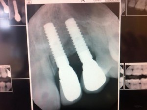
I am a Dental Hygiene student. I believe there is something not right with my dental implants. I was born with five congenitally missing teeth. I have three implants- #6, #10, & #11 [maxillary right canine, left lateral incisor and left canine; 13, 22, 23] . These implants are just over three years old. I first realized there was something not right about my implants when I looked in the mirror and could see the abutments through my gingiva, not so much by #6, but pretty bad by #10 & #11. The tissue is overall healthy in my eyes. There does appear to be a fistula between # 10 & #11, but no supparation, bleeding or tenderness, in fact there is not tenderness or sensitivity at all.

In my radiography class we took full mouth series on each other. That’s when I saw the bone loss around my all of my implants. I made an appointment to see the periodontist who installed my implants. He just said everything looked find and to check back in 6 months. I did some research on my own. Bone loss around implants seems pretty common, but not to the extent that mine have experienced and not as quickly as mine occurred. I tried to do some more research and I asked the periodontist who installed my implants to send me the radiographs taken prior to my implants being installed. The receptionist told me they didn’t have any on file and that I should contact the general dentist who placed my crowns. I called his office and they also had nothing. Finally I called the periodontist again and demanded answers. The receptionist told me she found some radiographs from 4 months prior to my implants being placed in their paper file. I ask her to please send them to me. She said she would, but they were not the best quality. I asked her to let me know who took them so I could acquire a better original copy. She had told me they definitely were not taken at their office because they did not have that type of machine. She said there was no name on them, and she had no idea where they came from. I received them in the mail. As I expected, terrible quality, and not able to reveal much useful diagnostic information. I would assume my bone at the implant sites was healthy prior to implant installation or the periodontist would have said something or done something.

Since my teeth were congenitally missing and not missing because of infection the bone would be at its normal level. I am almost positive that I do not have peri-implantitis, and this is due to inadequate surgical preparation, incorrect installation, fracture of the fixture, or inappropriate prosthesis design. Can you help me with advice?


![]implants](https://osseonews.nyc3.cdn.digitaloceanspaces.com/wp-content/uploads/2014/02/implants.jpg)

35 Comments on Rapid bone loss around maxillary anterior implants: advice?

New comments are currently closed for this post.
Board Certified Periodont
3/3/2014
My problem is not with how the implants look like on the x-ray, because they might be ok after all, and you may not need any treatment. I can't give a full opinion without a thorough clinical and radiographic examination. But here is where I see a big problem in your story: If the periodontist placed your implants without having sufficient pre-surgical x-rays in good diagnostic quality, and if a thorough clinical and radiographic exam is done by another independent board certified periodontist to get a second opinion, and if the second opinion results in diagnosing your implants with problems that could be the result of poor treatment planning due to the lack of good pre-surgical x-rays, and if the said problems require additional treatment such as grafting etc. then you may be able to pursue a malpractice complain in two separate avenues: 1) file a formal complaint with the dental board for a professional misconduct (substandard care), which may result in some kind of disciplinary action. 2) if you need additional procedures to correct the situation, then you may also file a law suit in the civil court for financial compensation to cover the needed procedures. Always look for specialists who are board certified by their American Specialty Board, and always look for independent practitioners in their own practices, and always read online reviews when they are available. Regards,
Dr SSenGupta
6/3/2014
Relax about the encouragement to patient to file complaints ....a lot more info is required before that road is taken.There is good clinical advise and comments on this forum.That is the whole point of this site.
ttmillerjr
3/4/2014
Hello. I'm sorry your stressed about your implants. Some more x-rays (include bwx) and photos would be helpful. What leads you to believe you have a fistula? Assuming you do not........ Going off the info we have now I have a few thoughts. The bone adjacent teeth will never be closer than 2mm from the cej. So we can't expect to see any bone until we measure at least 2mm apically from cej of #12. I can't tell for sure, but it looks like the restorative platform of the implant (#11) is about equal with the cej of #12. If so, a deeper placement would have been ideal. From this pa it also looks like #11 may be a little close to #12 . That could account for ( low blood supply)the vertical bony "re-modeling" distal #11. It would have been helpful to have seen the post-op x-ray too. If the metal showing through doesn't show when you smile and you are not getting more recession and or losing bone as time goes by, you might just try maintaining them. I'd recommend a Water Pik type irrigator and Super floss. I hope those were cogent thoughts cause it's late, too tired
Richard Hughes, DDS, FAAI
3/4/2014
This could be a case of pressure (avascular) necrosis, which is most unfortunate! This does need a surgical revision.
CRS
3/4/2014
My qualifying question is Did you have any preoperative grafting prior to implant placement? The place where one can get burned with congenitally missing teeth is the lack of bone development due to the missing permanent teeth. It has not much to do with a board certified periodontist placing the teeth or avascular necrosis. It is about understanding the concept of congenitally missing teeth. Sounds like the person who placed the implants did not plan for this and now these implants need aggressive grafting, removal of the crowns and burying the implants and wearing an Essex to keep pressure off the grafts. This is a tough case to restore, chasing the bone. at three years I would consider these failing not due to perio but lack or site preparation which is key in these cases. Find a good surgeon, consider an OMFS ,to fix this who has experience with these scenarios sorry to be so harsh and bruise egos but I see this often in my practice. It is just the background and training and understanding the cause. It is not as easy as it looks. I don't have a clinical photo for soft tissue or a CT for buccal plate so please take my comments in moderation.Good luck!
Richard Hughes, DDS, FAAI
3/4/2014
CRS, Thank you for the information about congenitally missing teeth! We can all learn from each other.
DrT
3/4/2014
I agree with CRS....however, I disagree that an OMFS is the only specialist who can help you. I am a periodontist and I often see implants which have problems that were placed by OMFS. It is not really the speciality certificate of the individual that is important here, but rather what is paramount is the experience and expertise of the individual practitioner. I think Dennis Tarnow is a periodontist, and I would trust my implants in his hands in a heartbeat. I also agree with the first few postings....it sounds to me that the standard of care protocol was grossly not followed when these implants were originally placed. I wish you all the best. DrT
CRS
3/5/2014
Dear Dr T, I do agree with you and I have Dr Tarnows original sinus lift videotape of sinus lifts yes I said VHS! So you know I respect periodontists, but my background in orthognathic, fracture and cleft grafting is based on experience on bone physiology based on grafting so please respect where I'm coming from. I also respect periodontal pathology and soft tissue grafting which is part of my background. I just want to state that site preparation is key and it has been my experience in my practice to see how this is sometimes neglected prior to implant placement. Congenitally missing teeth are tricky! But least not let's not for get the most critical element the restoring doctor who has the part that shows and is the most important team member just because an implant can be placed doesn't mean we should. And I have also come across some pretty poor OMS placed implants when the principles have not been respected so my main point is that in congenitally missing teeth the developmental framework is not present as seen in orthodontic cases with congenitally missing teeth Thanks for the comments.
Mark Montana
3/4/2014
Good post! Too often we see these results characterized as peri-implantitis. My experience is that bone loss in the absence of inflammation is often a sequela of stress to the bone's blood supply during implant placement. If the ridge is too narrow to accept the implant, it becomes thinned and the vascular network breaks down resulting in remodeling. I have received patients from very good oral surgeons, periodontists and general dentists with the same problem. There has been much recent discussion in implant/restorative groups about using narrower implants in the anterior maxilla because of these issues. If it is remodeling, then hopefully this is where the bone is happy and it stays put. Unfortunately, once the roughened surface is exposed, the bugs have a place to hide and inflammation is more likely to develop. An option is to seek more esthetic restoration and live with the loss. Surgical correction is really unpredictable. The disappointment is the lack of proper records from the parties involved. There really is no excuse for not having the minimum of a diagnostic pre-op image and a post-restoration image. Any attempts at diagnosis of future health or disease are limited without a baseline.
Michael Huynh, DDS
3/4/2014
Hey 'board certified periodontist', I cannot help but to say shame on you to tell this nice lady to go complain to the dental board. I am sure you place perfect implants in your office at 100% successful rate, you may go to other website for that. She wants some answers and i would tell her to seek another dentist to get it treated instead of trying to talk bad about other dentist pal!
Vipul G Shukla
3/4/2014
1) Please also post the X-rays of the other three implants placed by this periodontist and restored by your dentist that have no problems. 2) Placement appears to be perfectly placed, sinus floor expertly engaged by one of the implants, both appear to have 'platform switching' built-in. 3) Crowns appear to be cemented type. Correct me if I am wrong. 4) Bone loss was only noticed in the third year and that too accidentally. Are you not going for regular recare/follow-up appointments with your dentist/periodontist? As a dental hygiene student, you probably understand the importance of re-care more than anyone else? I'm sure you did not assume that implants do not need maintenance? 5) Horizontal bone loss after the first year around an integrated implant is either due to incorrect occlusal load or large gobs of cement overhangs (not always seen on X-rays) or very poor periodontal hygiene. Definitely not a failure of osseointegration. 5a) An implant does not integrate fully first and then start to lose bone because of avascular necrosis from incorrect placement torque. If that were the case, it would simply not integrate in the first place. No question of the restoring dentist proceeding for a crown. 6) It does appear that the periodontist's record-keeping is inadequate, from your story. 7) yes, usually congenitally missing teeth areas typically do not have a lot of vertical bone loss, but horizontally, bone is always less than if a tooth had erupted there. This morning, I placed an implant in a 1.3 location (upper right canine) where an over retained primary canine was removed 2 years ago by her previous dentist. X-rays showed beautiful bone, and we planned a 3.75 diameter (No, we don't always insist on a CBCT), but guess what? Raised a flap and saw a very thin ridge bucco-lingually. Barely could place a 3.3 mm Narrow Platform with bone grafting to bulk up volume. Will see how it goes in next 3 months. 8) Have you learned about 'probing depths'? Can you post some numbers along with the other information? Also mention if you (or your classmate) finds pus in the pocket around the "failing" implants. 9) Have faith in the profession and those who will eventually be employing you. Your summary in your post "Since my teeth were congenitally missing and not missing because of infection the bone would be at its normal level. I am almost positive that I do not have peri-implantitis, and this is due to inadequate surgical preparation, incorrect installation, fracture of the fixture, or inappropriate prosthesis design...." shows half knowledge of the subject and your hurry to rush to a diagnosis when you obviously are not trained for it and your need to blame it on someone other than yourself (and I'm not saying this is your fault either) tells me you have not even gone to see your restoring dentist or any dentist for that matter for your current concern. Have you?
CRS
3/5/2014
Dear Dr Skukla, I see your points but this is quite a bit of bone loss in three years in what I assume is a young patient with reasonable hygiene. I just want to caution on the congenitally missing laterals which are not a slam dunk. I don't think this could be blamed entirely on the patient I would have grafted preop. Her history seems typical for inadequate site prep but of course my opinion is 20:20 hindsite. Thanks for reading
Sarah
3/5/2014
At the time the implants were placed I was not a student. I am only two semesters into dental hygiene school, But yes, I obviously go for regular prophylaxis appointments and check ups with my regular dentist. Considering all the money that was put into my dental work of course I am keeping up on my dental hygiene. There was never a recall appointment made after the implants were placed with the periodontist. I did see the dentist who did the restorative work six months later. I do not remember exact details, but I do remember he did not take any x-rays and he did say everything looked like it was healing nicely. I moved about three hours away shortly after this (back to my home town) so I started to see my regular hygienist and general dentist again. They had no comments or concerns with the implants. About a year ago, I noticed the tissue pulling away, and the implants became visible through the tissue. I made an appointment to see my general dentist.He said there wasn't much he could tell me, other than to make an appointment with the periodontist who placed the implants, so I did. I went to see him about 6 months ago (about exactly three years after the implants were placed) he told me I had a fistula, although the tissue looked healthy, and as I previously said, no suppuration. He wasn't too concerned with the bone loss because the tissue seemed so healthy. I do remember him probing the implants but I don't remember the readings. I don't think they were of concern, as he did not mention them and said the tissue seemed healthy. He did not take any x-rays at this appointment, even though I suggested he take some. He just looked at the x-ray photo I had on my phone. He told me to come back in 6 months and he would see how things look again. And as far as faith in professionals go... I do have faith. I just don't appreciate the manner in which my records were kept, and the lack of answers and information I am being given. I feel I have been extremely patient and polite with the situation, it just makes me uneasy with the amount of money that was spent. I realize things happen, I'm not looking to blame anyone. No one can predict exactly what happens when it comes to the human body.
Periodrill
3/4/2014
Quick questions 1. How often do you get a routine prophylaxis? 2. Do you get any food impaction around these implants? 3. Do you smoke cigarettes? 4. Any medical Issues? Ie diabetes, overweight?
ttmillerjr
3/5/2014
Encouraging a lawsuit and a board complaint right out of the shoot?! Most people are pretty reasonable, talk more with the periodontist. Problem solve with him, maybe show him this discussion. Without more info, photos and x-rays, it's hard to give much more advice, but it may be worth considering smoothing the threads making trans-mucosal segments, then redoing the crowns for esthetics. I have a feeling that the bone will stabilize.
Dr Tooraj Moravej
3/5/2014
i think the best person who can help you is your own peridontist , he knows what he has done and i am sure can solve the problem in a better way
peter Fairbairn
3/5/2014
CRS is right , congenitally missing teeth cases are a lot more complicated due the lack of development on the cortical bone and as a result you have a deficiency bucco lingually with a concavity . Maybe this case was treated with expansion and the Implant placement closer to the adjacent teeth would suggest that the surgeon placed where the bone was the thickest .... But grafting is critical in these cases , particulates at the time of expansion and placement or else this can happen.... A challenging case to correct ... As they say the only "failures " are other Dentists Implants.. Peter
RAK
3/5/2014
I like to see this interest in this problem case. In my experience, 25 plus years, the labial-lingual dimension is often very thin with congenitally missing incisors. Since most of the remaining bone is cortical rather than cancellous the repair potential is compromised. Often a ridge split procedure can gain the necessary bone dimension. There is always the risk of losing vertical bone height with any procedure in that area. Of course any well done grafting technique can increase horizontal bone, but many patients are strongly opposed to grafting, so it doesn't always get done. Vince Kokich had several cases where he moved incisors into congenitally missing spaces to sort of force the increase in bone dimension, then he would move the tooth back to where he needed it and an implant was placed in the widened space with good long term success before the site could narrow or relapse to former dimension. These implants appear to be placed by an experienced surgeon.
Peter Fairbairn
3/5/2014
Yes sorry meant cancelllllous bone ......... speed typing between patients ..
DrT
3/5/2014
I agree that there are some anatomical limitations in this case. I also agree wholeheartedly that in spite of our best efforts, thing don't always end up the way we had hoped. I think that a critical issue in this instance is whether standard of care protocol was followed both during the diagnostic phase of treatment as well as in the post operative phase. Based on the information that we have been given by the poster, it seems to me that the answer to both of these questions is No. If this was a close relative of mine I would advise her to request copies of all clinical notes relative to these implants including all x-rays and digital images from both the surgeon who placed the implant fixtures as well as the general dentist who restored them. I would also recommend getting copies of all of the maintenance appointments that have occurred since the implants were placed.
Kaz
3/5/2014
Good to see that Board Certified Periodontist Megalomaniac has never encountered a loss. The young lady did not ask how best to attack her periodontist and inflaming the situation with such blatant comments is not warranted. The problem may be that the crestal bone was very narrow and there was not enough bone for proper blood supply to the system. The bone has resorbed possibly to a stable position where there is enough bone to help maintain the blood supply. Taking off the crowns and placing cover screws and then grafting over the implants and defect is a very risky proposition. Very difficult to get vertical bone height over the threads. There is no research that shows that bone actually will grow to the implant after this sort of repair.and realistically there is probably a fibrous connection and not bone. The system may be stable now because of the potentially greater amount of bone around the implants than in the beginning of implant placement. I would recommend to wait and see if there is more bone recession over time and make sure that the area is irrigated well with chlorine solution with a water pic device. If the bone loss progresses then remove the implants and graft.
Kaz
3/5/2014
Richard Hughes, could you site some research for the avascular necrosis issue? Thanks
Richard Hughes, DDS, FAAI
3/5/2014
The best info is in the orthopedic lit but you can go on line a google avasculae or pressure necrosis and dental implants.
stephen travis
3/8/2014
These are cemented crowns with marginal openings and most likely excess cement. The abutments are more than likely over contoured given current thinking. Rather than rushing to litigation, remove crowns, place ideal contour provisionals, see what the soft tissue response is and make decisions form there. Any surgical decisions at the moment are clouded by possible prosthodontic problems.
CRS
3/9/2014
Here is my treatment plan flap open site graft hard and soft tissue may need to remove crowns and leave buried. Then in four-six months evaluate if continued bone loss remove the implants and start over that will minimize the amount of total bone loss. I don't have a clinical photo to evaluate clinical placement or soft tissue but based on the film they are failing. Usually the placing doctor fixes this vs allowing the continued bone loss or they need to refer appropriately all this speculation won't solve much. That is how these cases are managed.
Gregori Kurtzman, DDS, MA
3/11/2014
I think you need some comparative readiographs to compare bone levels to, purhaps one taken after restoration of the implants. there are two schools of thought on bone loss on implants 1. watch and wait and 2. intervene early to limit further bone loss, in this case I would intervene flap the area and do an ailing implant treatment of the implant surface that is exposed (after granulation tissue removal, condition the implant surface with doxy paste then bone and implant surface with citric acid then place graft material and a membrane and close it up and let it heal
DrT
3/11/2014
How much do you want to put this patient through? and what about the financial aspect, unless you will be doing this non gratis. Also, do you really think this is going to regenerate the lost vertical bone in this case?
Gregori Kurtzman, DDS, MA
3/11/2014
Gratis no even if my case which I placed its been 3 years we dont give lifetime warranty on these do we? the poster is the patient and her complaint is that she can see the abutments at the gingiva due to the recession associated with this so it seems to be an issue for her and being in the esthetic zone it will worsen over time but if you intervene now we can improve it now and long term make it more stable. I would use a non resorbable HA so we dont have to deal with future resorption and mix that into a resorbable putty so we can get some reattachment to the implant. if treated properly you can get 100% or very close to it correction wise
CRS
3/12/2014
Agree with Gregori, however since you did not place the implants there is a considerable fee. If the initial surgery dies not work then remove before the loss is worse, with the remover kits available we don't have to wait. Also may I mention ruling out occlusal factors!! The key to this bone loss is usually multifactoral. Since the original "surgeon" is not helping I am suspect on the follow up maintenance perhaps they don't want to fix this appropriately. I thought this was part of managing peri implantitis. We don't know how an implant will fare until we restore it, but I would intervene here. It would be interesting to see a photo or study casts to see labial-lingually if the implants are in the alveolar housing also a ct to view the labial plate!
David Vaysleyb
3/21/2014
@CRS- Your ideas are ALWAYS on point. Here are my thoughts - Tough case to repair. Ideal course of action is remove crowns, untorque abutments, place cover screws. Thorough curretage of affected areas. Flush affected areas (I use hydrogen peroxide on sterile gauze). Place bone graft. Lay over PRF membrane( autogenous fibrin membrane from blood centrifuge), then cover tightly w/sutures (preferably PTFE) - Congenitally missing teeth area always problematic. When teeth fail to come in, bone shrinks (or perhaps never grows in). Bone growth follows teeth growth. - I DISAGREE W/CRS regarding bone graft pre-implant placements. In my experience, you can often skip this step. SPLIT RIDGE+ SPREADERS are a godsend. Very important for these borderline cases to flap it very wide so you have good buccal/lingual visibility. 2 buccal releasing and lingual envelope flap--- If you do indeed perforate, you can visibly see it. At that case, remove implant and place bone graft. - Less aggressive option- It looks like you have 3-4mm of bone-loss on a 11.5mm implant. While this isn't ideal, it often can work for a good amount of time. I would get have them flap the area, curettage, and flush w/antibacterial solution (hydrogen peroxide or CHX), the suture up. Maintain hygiene and regular check-ups/Xray. Treat it like as if you were a perio patient w/6mm pockets. As long conditions doesn't worsen, do nothing.
THWDMD
4/18/2014
Having 35 years of implant placement and restorative experience, I would suggest you take full arch impressions, facebow transfer, cr bite, and mount the case on a semi-ajustable articulator to evaluate your occlusion. As we know, anterior guidance and the excursion pathways if not properly worked out with the physiological harmony of the patient can destroy bone around implants or natural teeth. I personally would have splinted #10-11 and require an essix type nightguard in your case. Improper occlusion or excessive loads can make bring any of us to our knees.
Gregori Kurtzman, DDS, MA
6/3/2014
I do not agree that this needs mounting on an articulator as its a finished case, check the occlusion in all positions intraorally (mouth is the best articulator) and adjust accordingly. With the horizontal bone loss I am not convinced its related to occlusion and we need to see a radiograph at time implants placed to see the changes if any in the bone levels
Barry
9/13/2014
What I see here is 2 implants placed adjacent to each other in the maxillary anterior which never works out cosmetically except for central incisors because there's no contra lateral papilla for the eye to compare to. You just can't maintain interproximal bone between implants to support papilla well. Everybody is trying to do it. Even with crestal bone preserving implants (platform switching, Astra). Granted there's more bone loss than the norm here but so much we don't know. 1) what was the bone levels at the time of healing abutment placement? And prior to final abutment placement? 2) what's the buccal-lingual inclination of the implants? 3) retained cement??? Implant the cuspid and cantilever the lateral is always my first plan.
LSDDDS
10/26/2016
I agree with Barry and others, "So much we don't know." Full work up needed first for more accurate determination of current status and remediation, hopefully minimal. But great commentary on potential scenarios.
Bill M
5/30/2017
I'm sure we all know a 3d evaluation would be the first thing to have done. Everything else is conjecture at this point. 2d imaging tells us nothing except if we missed the neighboring roots. Occlusion , cement, buccal placement with no facial protection, angulation ,explant or graft ? -- Personally, I think Post op conebeams should be the standard of care, especially if you are going to restore or "fix" an implant placed by someone else. I would want to know it is in bone and how much buccal bone is there. The fact that she can see the abutments through the gingiva could mean too far buccally . If outside the boney housing- particularly # 11- you can't graft and gain anything over the implants due to the lack of neighboring horizontal support in the arch since the cuspid is the most prominent of the teeth.

Featured Products

OsteoGen Bone Grafting Plug
Combines bone graft with a collagen plug to yield the easiest and most affordable way to clinically deliver bone graft for socket preservation.
CevOss Bovine Bone Graft
Make the switch to a better xenograft! High volume of interconnected pores promotes new bone. Substantially equivalent to BioOss and NuOss.