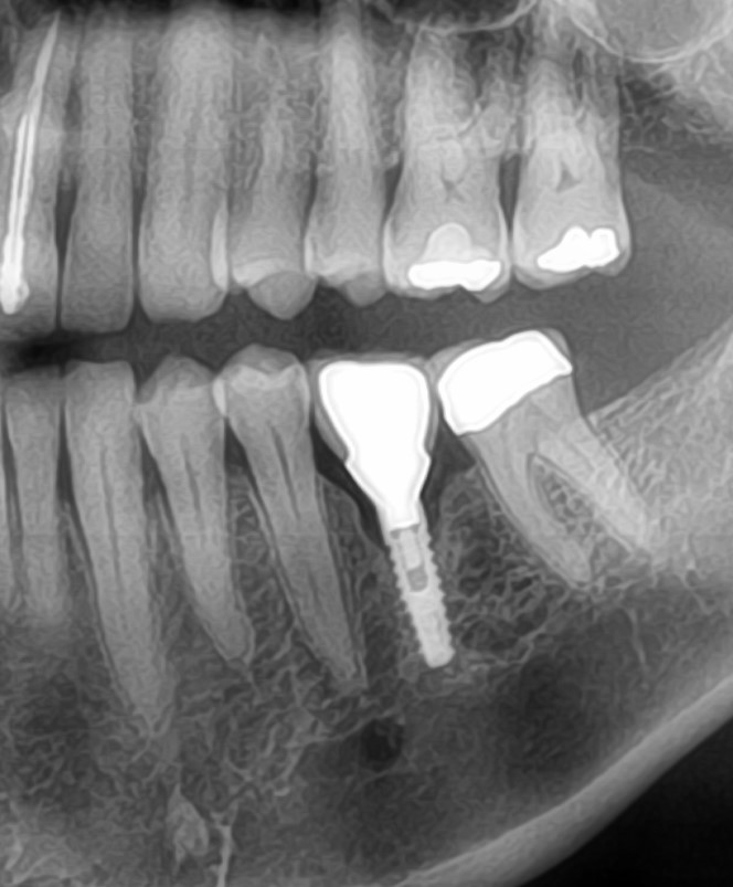
Re-endodontic therapy and place implant or place immediate implant after extraction?

Dr. R asks:

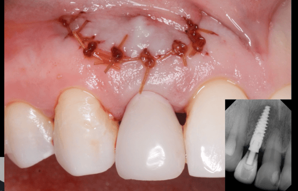
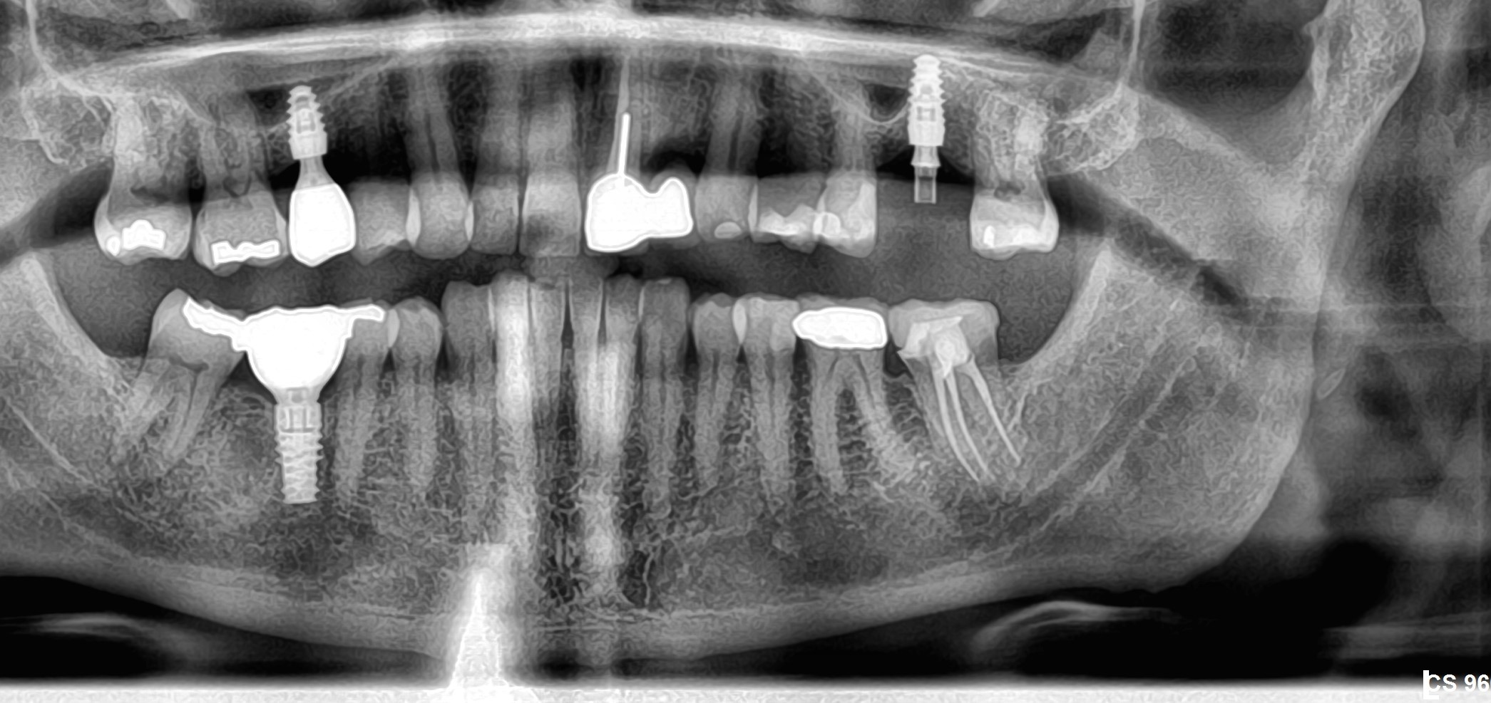
Male patient 36 years old, chronic smoker. Presents the tooth 22 with grade 2 mobility and periodontal examination distal bag of 5 mm, radiographic periapical lesion presents and loss of alveolar ridge in distal loss being in greater proportion. Poor endodontic treatment and poor prosthetic too. No fistula or drainage present. I have two treatment options: a) to repeat the endodontic and prosthetic treatment to rehabilitate the adjacent bone, with appropriate antimicrobial therapy. Once healed the bone, making extraction and place an immediate post extraction implant. b) Make the extraction, alveolar bone curette, antimicrobial therapy and placement of a provisional Maryland type bridge. Once healed the bone, implant will be placed. Which option do you think is better?…Do you have any other suggestions?

30 Comments on Re-endodontic therapy and place implant or place immediate implant after extraction?

New comments are currently closed for this post.
Danupas JS
4/17/2011
This is root fracture due to post crown. I think extraction this tooth and preserve socket graft and wait for 3 month implant placement.
Thomas Cason MFOS
4/18/2011
I would remove the tooth as atraumatically as possible and debride. Allow to heal and then assess for implant and grafting as needed. I am very wary about placing "socket presevatives" into potentially septic areas and I dont like to preserve sockets - I like bone to fil them!Then you can also assess the soft tissue situation.
Truth
4/18/2011
Do you like to fill it with resorbable or unresorbable bone substitutes?
DR MILAN
4/18/2011
hi, i would rather agree to go along ur way, as its clearly a # due postcrown load, do endo treatment again n allow healing, wait for 4 weeks osteogenesis time. then do extraction n allow healing in natural course n load implant , do atraumatic extraction, n debride throughly, i have similar case on 14, 15 loaded implant imtec-,without preserving socket n loading crown after 3 months n quite sucessful after 1 year follow up.
Simon Milbauer
4/18/2011
I think that it may prove to be a challenging case particularly from the cosmetic aspect, mainly due to the bone loss distally. After xla therw would be even more bone loss and since it is vertical could be very difficult to regenerate. I would remove the tooth with peiotomes and in this case I would not place any bone substitute or even collagen plug to the socket as it may retain the pathogens from infection. The reassess after 3-4 months but I would keep my mind open about bridge option.
Greg Steiner
4/18/2011
Due to the angle of the defect interproximal 10 and 11 this is a periodontal lesion on #11. Looking at the periapical lesion on #10 you have a very high likelihood of loss of the buccal plate on #10. A satisfactory result will require treating the periodontal lesion to regenerate the interproximal bone and any bone lost on the buccal and crest. I would remove #10 and debride the socket and root plane #11. Treat the surface of #11 with prefgel then do a ridge augmentation using Osseoconduct granules and Regen Biocement and cover with the periosteum. Place implant in 3 months. You can use any graft and membrane you find works well in your hands. The Doctors that are advising against grafting with pathogens in the site are using graft materials with macropores that become infected (allografts, xenografts and some synthetics). They are correct. Placing graft materials with pores large enough for bacteria to colonize will often result in an infected graft that will not respond to antibiotics. Place the patient on antibiotics and use a graft material that is resorbable without macropores and you will not have to deal with the potential of infected bone grafts. Greg Steiner Steiner Laboratories
Truth
4/18/2011
Schlegel und Donath [25] konnten bei 126 klinischen Biopsaten mit einem Nachsorgezeitraum bis zu sechs Jahren keine Resorptionszeichen nachweisen. BIO-OSS--a resorbable bone substitute? Department of Oral Maxillofacial Surgery, Ludwig Maximilians University, Munich, Germany. Abstract BIO-OSS is an allergen-free bone substitute material of bovine origin, used to fill bone defects or to reconstruct ridge configurations. Seventy one patients (39 female, 32 male) received 126 BIO-OSS implantations. Some health parameters or habits were documented to eliminate possible risk factors of influence. The diameter of jaw defects filled with BIO-OSS was measured. There was a significant influence of the defect size on the healing result. In X-ray controls, BIO-OSS served to identify the surrounding native bone. The density of the BIO-OSS areas was higher than in control sites. These radiological results were supported by bone biopsies. Histologically, the permanency of the BIO-OSS was still recognizable after 6 years and longer. The ingrowth of newly formed bone in the BIO-OSS scaffold explained the increased density of the implanted regions. There were no clinical signs of BIO-OSS resorption. Therefore, we can assume that form corrections achieved by BIO-OSS insertions will last. PMID: 10186966 [PubMed - indexed for MEDLINE]
Truth
4/18/2011
# paul carie May 31st, 2009 I can’t believe you guys. I had bio oss used on me 5 years ago. Never resorbed, still having chunks that have migrated everywhere taken out, gross sinus problems because of migration into the sinus. Dysguesia also. Why would any of you use this product? My dentist has photos of chunks he has been taking out. The company should be sued as well as the people who use this garbage.
Sb oms
4/18/2011
Once again, a perfectly decent question ruined by truth and the bio-OSS chronicles. Man, I'm sick of your comments. Here's an idea, start your own blog and leave us alone.
Simon Milbauer
4/19/2011
I fully agree with Sb oms.Out of 15 comments 7 are truth's conspiracy theory thoughts about bio-oss. It pushes us away from Dr R's original question.
John Manuel DDS
4/19/2011
A recent Bicon webcast showed just such a case. Dr. Shadi Daher removed the lateral incisor,curretted the socket, soaked the cleaned socket with sterile water to lyse the bacteria (antibiotics could alter the resorption of the Synthograft material), feeling out the size of the missing buccal plate, cut a Collagen membrane slightly wider than the buccal opening and long enough to go from the apical area up and across the open socket palatally. Without a flap, and through the open socket, the periosteum was gently loosened just adjacent and apical to the buccal defect. The Collagen Membrane was cupped a bit and slid down to the apical sound buccal plate in one fluid movement (going slow would cause the membrane to soften and it would curl as one pushed it apically). Then a mixture of the patient's blood was violently and thoroughly mixed with Synthograft crystals and that mixture injected into the socket with a graft injection tube. The collagen membrane was then folded over to the palatal to encase the graft crystals and covered with a section of Collaplug. The Collaplug cap was then held below crestal ginival level with a reverse cross suture of resorbable chromic gut. While the healing time may vary from 2-6 months, most cases would be able to have a Bicon implant in 2-4 months after this procedure.
John Manuel DDS
4/19/2011
If you go to the Bicon site right now, you will not be able to see this new video since there is about a month's delay between the initial webcast and the "webcast replay" listing. However, you could see some similar cases from the past.
mike stanley, asst.
4/19/2011
"truth" should at least not reuse the old post date, unless he/she/it wants us to refer back to the original post. Oh, and no-one mentioned Bio-Oss. Perhaps the moderator can block these repetitive posts?
dr.hosein akhavizadegan
4/19/2011
1- cast post removing in this case is close to impossible 2- what warranty the success of RCT ? 3- what warranty the success of periodontal therapy ? 4- if periodontal treatment is not completed , after fresh socket implantation the bone loss around the implant will happen after few month. i think the EXT and waiting 3 month is better.
Glory
4/20/2011
hi, my opinion: 1.- Periodontal treatment, because the other teeths have periodontal disease. Vertical defect is evident. 2.- Antimicrobial therapy. 3.- Atraumatica Extraction with bone alveolar preservation using bioss with autograft injert. 4.- colagen membrane and colaplug. (protect colagen membrane) 5.- Control RX 6.- after 5 months implant placement.
jg
4/20/2011
Right on SBoms!!!!!!...And, saying that...And without trying to offend any of the good opinions about re-treating ect, ect....This, clearly is a fracture root and agree 100% with Danupas...don't matter what technique you use,it must be ext. immidiatly, graft,wait then place your implant....
DrO
4/20/2011
I have had issues with heavy smokers and implant placement in prior infected upper anterior locations. This would sometime yield a cosmetic deficit or a failed implant. I now prefer to just place a bridge in such situations. An extraction, a graft if needed, a transitional and the final restoration several months later. In this case a 2 unit cantilever would likely work well. Quick easy and cost effective.
KPM
4/20/2011
It's curious that no one has talked about atraumatic extraction and immediate placement of an implant and bone graft as an option. Why? While it's obvious that the tooth has a recurrent apical infection due to failed RCT,if there is indeed a root fracture, and I tend to think there is, then the cause of the bone loss between 10 and 11 is due to that fracture and not any active periodontal infection. "Remove the splinter, remove the irritant". I see no reason why this tooth could not be extracted, the area thoroughly debrided and an implant immediately placed, with initial, solid stability and a bone grafting material placed. Preferably one with some body so it stays in place. Although, certainly, a granular graft with a membrane could be used. Of course, if depends on the patient and circumstances, but I say push the envelop. This, considering the coatings now on implants that resist infection and promote integration, the myriad of good, predictable grafting materials and the better understanding and experience of we, the clinicians. If we had not and do not push that envelop, at least on occasion, we would still be placing 14mm implants and waiting 6 months to restore them! Inform the patient of the positives and negatives of your plan. I have found that most patients are receptive to trying something that will lessen treatment time and/or number of visits. Within, reason, that is. I would not propose this if it were a circumstance which could not be reversed or remedied.
King of Implants
4/21/2011
I would not extract and immediately place an implant. Considering the size of the lesion, the buccal bone, most likely, has been compromised. You will get a much more predictable result by extracting and performing bone grafting, then placing your implant. Last thing you want is to immediately place, and graft and have titanium showing through the gingiva or having a result where there is severe apical recession of the gingiva. I'm all for pushing the envelope but not on an anterior tooth that is already esthetically compromised.
Dr. B
4/21/2011
I agree with those who say: 1. Extract, curette, socket graft. 2. Implant (four months post grafting).
K. F. Chow BDS., FDSRCS
4/22/2011
Extract and currette thoroughly. Screw in a mini dental implant and restore temporarily with composite, bonding it to the adjacent teeth for stability. Cover with antibiotics and wait for 6 months. The bone will have recovered and you can either do a PFM over the mini or unscrew it and place in a conventional and finish accordingly. The problems of infection and temporization are solved in a simple reliable way.
K. F. Chow BDS., FDSRCS
4/22/2011
It sounds simple but a narrow diameter that is stable is very conducive to healing especially in a compromised smoker's tissues. It allows immediate temporization and obviates the tedious process required to make and then remove a Maryland.
ttmillerjr
4/23/2011
Dr. R, This looks like a root fracture, I could use more info, but my first thought is not perio disease, it's root fracture due to inadequate post placement (too short). Does the patient have periodontal pocketing elsewhere in the mouth? In any case this tooth needs to be removed, no re-treatment. The gum will ride low around this implant and crown. This may be acceptable if the patient has a low smile line or is not esthetically concerned. As to the tx sequence, since blood supply is so crucial to bone grafting,and smoking effects this, graft first then implant later. ;-)
JAV
4/23/2011
It appears that someone tried apical surgery on this tooth. It would be difficult for anyone to do that bad of a job with the RCT. There will be no buccal plate to get stability. You would have to graft this type of case first. Yes we can graft, but chronic smokers and bone grafting does not go together. This is a case where a 3 unit bridge would be the best solution. You will not end up with a very good esthetic result. Chronic smokers have minimal blood flow to their tissues. I do not see good results with any type of bone grafting with a chronic smoker. This is not a situation to push the envelope. A low lip line could make this acceptable. But it will give you problems in the future.
Dr FCampos
4/27/2011
It might sound like a long shot but how about having your endodontist retrofill that root canal/bone graft defect,after that with ortho bring down the crown to regain some of that vertical bone especially on distal at least to even up as much as possible the ridge mesial/distal once you are there then extraction and implant placement.like i said before a long shot. Good luck
sajjad pezeshki
5/7/2011
hi,i agree that: 1)ext tooth 2)socket preservation 3)4 mounth latter insert implant
Landon
6/7/2011
Check out recent article from JOI Volume 37 March 2011. "Immediate Implant Placement into infected Sites:Bacterial studies of the Hydroacoustic Effects of the YSGG laser." by Edward Kusek, DDS Synopsis 1.Extract 2.Laser Site 3.Place Implant and Graft
RS
7/18/2011
i dont agree with some comments. Its not a question of achieving primary stability because i'm sure experience surgeons can lock into the palatal bone and into the apical 2mm beyond the existing apex. The concern is the active lesion present. Im my opinion, its best to extract and clean the site thoroughly. A bone graft will maintain the socket shape. After 4-6 months the implant can be placed with a further GBR technique should result in a good aesthetic outcome. The soft tissue can be advanced from the flap design by moving palatal tissue into a more buccal position if needed.
W ORoark, DDS, DABOI
7/18/2011
I will routinely extract and place the implant. Two absolute protocols, aggressive curettage, and since grafting after the implant is placed (assume a tapered threaded implant) is very difficult so...when ready to place implant, insert into blood in socket to coat implant with blood then roll the implant in HA graft particles. The blood will pick up a layer of graft that can be carried to the socket. As the implant is screwed into the bone, the graft will be displaced as necessary coronally, or remain in the defects of the socket. The implant must not be in just graft, it should be at least 2mm longer than the socket and engage the walls of the socket at some point. Must be stable. If the socket is grafted first after prep, before the implant is placed, tremendous pressure will be created as the implant is seated and post operative pain will result. If buccal plate is gone, a GTR membrane can be placed to the buccal prior to implant placement, see technique by Dr. Manual. Also agree with KPM. Patient should be on an antibiotic at least 48 hours pre op.
Richard Hughes, DDS, FAAI
7/19/2011
Wayne O'Rark: good job.

Featured Products

OsteoGen Bone Grafting Plug
Combines bone graft with a collagen plug to yield the easiest and most affordable way to clinically deliver bone graft for socket preservation.
CevOss Bovine Bone Graft
Make the switch to a better xenograft! High volume of interconnected pores promotes new bone. Substantially equivalent to BioOss and NuOss.