Retained Root Tip in the Sinus: How To Proceed with Dental Implant Placement?

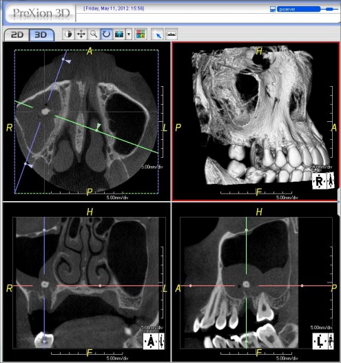
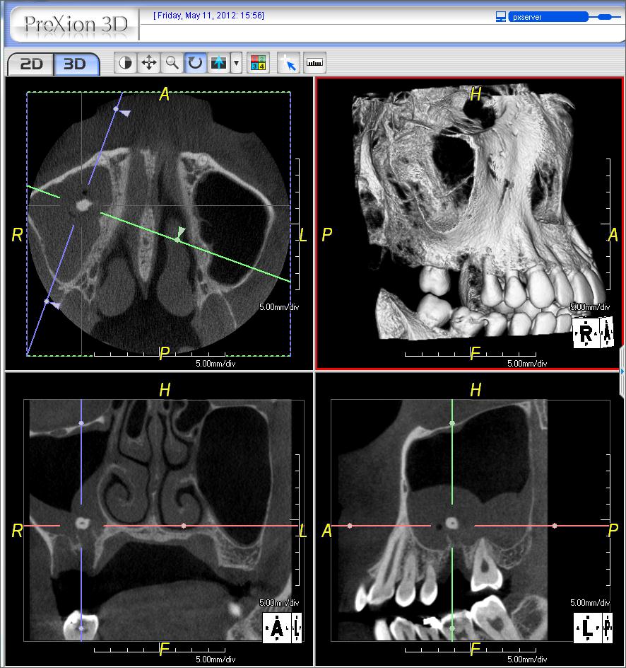
I am a periodontist and recently had a case referred for installation of a dental implant in #3 area [maxillary right first molar; 16]. Â The patient had #3 extracted 2 weeks prior and there is a root tip — probably from the palatal root — in the maxillary sinus. Â This is clearly visible in the CBVT scan. There is also an apparent oral antral fistula between the maxillary sinus and the oral cavity. Â I prescribed Biaxin [clarithromycin]. What is the recommended course of treatment to manage the root tip and graft the area for a future implant? How do you recommend I proceed? Â
(click image to enlarge)

![]retained root tip in sinus](https://osseonews.nyc3.cdn.digitaloceanspaces.com/wp-content/uploads/2012/05/Root-tip-in-the-sinus.jpeg)

45 Comments on Retained Root Tip in the Sinus: How To Proceed with Dental Implant Placement?

New comments are currently closed for this post.
Leal
5/13/2012
That is a preaty well unilateral inflamed sinus. I would prescribe Ciprofloxacin 500mg DID and some Fluticasone nasal spray DID for 15 days (OD for the following +/- 15days). Remove the root tip crestally during the antibiotic therapy, do a very good wound closure and stick a couple of Gelatamp with povidone-iodine. Wait 3 months +/- and rescan. If everything's OK sinus lift, wait 6 months and place implants. Let's hear some of the experts' opinions.
Baker vinci
5/16/2012
Leal, if the sinus is actively infected, getting wound closure, is the last thing you want. The wound will close, when the infection resolves. Bv
Leal
5/16/2012
Will the wound close 100% sure? Not even 50% sure. The oral cavity is a dirty place and an oral-antral communication does "quite" difficultly resolve by itself / "...will close, when the infection resolves". Fat pad closure is a common procedure for an oral-antral fistulae. Again: GOOD antibiotic coverage, good disinfection/degranulation and wound closure. Just my opinion.
Baker vinci
5/16/2012
These are all just opinions! In my experience getting closure on an infected sinus is contraindicated. Just as a general surgeon doesn't close an infected belly, we have no business closing an infected sinus. The typical oral antral communication will close itself, if all the infected material is removed and if the osteum is patent. Yes the transposed fat pad is a great way to tx the unusual, persistent communication. This particular patient, would be best served , in my opinion, by removing the source of the infection and letting nature take its course. This patient will regenerate 5 mm of alveolar bone, with no graft. Just because you have a bunch of tools, doesn't mean you need to use them. Bv
Gregori M. Kurtzman, DDS,
5/16/2012
Couple of points here.... 1. a sinus communication doesnt close on its on as the pressure in the sinus due to air flow thru the opening prevents this and the bacteria from the oral cavity keeps the area infected as the normal flora in the oral cavity is not the same as found normally in the sinus. So one will need to close the communication and various methods have been describe. 2. am sorry but strongly disagree that 5mm of bone will regenerate without some graft. if you have some cases to illustrate this please post them. 3. at this point the communication has possibly eplithelized which will also keep it open unless some surgical means is taken to close it to allow healing
David Nelson DDS
5/18/2012
Nice post BV. I am in total agreement!
Guy Carnazza DMD
5/14/2012
If you are not comfortable treating then ent/omfs should be consulted.
Baker vinci
5/17/2012
Dr kutzman, give me your email address and I will send you several images( cbct ) showing first molar sights that have generated 8-12 mm of bone, spontaneously. If there is a solid non- diseased tooth on either side of the defect, your gonna get enough bone, most of the time, to come back and do a standard sinus lift. Bv
Baker vinci
5/18/2012
Dr. Kurtzman, the 5mm or less rule has been proven in several studies. The communication from this small root tip, should leave less than a 5mm hole. Unless the patient is immunocompromised, it will close, once the infection resolves. Just because we go in and close these things before it happens spontaneously, doesn't mean it had to be done. I did a case ten days ago, where a radiologist refused a graft or an implant at an nonrestorable, perio involved #3. 4 months later he changed his mind and wanted an implant. I placed a 5.7x9 implant, without a sinus lift. Like I said, send me ur email and I will send you the ct slices, with the progress notes. Bv
Gregori M. Kurtzman, DDS,
6/28/2012
In a case like this I dont think you can easily retrieve the root tip via the socket as its floating in the sinus without making the communication much bigger. so to retrieve it a lateral window approach allows that and can then seal the communication at same time internally
Greg Steiner
5/14/2012
I would suggest that you attempt the removal of the root tip, closure of the oral antral fistula and sinus augmentation at the same surgery so that the next surgery the patient receives would be the placement of the implant. I suggest opening with a flap and removing all of the tissue from the socket and attempting to remove the tip through the hole into the sinus if it can be done without displacing the root tip further. If you are successful in retrieving the root tip then I would proceed with sinus augmentation and oral antral fistula repair as outlined on our web page. The only difference in your case I would use a flat cortical plate placed over the alveolar extraction socket as a cap. If you cannot easily retrieve the root tip through the extraction socket you need to be prepared to use a lateral wall approach but then proceed with the sinus augmentation and oral antral fistula repair. Then wait three months and place your implant. Make sure the patient is not using a cpap machine as this can make closing the fistula very difficult. Greg Steiner Steiner Laboratories
Baker vinci
5/21/2012
Yes, the c-pap machine would be the only contraindication to this suggestion. Oh, and the bat dung! Bv
Levon Galstyan, MD, DMD,
6/28/2012
Great, you reading my mind!
Levon Galstyan, MD, DMD,
6/28/2012
But only after course of anti-imflamation therapy with AB and topical steroids.
Gregori M. Kurtzman, DDS,
5/14/2012
Appears to be a large mucocyst in the sinus and with the oral antral communication and the root tip would suggest a lateral approach lift the sinus membrane remove the cyst and root tip place a mrmbrane over the floor where the communication is and graft the sinus, fill the socket then place another membrane under the periosteum so that the socket is sandwiched between membrane with graft between them. wait 4 months then place implant and then allow 5-6 months before loading
Dorian Hatchuel
5/15/2012
What are all those titles behind your name?
CRS
5/15/2012
It is not a mucous retention cyst(mucocele) but an inflammed sinus membrane. The suggested treatment is very risky. It would be appropriate to refer to an Oral Surgeon who will probably work with an ENT to use an intranasal approach and spare the maxillary lateral wall. Once the inflammation and infection is under control then the grafting/implant can be performed. Or the ENT may use a course of meds instead. How many infected sinuses have you managed? This is a lawsuit waiting to happen, it is appropriate to refer.
Baker vinci
5/16/2012
With a bit of respect. How do you know the root tip wasn't pushed into a sinus retention cyst. With less respect, send it to an ENT? THIS IS A DENTAL PROBLEM!!!!!!! Bv
Peter McKenna
5/15/2012
How interesting that two very competent surgeons prescribe a performance procedure which tick the boxes and others say hold on hold on a referral is what you should do. With some confident positive advice it's up to you Mr Opinion seeker where your confidence lies?
Cristian Lagos
5/15/2012
I read some articles of the access to the maxillary sinus, for consideration, and graft with endoscope technique using a minimally invasive flapless surgery. what could be removed the tip, and leave,intra sinus, antibiotic treatment, and later for the same via grafting.
Dr. dan
5/15/2012
This is too funny. I had the same situation happen to me also. Well, guess what I did? I treated the patient as if I were doing a lateral window and took out the root tip. It was not easy to do, but got it out. Generally speaking, if the patient is not allergic to penicillin, what has worked best for me is to have the patient on augmentin to lower the amount of membrane inflammation in the area. and if you are not comfortable with removing such a root tip, refer it out.
Vipul G Shukla
5/15/2012
The CT clearly shows a very inflamed and (possibly) infected right sinus membrane, secondary to a retained root tip and oro-antral fistula. This is definitely not a mucus retention cyst. The thickened lining can be a blessing in disguise when you do a lateral approach, as it is thick enough to not tear easily during lifting with a blunt instrument. Having said that the palatal root tip is usually close to the median wall of the sinus, so be prepared to lift a good amount of the tissue before you can visualise it. After the root is removed, irrigate well with saline and then a slow-resorbing membrane can be used to separate the lining from the punctured floor, then graft the socket and layer with membrane again on top. Wait a solid 4 months before placing an implant at this spot, and then too preferably use a bone compression kit for your osteotomy. I'm not a specialist, so consult with an OMFS if you wish; they love playing in this neck of the woods. Good Luck!
osurg
5/15/2012
Having treated many oral-antral fistula I can tell you that your first attempt is you best chance to close the defect. If you screw it up you can end up with a chronic condition that requires multiple procedures, and may be next to impossible to close. OMFS do not "like playing in this neck of the woods". We do it because we have to. Often after someone has mucked up the situation.weigh the advise your are given. You are the one who will have to answer for the results. I think there has been some advise given that is not really in your or the patients best interest.
Baker vinci
5/16/2012
I take offense to the statement, omfs's don't " like playing" in this area. A lot of us make a good part of our living doing sinus/nasal surgery and I rather enjoy it !!!! To your defense, a lot of programs don't focus attention to this area, but most do. Bv
TW
5/15/2012
I agree with previous comments made, start with ABx, and some topical/nasal spray steroids to bring down the inflammation - may result in less bleeding and better visualization when retrieving the root tip. Augmentin as suggested is a good course, but Biaxin is an appropriate second line therapy. Also, saline nasal rinses may help the patient out. From the CBCT, a lateral window will probably be needed to get good access to the root, and permit proper debridement of the subantral space for grafting. In my opinion, vertical augmentation is needed if an implant is to go into that site. I would leave the decision to graft the sinus only after the root is removed, and the site well debrided and irrigated - last thing you want to do is place graft material in the site and get that infected! If you do graft, you will need a barrier on the sinus floor, so that your graft does not fall out through the socket (obviously, you want to curette out the socket before any grafting). Regarding the socket, that should also be grafted, and you can cover it with a dPTFE membrane for healing, or cover with a pedicle CTG. I would place the patient on systemic steroids short term for the sinus augmentation, and extend the antibiotics after the augmentation for an additional week. Last but not least, if you don't feel comfortable, refer out.
SBoral surgeon
5/15/2012
This case screams referral I probably do 8-9 cases like this a year, I I thank god for everyone I can help. One miracle at a time folks, Get the root out and close the fistula. Once this is accomplished and you have a few months healing, then think about grafting. Some very bad advice here guys... This is a single tooth defect. It's not like you have a patient in a denture, there is no rush. If you graft this and it falls apart it will be a mess. A healthy maxillary sinus is a very resilient place. I know, I'm shoving implants in there all day long and getting away with it. This is not a healthy sinus. This should be treated as a compromised site. One step at a time. Once the fistula is closed and the sinus is healthy, this is routine stuff. Again, some very bad advice here...
SBoral surgeon
5/15/2012
Greg Steiner--- Are u refferring to an alloplastic cortical graft when u say "flat cortical plate"??? This sounds like a horrendously risky concept in a site like this. An oral antral fistula is both a chronically and acutely infected site. Where do you plan on getting tissue to cover this? This area is loaded with granulation tissues and has a compromised vascular bed. Have you done this with good success rates??? Sounds like something I may have tried in my residency... Again- if you just get the fistula to close this is a routine case for someone with experience in sinus grafting. You guys are turning this into a science experiment and pardon me for saying so, but giving terrible advice with zero biological rational.
Baker vinci
5/16/2012
This defines mismanagement . Yes we all make mistakes, but are you really going to treat this patient based on the responses generated here? If this goes to an ENT, the patient is going to get a FESS procedure, wether they need it or not. Send this to an omfs that understands that you simply remove the source of infection and wait. Augmentin has always worked well for me. Once the root is out, the communication will fix itself, in the non compromised patient. Bv
Gregori M. Kurtzman, DDS,
6/28/2012
How do you purpose removing the root?
Baker vinci
5/16/2012
FESS= functional endoscopic sinus surgery . I have removed a tooth fragment this way, in a brittle diabetic, but don't send this to the ent please. I'm sure the doctor that broke this thing off, has already scared the patient to death. Your patient needs " comprehensive care", from a single doctor, that can treat the complication and ultimately do the sinus lift and implant. If this sinus infection " goes south ", a trial attny is going to find this question and " burn you down"! Bv
Baker vinci
5/16/2012
How do you like your cbct unit? With respect to the rules, regarding proprietary statements, I'll let you say the name, but I looked at several units and almost bought the I- kitty, but fortunately got the one you have. What a fantastic piece of equipment, eh? Bv
Dr BJ
5/16/2012
IMHO this surgical complication is best addressed by removal of the root tip through the original site. I would examine the condition of the soft tissue at the surgical site. It would be ideal to have some healthy tisssue to be able to extend and close over the secondary entry wound. If this was a destructive extraction perhaps this pause is beneficial. The root tip appears to be placed beyond where I expect the original socket to be. The position may be adjacent to a boney prominence of the socket wall that may project slightly (think stalagmite) in the sinus floor possibly (hopefully) under the membrane against the superior wall of the maxilla. Hard to tell from the scan. Going on thios assumption, delivery of the fragment through the socket seems a better avenue for removal. I am speculating that the sinus lesion (possible mucous retention cyst) was present long before this surgical complication occured two weeks earlier and would be best addressed with a separate removal following the intraoral root tip extraction. The removal of the possible cyst and conventional sinus lift graft could be elected to be done concurrently after the OAF was closed and the area stabilized. Please consider my comments as a guide to you choices in managing this case. Thank you for posting the case and allowing us to review our own strategies of management. Thanks to all who weighed in on this.
Baker vinci
5/17/2012
Hard to tell from the scan? Just three of several thousand slices, tells us exactly where the root tip rest. Maybe you didn't get images, before you responded. Bv
peter fairbairn
5/17/2012
Hi Dr BJ , I occasinally get asked to removed things from the sinus and trying through the socket would be too difficult in my opinion. So usual lateral window to remove which should be easier to find as not floating in the sinus using a surgical suction. I would then graft the socket site after de-granulation with a synthetic fully bio-absorbable material close up and treat with ABs , Dexamethasone and a decongestant. Monitor with possibly another CBCT and re-enter a few weeks later as the window will still be patent but the lining would have healed and graft sinus again with synthetic graft. We have done over 20 cases where all the procedures including implant placement where done in one surgery with 100% success , BUT this is not protocol . It merely shows the bodies ability to heal . Peter
Dr Campos
5/17/2012
On the sagital view the root tip appear to be slightly above the socket, and the others views shows tear of the membrane but doesn't clearly shows the root tip into the sinus cavity is just possible that is between the socket and the membrane .The thickening of the membrane will probably help on keeping it in a good place for retrieving .Crestal approach remove root tip ,repair membrane, sinus and graft if you feel comfortable doing so at that time, otherwise come back at later time for the lifting. Best Wishes and please let us know the outcom
Art Greenwald
5/17/2012
I couldn't agree more with sb oral surgeon . It is in the best interest of the patient to refer this patient to the appropriate oral surgeon for treatment. You are in over your head with a lack of training and experience in this area. The proper treatment will be determined by the treating oral surgeon and he/she will know how to treat in the most efficient manner. To become a better periodontist you need to know when to say NO.....
DR S
5/18/2012
Just keep it simple. Get the root out first and focus on the sinus healing.(crestal or lateral….just be sure whatever you choose you can get it out!) What ever happened to one miracle at a time. Im not saying that grafting cannot be done at the same time, and im sure in the right experienced hands it can. But what is the rush? What harm can come from re entering this site in 4-6 months when all has healed, rescan, and then all you have to do is focus on grafting and implant placement. By the way love all the opinions here Sav
DrOMS
5/19/2012
Let's go back to basics and sound surgical principals and not debate the surgical approaches not instrumentation. Problem(s) 1. Infected foreign body in sinus 2. Acute and probably infected sinus 3. preoperative radiographs should indicate if pre-existing mucouc retention cyst was present (?clinically significance) 4. Oro-antral communication. Size of bone dehiscence and not mucosal defect is the salient point. a.A mucosal defect is in fact much smaller that the bony opening. b.removal of the epitheliazed or connective tissue fistula is imperative if bony healing is anticipated. c. The infection should be treated to eliminate any acute infection and decrease bacterial load in the granulation tissue. This can be compared to a bioflilm where you cannot sterilize by chemotherapy. Treatment recommendation: Abx, possible need for saline irrigation of the sinus, this may demonstrate a complete communication through the ostium into the nose. If the ostium is not patient an antrostomy should be considered. Removal of the root with your favorite approach-du-jour. Removal of the fistulous tract. with double closure, again with clinician's choice based on clinical evaluation. Once this is accomplished and the pathology has been eliminated, then and only then should you consider moving forward to reconstruction. There are so many variables present that you cannot control them all. This is a conservative approach however remember that unless you have evidence that a particular treatment has proven efficacy some might view complicated multi modal care as experimentation. A position you must be able to defend.
Richard Hughes, DDS, FAAI
5/20/2012
DrOMS, you made excellent points. Good work!
Jace
5/20/2012
Expose and remove the root tip. Graft the socket. Wait four months. Place an implant with an osteotome sinus lift.
Gregori M. Kurtzman, DDS,
5/20/2012
I dont think this root fragment is removable via the socket without making the communication larger better to retrieve it via a lateral window
Allen ong
5/23/2012
Take the root-tip out is first choice.
CRS
6/5/2012
You know it's funny but it seems in the dental community there is such a reluctance to refer. MD's do it all the time, it's too bad we are not trained to refer. Good Luck!
CRS
6/5/2012
Again it's a lawsuit waiting to happen if you have not routinely managed sinus infections.
Shd
6/19/2012
Hello again, everyone. Thank you for all of your wonderful suggestions. I referred the patient to an oral surgeon for the removal of the root tip and he recommended leaving it alone and proceeding with bone grafting after 2 months. Any opinions? Thank you.

Featured Products

OsteoGen Bone Grafting Plug
Combines bone graft with a collagen plug to yield the easiest and most affordable way to clinically deliver bone graft for socket preservation.
CevOss Bovine Bone Graft
Make the switch to a better xenograft! High volume of interconnected pores promotes new bone. Substantially equivalent to BioOss and NuOss.