Secondary mental foramen: recommendations?
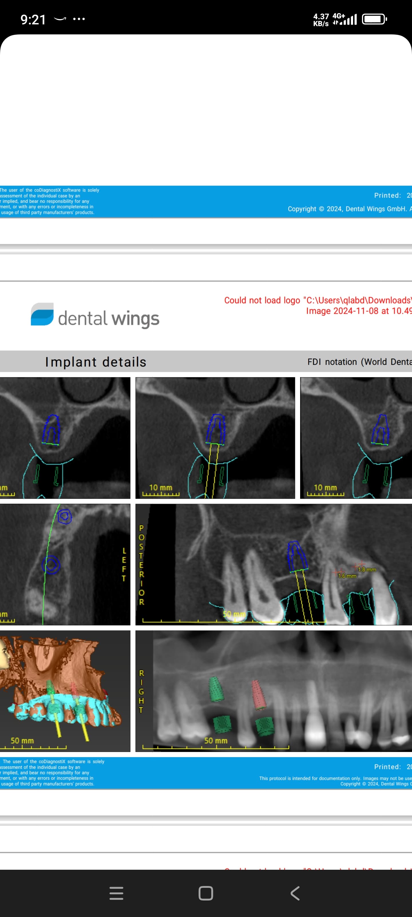
I have treatment planned this patient for implants in the 46, 45 and 44 sites. I have identified the mental foramen but it also appears as though there may be a secondary foramen just mesial to it. I am seeing the shadow just mesial to the mental foramen and am wondering what it could be.
Because of the loss of vertical bone height I am planning on using 6 or 8mm length implants. Maybe when I lay the FTF I will try a dissection of the mental nerve to see whether there are additional branches or if this is part of the anterior loop of the mental foramen nerve. Any recommendations on diagnosis and how to proceed?


13 Comments on Secondary mental foramen: recommendations?
New comments are currently closed for this post.
rsdds
6/19/2017
please double check because I think you're not mapping IAN correctly it looks like is way lower and no need to use short implants
Vlad Reznikov
6/19/2017
Totally agree
Oleg Amayev
6/19/2017
Agree with previous comment, I don't think you are marking correctly. It will be good to see image without marking.
I also don't see second foramen, I think you didn't marked all way. Also, wouldn't recommend to dissect mental nerve.
Dralfdel
6/19/2017
It's likely the anterior loop of the IAN.
Not only do you need to identify the mental foramen but the anterior loop too.
Vipul Shukla
6/19/2017
I agree with comments above. Your mapping of the IAN canal seems incorrect. The second image where you have shown a red dot is where it exits at the mental foramen, but the canal itself is the dark region below it.
Agreed there is vertical ridge loss here, but the IAN Canal does not change its position, it is usually just above the dense corticated lower border of the mandible, reaching there from the mid-ramus lingual aspect where it enters the mandible.
I think your CBCT is too dark (overexposed or high kvp), hence you cannot clearly see the difference between bone trabeculae and canal wall.
Use a low kvp/low exposure pa X-ray to guide you for vertical height.
And yes, I would avoid touching the mental nerve.
Good Luck
Fereydoon
6/19/2017
I have two Articles about anatomical variations in mandible, the nerve sometimes counties to the mid part of the mandible,there is also two foramens in some patients.I strongly recommend considering all 3 views of CBCT.
my articles are:
1.TMOGRAPHIC VOLUM EVALUATION OF SUBMANDIBULAR FOSSA
2.Characteristics Of anatomical landmarks in mandibular inter foraminal region
F. Parnia
FES DMD
6/19/2017
Do not dissect the mental nerve. Likely to cause irreparable and unnecessary trauma to the nerve. Perhaps more surgical training should be undertaken before you jump off the occlusal table.
Dr. Vishal N Sahayata
6/19/2017
One more option is real time evaluation after reflecting the area, identify the mental foramen clinically. Drill short with first drill and take radiograph to locate exactly.
Nerve extension anterior to mental foramen which is not anterior loop will not trouble much. good luck.
MKG-Westend
6/19/2017
Dear colleague,
This is so called canalis incisivus mandibulae. This is the vascular and nerval bundle for the lower incisivi.
Wesley Haddix
6/20/2017
I agree that the IAN mapping does not appear accurate, although I am only looking at an image on my tablet. In my image, the true IAN appears to be inferior to the trace appearing on the radiographirepresentation. If what I see is actually the IAN, this patient has an "anterior loop", an anatomical variant of the course/bifurcation of the mandibular nerve into the mental and incisal nerves. In this variant, the IAN actually proceeds inferior and anterior of the mental foramen before turning superiorly and posteriorly to bifurcate and then exit the mental foramen. The average loop extends 5-7mm (up to 12 mm in extremes) anterior to the mesial wall of the foramen. It is of concern if not identified placing implant between the forameni, assuming that the normal 5 mm +1/2 implant diameter distance safety clearance anterior to the mesial wall of the foramen will prevent nerve damage from a drill. It is positively diagnosed by exposing the mental nerve and foramen with blunt dissection (saline soaked lint-free gauze wrapped around a judicious index finger) and a blunt probe inserted into the POSTERIOR aspect of the foramen (why posterior?). Encountering a hard wall at the posterior of the foraminal orifice confirms an anterior loop. Implants anterior to the foramen should be placed a distance equalling the sum of the loop distance anterior to the mesial wall of the foramen + 5mm safety margin + 1/2 implant diameter; conversely, implants may be placed above the loop similar to placement of implant in the posterior mandible above the IAN. I am sorry I do not remember the incidence of this loop; I remember it being quoted in the high single digits, but I always considered the incidence 100% until proven otherwise. This should be taught as a normal surgical consideration in any implant externship. Bravo for the catch and prudence, best of all to you and your patient.
Dr Bob
6/21/2017
Do the full thickness flap and locate the canal opening so that you can stay above it with the implant placement. There will then be no doubt. This is what I did before the cone beam CT. Do not manipulate the nerve unless you have been well trained to do so and have done this with great success in the past. There is a high risk of nerve damage in doing so. Short implants work but load accordingly.
drds
6/21/2017
Thank you very much for suggestions.
I' ll use 10 mm implant and avoid suspicious shade in the 44 area, and probably 8 mm implants in the 45 and 6mm or 8mm implant in 46 area. Have enough bone in horizontal aspect.The three implants will be in monolith construction.
I put some images without mapping.
Wesley Haddix
6/24/2017
After viewing the unmapped images, I retract my statement that the IAN was not well mapped. Comparing the mapped images with these, it appears that you did an excellent mapping and that you have a firm grasp of radiographic planning . Again, best of all to you and your patient.


