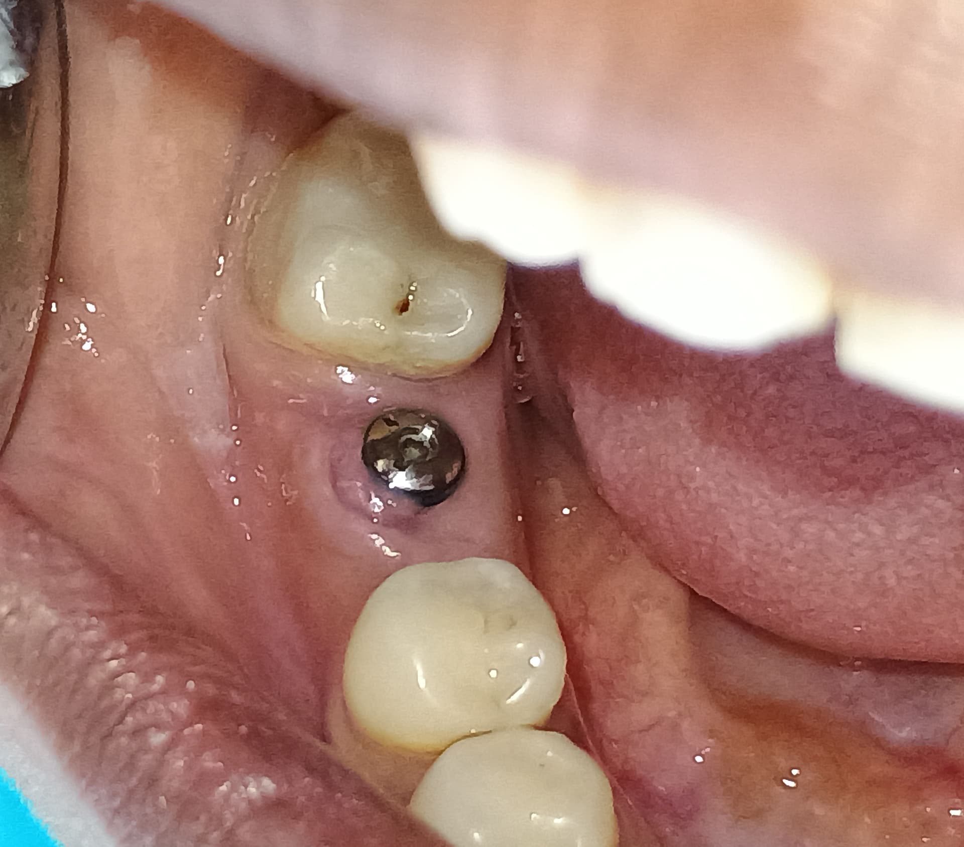
Severe bone loss 10 years after placement: advice?
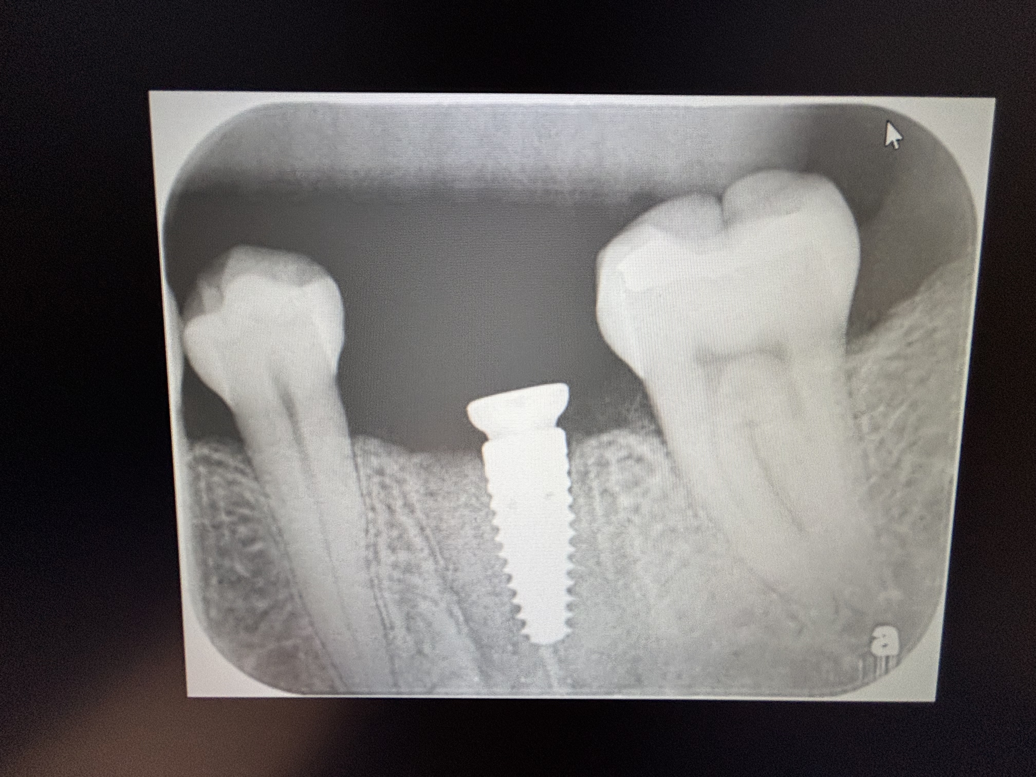
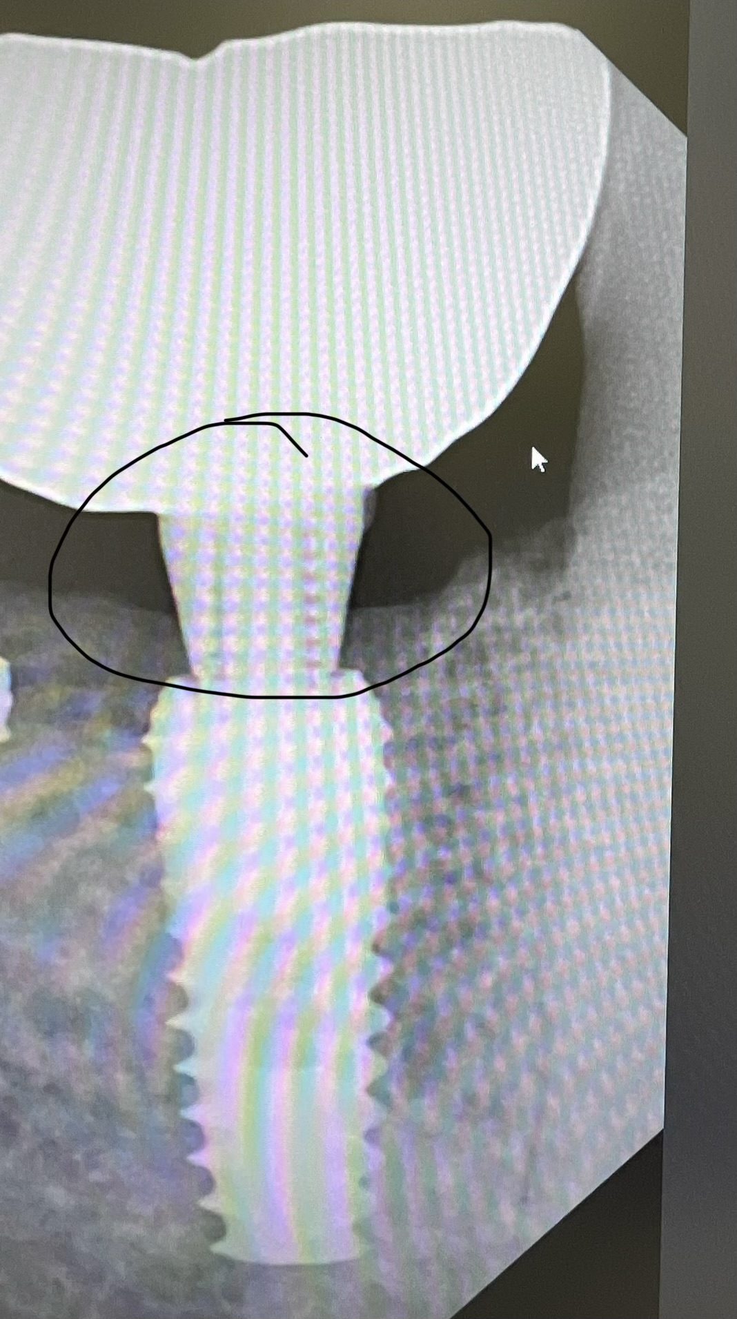
I placed this implant about 10 years ago. It was at a different office. About 2 years ago the patient started coming to my practice for followup. #18 implant had severe bone loss but is still osseointegrated. The gingiva is firm and appears healthy without bleeding or purulence. The patient is asymptomatic. I believe the bone loss was from a TMJ appliance that was overloading the implant. Initially I adjusted the occlusion in 12/2018. I then found out in 8/2019 that she has been wearing a TMJ appliance that was putting pressure on the implant crown. Is it acceptable to observe or be proactive? Any advice would be appreciated.


34 Comments on Severe bone loss 10 years after placement: advice?
New comments are currently closed for this post.
mark
3/11/2020
is the implant crown splinted to the first molar?
remove the implant and leave it out. I don't know why so many dentists place implants in second molar sites
Dr Zoobi
3/11/2020
To prevent the max 2nd molar from supra-erupting. Especially on younger patients.
S. Hunt
3/11/2020
Is placing fixture at 2nd molar sites contra-indicated?
Dr. Nguyen
3/12/2020
It's not splinted.
Xeee
3/12/2020
I'd just clean, adjust occlusion if required, modify that TMJ appliance so no parafunctional vectors are in play and observe.
mark
3/12/2020
no, the implant has failed and should be removed
Mike
3/12/2020
It’s not contraindicated, but electing not to replace it, likely won’t result in occlusal disharmony so many people don’t. Plus it’s usually the closest to the alveolar nerve which can also be a deterrent.
Fernando A
3/15/2020
This Rx have the image of oclusal patologic trauma.
Dr. Gerald Rudick
3/11/2020
Looks like a classic case of Peri-implantitis...… the bone just ran away from the implant, I do not believe occlusal factors had anything to do with the bone loss.....do not blame yourself, we all see this from time to time in our practices..... we are all looking for a cure for this dreaded curse.
Dr Beckwith
3/11/2020
Remove the implant, graft and reevaluate the need to replace after healing. Review medical history and recent CBC
Dr Zoobi
3/11/2020
For the sake of #19, I would remove implant, bone graft and replace 4-6 months later if necessary. You may start having caries and more bone loss on distal of 19. Happens to all of us.
Robert
3/11/2020
Is this a SteriOss Replace Select?
Dr. Nguyen
3/12/2020
Nobel Biocare Replace.
Neil Zachs
3/11/2020
There are a few things that can cause this type of bone loss after an implant is well integrated....occlusion/parafunction is a primary cause, cement extrusion (assuming the restoration was not screw retained) and lastly poor hygiene. This is a classic example of why implants are so difficult as some like myself who solely does the surgical aspect. As a specialist, we are so dependent on the restorative dentist as well as the patient for the long term success of the implant. It could be the most beautiful surgical success, all can go downhill if a number of things are not done or not addressed.
As for what can be done....if you want a definitive answer, remove the implant, graft the site and wait for good bony regeneration. After that, replace. If you want to try to be a hero, remove the crown and place a cover screw. The tissue would have to be flapped and the defect debrided followed by disinfection of the implant surface. The defect could then be grafted with Bio-Oss with a membrane placed over the graft to act as an exclusion barrier. Now I realize that I will hear negative comments on this. But listen carefully...this is heroic and I would tell the patient that the likelihood of results with this option is poor. If you want a definitive answer with a significantly better prognosis....REMOVE THE IMPLANT
Neil Zachs Periodontist, Scottsdale AZ
Dr. Nguyen
3/12/2020
Thanks for taking the time to respond.
RRO
3/11/2020
You can try to be a hero, flap, remove granulation tissue, decontaminate the implant surface, graft etc, but access will be difficult. The one thing that has not been mentioned as of yet is the zone of attached gingiva around the implant. If inadequate, it can make the difference between success and failure with some patients. Best chance for long term success is removal, graft and do over as many have stated. We have all faced this scenario. I hope it goes well for you.
Dr. Nguyen
3/12/2020
Plenty of attached gingiva. The two radiographs where taken two years apart. The right one is the recent one.
mark simpson
3/12/2020
It happens to us all. There are many factors that contribute . All of the previous comments cover those . repair at this point is unlikely but giving the patient that option makes sense.
I think removing and grafting is more predictable .Blaming it on the surgeon or restorative dentist is counter productive .
Timothy Carter
3/12/2020
Looks like a Nobel Replace (Tri-Lobe connection). We placed hundreds of these in the Army and a friend of mine who is a prosthodontist working for the VA now reports seeing this all the time. I now practice in a military community where I placed a lot of these 10-12 years ago and I too see this fairly often with the tri-lobe connection. I think it is a function of the simple to restore yet sloppy prosthetic connection with this system. To your point though I would not remove it at this point and I do not think the appliance was the issue.
Timothy Carter
3/12/2020
We think the problem initiates at the sloppy flat-flat tri-lobe connection and then once threads are exposed the bone loss happens quickly. The Nobel ti-Unite surface is great when covered by bone but it is an extremely rough surface and does not tolerate exposure well at all.
Andre
3/12/2020
This is a classic example of anodised implant mode of biofilm-mediated implant failure.
go to
DOI: 10.3844/crdsp.2019.1.17
open " On destructive peri-implant bone loss."
All your answers are there!!
And be afraid, be very afraid!!
Richard Hughes DDS
3/12/2020
Looks like occlusal overloading.
Follow a periimplantitis protocol and properly address occlusal overload.
Peter Hunt
3/12/2020
This might well be a case of Peri-Implantitis initiated by “Avascular Necrosis”. This is a syndrome that is just starting to be appreciated.
The cortical labial bone in this region can be very thick. It tends to have low vascularity. When an implant is screwed down into the implant channel it can come up hard against the bone, cutting off the blood flow to the cortical plate. So, there is very poor potential for healing and OsseoIntegration in that immediate region.
In time, the bone necroses and the gingival region above loses its attachment. Infective peri-implantitis then becomes established and bone loss about the implant increases.
It may take considerable time to get to the stage seen here. When as well established as seen here it would be extremely difficult to treat and recover. It would be easier and more effective to remove the implant, to regenerate the region and then to come back and place a new implant.
In replacement of the implant it would be sensible to place the implant into a region with more vascularity. We tend to place the implant into a region where the graft is relatively immature and with lower torque than usual.
DrT
3/12/2020
I would try LAPIP with BioLase...minimally invasive and nothing to lose
John dickinson
3/12/2020
There can be several causes for this failure. I see oral hygiene mentioned and yet as I look at the radiographs it appears the implant was placed with the platform at or near the bony crest. Due to biological width , bacteria living in the “Sloppy” micro gap will automatically cause a loss of 3 mm of bone. I hypothesize that this phenomenon will progress over time because the implant surface cannot be cleaned and the inflammation progresses slowly ahead of the infection.
You could remove the crown and laser debride the implant and place antibiotic into the defect. Then place a healing abutment with Implaseal but I think you’ll be better off to remove it and if you put another in there , keep the micro gap within a mm of the gingival surface or supragingival.
mark
3/12/2020
lots of great commentary above and I thank you all. No need to finger point, feel bad or blame, the previous dental work gave this patient 8-10 yrs of chewing service- irrespective of hygiene - remarkable really. So question, just because the implant is not mobile do we accept the 10 mm circumferential pocketing as being ok? Would we accept a 10 mm circumferential pocket anywhere else; particularly in these times of the dreaded oral systemic disease connection? Recommend to remove the implant, have her decide and move on.
Cognitive dissonance is a real human thing so let’s all be factual. And protect 19 with a class 5 glass ionomer on the buccal .
Wally Hui DDS. FAAID. DAB
3/14/2020
I would like to add little more about this case:
yes, most likely occlusal issue followed by hygiene issue; anyway already happened.
if you and your patient want to save this implant, replace the abutment by cover screw and see the gum can growth and cover the implant, and graft again.
if not, --> remove implant and graft...... keep an eye on occlusal loading, there is nothing wrong put implant at 2nd molar region, trust your fundamental dentistry.
Sergio Aliota
3/15/2020
That is a lesion from peri-implantitis, not from overloading.
Take the implant out.
The lesion is too severe to be regenerated.
Brly
3/16/2020
what was the torque value you placed this implant in at?
Dr. JLD
3/16/2020
Remove it ASAP. No bone graft. No replacement implant. I recently lost tooth #31 because of an unrestorable fracture and I have not missed having that tooth for one second.
mark
3/18/2020
correct , second molars are not important and I never argue with a patient who wants a prosthetic replacement
RRO
3/18/2020
Several comments indicate that second molars are not important. That is certainly true for All-on-x implant treatment. It is often true in the natural dentition except when it isn't. If a patient has severe wear or a bite so strong they destroy their natural dentition , then second molars are important. Missing a second molar in those circumstances will accelerate wear on the remaining posterior teeth and the surface area for chewin decreases. After a decade or two, such patients increasingly destroy their remaining molars. So I would say that second molars at times are important.
Sajjad a.Khan
4/9/2020
Steps to take
R/O medical issues
Make sure no occlusal load and enough clearnace.
Control and manage perio health then Flap use Yag laser with proper training follow established protocol . Then later time graft and enhence the bone .
Joe Orti, DDS
4/13/2020
Fernando is right. Pathological over occlusion. Great failure of fixed implants. All of them. Remove the crowns, design a new TMJ occlusal appliance and when the treatmen is completed, replace the crowns. Let us know what happens after that.


