Slight Bulge Over the Buccal Cortex: Is Intervention Necessary?
Dr. D. asks:
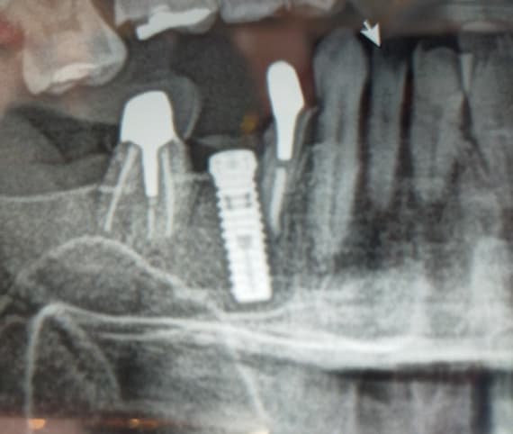
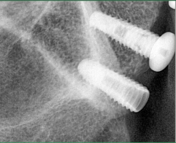
I recently saw this patient. The dental implants are well integrated and are asymptomatic. One unsettling feature is that the buccal cortex is expanded to form a bulge on the buccal aspect of the implants. Otherwise everything appears normal. The implants have been restored and are in function. Does anybody have a feeling for what has happened here and if intervention is necessary or should I just continue to observe the implants?

23 Comments on Slight Bulge Over the Buccal Cortex: Is Intervention Necessary?
New comments are currently closed for this post.
Gregori M. Kurtzman, DDS,
8/29/2011
Is the buldge soft or hard? has it been there since they came under your care or just appears recently?
Dr.B
8/29/2011
If you're concerned about pathology order a cbct.
Dr. Alex Zavyalov
8/29/2011
There is a definite bone destruction process here and if it is asymptomatic - at least occlusion correction is required to prevent overloading.
mike ainsworth
8/30/2011
Need more info.
look at the basics: colour, shape, texture, hardness, pain on palpation? pus from sulcus? Probing depths? ulceration, changes with time? etc etc.
some of my nicest cases have a bulge over the implant.
Steven
8/30/2011
Regardless of what the bulge is due to, I strongly suggest that you replace those "crowns" with ones that are not so grossly over contoured. The present ones are totally unmaintainable periodontally, and if there is any occlusal contact on the distal most aspect of the distal crown, this is in effect a cantilever, and will eventually lead to bone destruction. I have to wonder what the dentist who made these crowns was thinking about...if he was thinking at all.
Dr. H
8/30/2011
Interesting comments. There does appear to be some cratering around the distal implant and the level of the implant top is clearly above the bone at this point in time. The question would be what was the level of bone at placement? Only then can we tell if bone loss has occurred although it does appear that this is subsequent to placement. In regards to the contours of the crowns, it is difficult to tell if this negatively affects the tissue by looking only at a two dimensional x-ray. If these are laying on the tissue rather than under it, I would submit that this is acceptable. Placement of the crown margins deep subgingival also creates problems and that has to be taken into account. As for the bulge that the question is about, the x-ray and description is insufficient for any meaningful comment.
Dr. H
8/30/2011
One more thing: I would wonder why such short implants were placed since there appears to be much more bone available?
Dr G J Berne
8/30/2011
In answer to the original enquiry, the bulge is probably due to infection around the implant. There appear to be signs of bone loss around the implants, particularly the posterior one and if the gingiva is thick around the implants infection around the implant could cause swelling towards the crestal part of the implant. Looking at the bone levels around the implant it would probably be unlikely that infection was the cause if the swelling was much lower.
As to Steven's comments about the contour of the crowns, I'm not sure what he would do to correct the perceived problem. The crowns appear to be at or above the gingival margin, which is good, there is platform switching type abutments, which is good. Any attempt to use flared abutments to try and obtain a better emergence profile is in my view contraindicated. I would certainly not be attempting to replace the crowns, but attempt to get better oral hygiene. I'm not sure of the type of surface on the implants, but if there are exposed screws there may be ongoing problems.
Dr. Alex Zavyalov
8/30/2011
How about the other side of mandible? I think the cause of the problem-unilateral mastication function. Rarely do I meet asymptomatic infection with bone atrophy process.
SG
8/30/2011
I need to reconsider and in view of the small diameter of the implants, it would appear that the contours of the crowns are consistent with the size of the implant fixtures. I would still highly recommend that there be no occlusal forces on the distal surface of the distal most crown.
Keith VanBenthuysen, D.M.
8/31/2011
It could be the quality of the radiograph but there appears to be something going disatl to the implants. The bone appears to have some mixed radiolucency and opaqueness. Could this be pathology? And is what you are observing actually bone expansion due to this pathology?
Baker vinci
8/31/2011
Certainly some of these questions have been entertained by the treating surgeon, with regards to infection, comparison to the contralateral side, ect. . Assuming you have ruled out the obvious , an appropriate diff. Diagnosis has to be considered. First on my list would be metabolic processes such as paget's dz. . In a scenario where bone turnover has been affected , this could be an ideal case. It is more common to see this dz in the maxilla but it does occur here. Let your pcp draw some simple blood studies . Even as an omfs I have to agree that the crown form on these implants leaves a lot to be desired. Not sure why contact form, embrasure anatomy and hydrodynamics gets tossed out the door when we start restoring implants. Bv
Baker vinci
8/31/2011
This seems to be an excellent example as to why a cbct preop should be obtained. While we all need to respect the ia nerve. These implants could be 4 mm longer, thus allowing a more approriate restoration. Was the surgeon scared???? Bv
Dr. H
8/31/2011
Keith has a very important observation. It is easy to get focused and clearly he moved back and looked at the whole picture. Again, the description and two dimensional radiograph are insufficient to come to any conclusion, but it always is a good thing to step back and view away from the focused point. Good call, Keith.
Baker vinci
8/31/2011
Excuse me gentlemen , but bony expansion is a clinical finding. This was noted in the initial query. He stated the pt has expansion. You can't detect bony expansion from a one dimensional film. It is absolutely pathologic , if it's a new finding, wether it be infective, neoplastic or metabolic. A cbct would be helpful. I would encourage letting the surgeon that placed these take a look. The condition could be unrelated to the implants, although unlikely. Bv
Baker vinci
8/31/2011
While reading the initial question , it appears to me that this may the clinicians first time to see the patient. If this is the case , are you certain that the expansion isn't the result of lateral ridge augmentation that was performed to prepare for the case. It's not uncommon for a patient to have Tori or exostosis and not know it until it's pointed out. Just a suggestion. Obviously a little more history would be helpful. I had the daughter of a pcp come in with swelling and pain under her chin. She denied any surgeries in the past, and until the non opaque loose chin implant was discovered on MRI she continued to be less than candid. I was less than pleased. Spend some time and get a thorough history. Bv
d
8/31/2011
hello
additions ....
1 the swelling is bony hard normal colour minimally tender on palpation. no exudation / pus.
2 it has been restored pretty recently ( 2 weeks i guess ) so i doubt the bone loss i guess some threads were open right from the start.
3 the prosthesis seems to be slightly out of occlusion .
comments invited
Baker vinci
9/1/2011
Please remember, when you use the word tender, that means the area is "hot", and extremely painful. Typically the patient will not let you touch it again. The best analogy I have is when the right lower quadrant of a child is tender ,he/she has an infected appendix. If someone is sore, they typically have gas. It may seem like I'm parsing words, but accuracy when describing the condition is crucial. Bv
Baker vinci
9/2/2011
Please don't attempt to read something( ie. Mixed rad/opac.), from a duplicated panoramic film. Bv
d
9/3/2011
correction....
the area is sore and not tender .
Baker vinci
9/3/2011
I'm sorry dr. D , just thought it might be important . Really trying to not be sarcastic. I have seen quite a few patients in 5th thru 9th decades of life exfoliate a layer of dead bone for no apparent reason . Admitidly it happens more commonly on the linqual aspect of the mandible, but it is typically preceeded with slight discomfort , expansion and subsequent spontaneous exfoliation of a dead spec of bone. I have found that the best way to manage this is with aggressive oh and tincture of time. Bv
Greg Steiner
9/6/2011
Dr. D The trabecular pattern and density around the natural tooth is normal so I do not question the quality of your radiograph. In comparison the bone distal to the implants looks very abnormal. This radiographic image combined with an expanding ridge warrants further evaluation. I would either send the patient in for a CT scan with a report from a dental radiologist or refer to an oral surgeon(or both). I would also examine the tissue immediately distal to the implants and up the ramus for any fistulae. In my limited experience a fistula from a bone lesion is hard to spot as the surrounding tissue may have no inflammation to help you locate the fistulous tract.
Greg Steiner
CEO Steiner Laboratories
Member American Society for Bone and Mineral Research
Dr Ares
9/9/2011
Viewing the periapical x-ray you posted at a distance, there seem to be changes in the bone density distal to these implants. If you don't have a point for comparison because you where not the initial treating dentist, I suggest you consult the original dentist, because this might actually not be an expansion of the ridge but just an exostosis. Or perhaps as Dr Vinci pointed out, a ridge expansion was performed prior to the implant placement. If in doubt, I agree that CBCT would be useful in ruling out bone pathology.


