Tingling Sensation After Implant Placement
David, a dentist, asks:
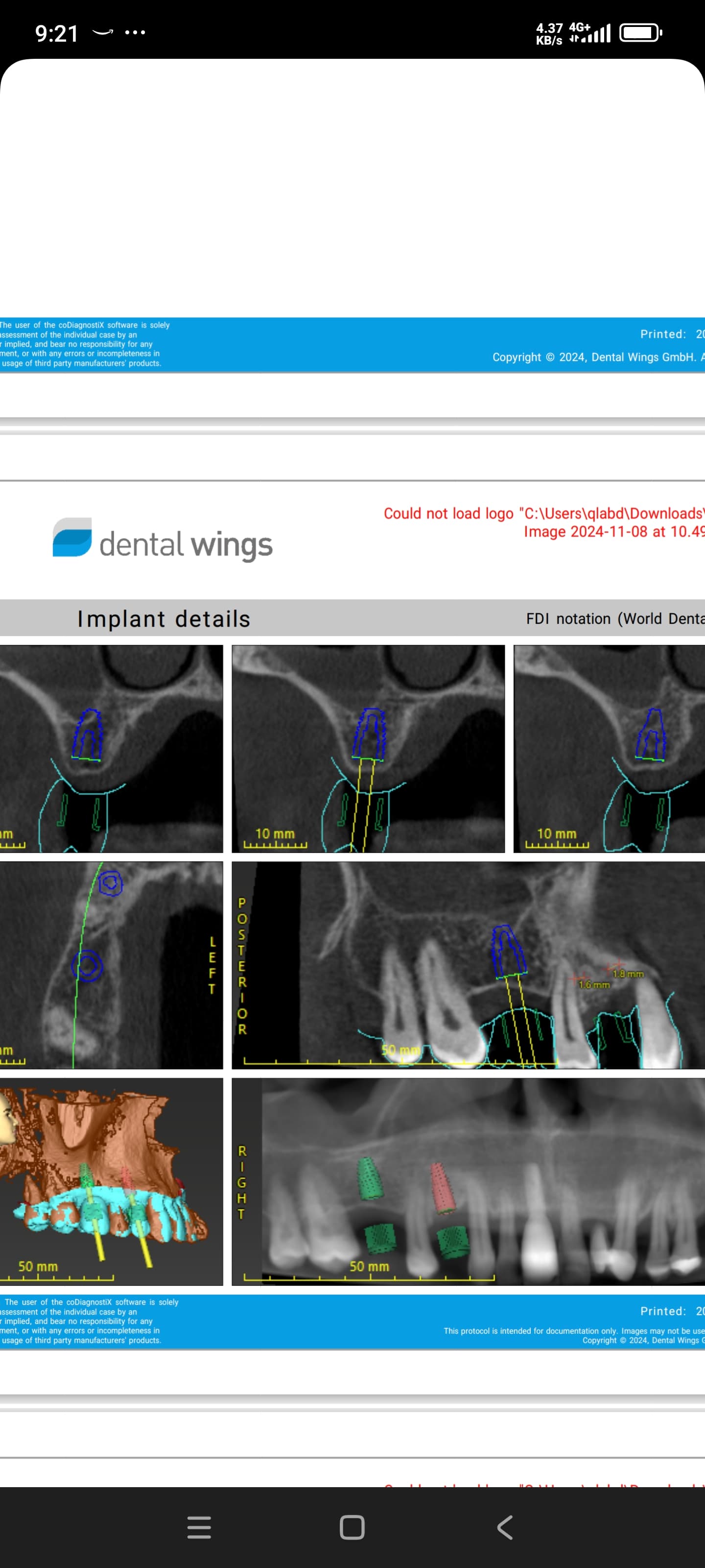
I placed dental implants in #18 and #19 region three weeks ago and the patient has developed tingling/burning sensation in the chin area. I sent patient for a CT scan at one week post op and found #19 very close to the inferior alveolar nerve.
I removed the implant fixture and placed a shorter fixture above the canal. I placed the patient on Medrol dose pack and the patient experienced a slight improvement at week two, but still has tingling/dysasthesia. Do I remove the other implant? Should I not have placed a shorter dental implant fixture at this time?
9 Comments on Tingling Sensation After Implant Placement
New comments are currently closed for this post.
sf
3/19/2007
Hi David,
You may want to check out:
Kraut RA, Cahal O. Management of patients with trigeminal nerve injuries after mandibular implant placement. J Am Dent Assoc. 2002;133:1351-1354.
In that paper the authors conclude:
"Placement of mandibular endosseous implants can result in damage to the lingual nerve, the inferior alveolar nerve or both nerves... Since the inferior alveolar canal can be seen on most panoramic radiographs and on all high-quality computed tomographic scans, it is easier to avoid damage to the inferior nerve than to the lingual nerve, which is not visualized on radiographs and whose relationship to the posterior portion of the mandible varies from person to person...These results reinforce the need for early referral and intervention when inferior alveolar nerve injuries occur. Failure to refer patients with trigeminal nerve injury before distal nerve degeneration develops prevents minimization of the injury through microneurosurgical repair."
Hank Tabeling, DMD
3/20/2007
Hi, sounds like Mr nerve is not happy to due drilling or implant position..I would remove both and leave until some kind of resolution. Paresthesia /dysesthesia needs to be mapped and referred to someone who can do appropriate neurosensory testing and follow-up/treatment if necessary. Keep the dialogue and good communication up and document every little spec. Hope you had a good informed consent covering this and the possibility of permanence. If you do refer to someone, you will be covering the expenses now or later. The lesson here is buy an ICAT if your serious about placing implants over vital structures so you'll know exactly the 3 dimensional space pre-op. I have done a big bunch with panporex and prayer without sequella, I usually light a candle on sun in church if I'm planning an over the nerve case. REFER NOW !!!
Alejandro Berg
3/20/2007
If there was no intrusion by the implant in the canal, the initial sensation was probably result of the burs going to far(did you use external irrigation? in those cases a heating of the tip may sometimes produces an inflamatory response that can be mistaken with a neurological compromise), if the CT shows that the canal is intactyou may be in luck.
The use of high dose B complex is indicated.
Without seeing the ct is difficult to advise a removal of the second implant.
The shorter one shouldnt be a problem the only thing with that is that you restarted the initial imflamation process.
You can prescribe a combination med of citidÃn 5'-monofosfato sal sódica (CMP) 2,5mg, uridÃn 5'-trifosfato sal sódica (UTP) 1,5mg, hidroxocobalamina 1mg.
Its a great nerve regen system and has been widely used for this cases with success.
LUCK
Dr. D.
3/21/2007
David, Most likely, if you have removed the implant the NN will regenerate. (Unless of course, you have drilled through it) I doubt that is the case. You have probably just encroached on it. There are some great regenerative NN drugs out there. I would suggest that you contact a neurologist and explain the situation to him and make the referral. These guys are usually great to work with and should be the ones administering these drugs. The last thing you need now is to have a drug interaction problem with this patient. Good luck.
Dr. Mehdi Jafari
3/24/2007
The reported incidence of dual canals varies considerably. In macroscopic examination of the mandibles, researchers detected substantially more anatomic variation than that observed in the panoramic radiograms. Because they examined dry mandibles, they could not identify the anatomic features that ran in the accessory canals. However, they assumed that nerves and vessels occurred in the thicker accessory canals and that nutritive vessels were present in the thinner canals. Knowledge of the anatomic variation of the mandible may be important in oral surgery, prosthetics, and dentistry, may increase the success of certain interventions, and may avoid surgical complications. In general dentistry and oral surgery, lower conduction anesthesia may prove ineffective in the event of divided canals, particularly when they start from two separate mandibular foramina that are distant from one another. In such cases, the anesthetic solution does not reach the inlets of both canals in equally high concentrations. Damage to the alveolar inferior nerve causes paresthesia of its supply area. Subsequently, a neuroma may develop at the site of injury, and atypical pain may occur. Damage to the vessels in the mandibular canal results in bleeding, followed by the development of a hematoma, which might lead to nerve compression and inflammation. Damage to the accessory canals can give rise to similar symptoms. There is a risk of damaging the neurovascular formations running in the mandibular canal during interventions such as surgical removal of the lower wisdom teeth or sagittal osteotomy of mandible. In the latter case, the risk of injury to the formations in the mandibular canal is high, and this risk is even greater for dual canals. The above-mentioned lesions may also be caused by damage to the nerve. The insertion of endosteal implants is now a routine intervention. When they are implanted in the lower molar region, care must be taken concerning the presence of accessory canals to avoid the unpleasant complications accompanying damage to the nerves or vessels running in these canals.The upper accessory canals running to the molars are of prosthetic significance; if the mandibular alveolar process were to undergo extensive atrophy with aging, the neurovascular formations may become subperiosteal. In such cases, the pressure exerted on the area by a removable prosthesis may cause severe pain. Accordingly, consideration of the possible presence of such interesting anatomic variations may help avoid complications during routine procedures, increasing their success.
Dr. Joeph Como
3/27/2007
I would leave the shorter fixture in, place the patient on neuorotin 300mgs qd week one , 600mg for week two. It sounds as if the patient has a compression parathesia. If it does not resolve within three weeks , it is advisable to get a consult from a microneurosurgeon,there are some OMS that specialize in this. This will give you some better insight along with proper legal coverage . An antiflammatory is good to reduce any inflammation in the canal.Now , regarding what occured ,there are some ways to possibly avoid this some are depending on the extent of reflection of the surgical flap on the lingual can cause damage to the lingual nerve, but the "tingling" sensation would be mostly on the tongue. If there was encroachment on the inferior alveolar nerve then there would be tingling of the lip on the treated side along with tingling on the gingiva and possible numbnesss to the teeth. One pearl I have used doing implants in the mandible is Not to give a block. Since 1993 I give inflitrations only in the mandible. The reasoning behind the is One: There are no nerve branches in the bone, so you are able to place your osteotomy as close as possible to the inferior alveolar nerve. Remember that a panoramic x-ray is the standard of care for placement of implants , periapicals are an additional advantage along with dentascans. If you do get to close to the I.A.N the patient will tell you that it is sensitive.At this point a radiographic measurement should be taken to see where you are to the nerve. Lastly, it is becoming very popular not to make incisions and flaps. Instead, using a 1.8mm trispade drill for your pilot, through the gingiva, then using your increasing size burs to get to the desired size. This allows for less post operative swelling, decrease chances to damage the lingual nerve , and no sutures. Of course this technique is only good if there is adequate buccal lingual thickness.
Wilson Rivera
5/20/2007
Hello,
On May 14, 2007, I had #22, 12, and 13 implanted. It has been close to a week. I had a follow-up. However, it seems I still have numbness to the left of the lower lip and teeth before and after #22. No issues with #12 and 13. The provider stated to give it more time. Swelling or pain is no longer present. Radiographic procedures followed as per standards. I'm concerned! Should I be? Possible lingual nerve compresion? What are your recommendations if any?
Sandi Derr
2/28/2008
In reading various articles here and elsewhere, it is clear to me I have experienced nerve damage which affects my chin, lower lip and bottom teeth which feel like they are in a vice. The pain is hard to decribe overall, but is very uncomfortable. My surgeon removed the implant he suspected was the problem. At first he indicated the implant may have touched the nerve then later said he feels he drilled too deep and touched the nerve. Where do I go from here. I have no more feeling today with the implant gone that when it was there. I am scared to say the least. I am female 56 years. He did upper bone grafting and lower bone graft and then 11 implants, 6 on top and 5 on the bottom, back side 2 and other back side 3.
Thanks to anyone who can give me some advice now. I do not know what to do next. I cannot eat/chew because I am so dead in the bottom. Soup and liquids only.
Lynnlou02
5/14/2021
Did this resolve?


