Tooth 46 large abscess and radiolucent lesion: recommendations?

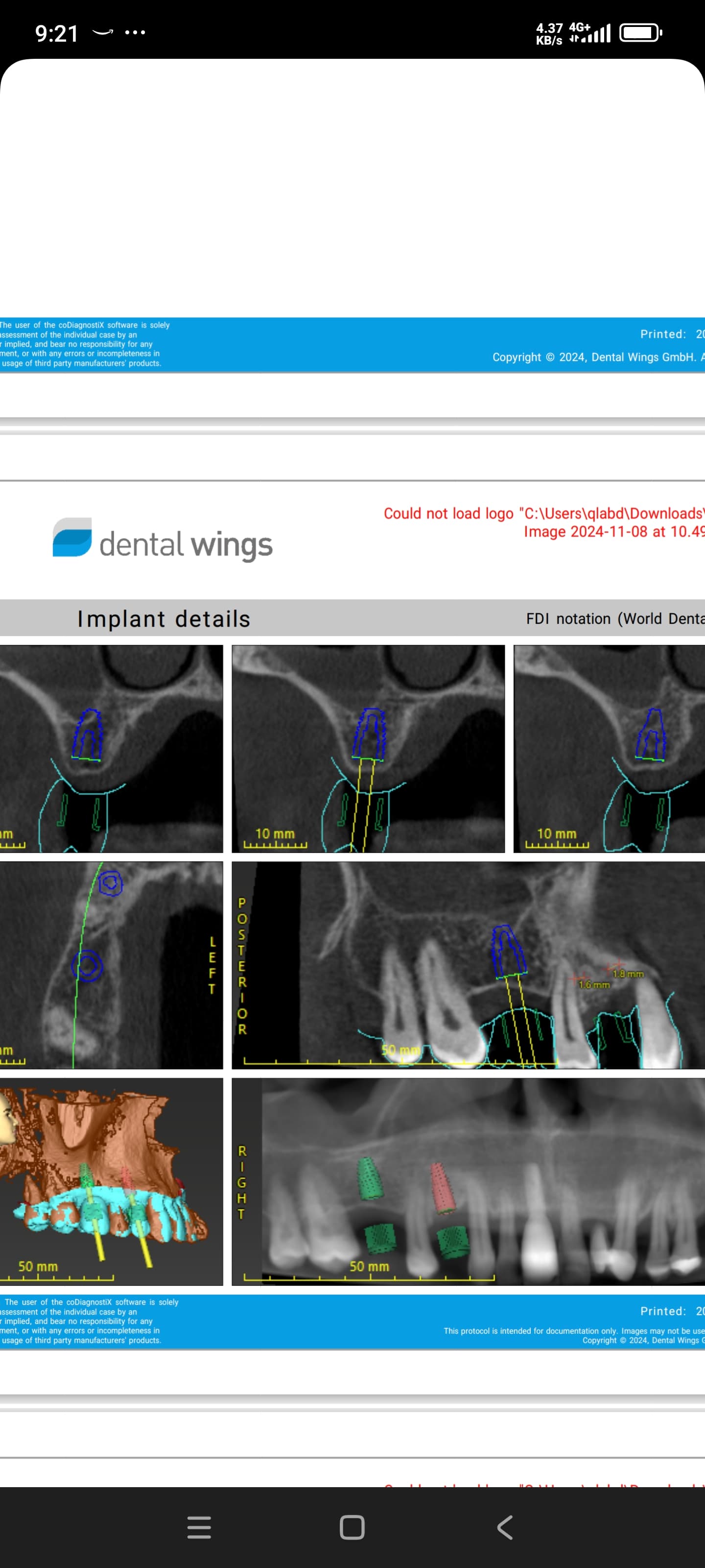
I extracted tooth 46 ( lower right 1st molar) exactly 6 months ago. The tooth had a large abscess and large periapical radiolucent lesion. I tried to curette as much as possible, but did not graft the socket. In my attached scan pictures, you can still somewhat see the outline of the radiolucent lesion. My questions are :
1) Is 6 months enough healing time if I had not grafted the socket? Would most of the infection have cleared up by this time?

2) Due to the buccal bone loss, I will need to place the implants more lingual. I am contemplating between a 5 x 12 or 5 x 10.5 mm implant. I do not want to perforate the lingual plate, so was leaning towards the 5 x 10.5 mm implant. Thoughts on this?

Any other recommendations on how to proceed with this case? I am relatively new to implants and have taken both the prosthetic and surgical courses at the Misch Institute. I have placed only 6 implants so far. I had been intending to start with very simple cases. Any suggestions would be much appreciated.


7 Comments on Tooth 46 large abscess and radiolucent lesion: recommendations?

New comments are currently closed for this post.
Peter Fairbairn
1/30/2017
At 6 months this will be fine and the case looks relatively straight forward as you have good diagnostic information .... so nice case to do , just take time and check with rads .. Regards Peter
JW Perio
1/31/2017
I would offer that you need to check your opposing occlusion before you pick your exact angulation. Check your study casts from which you will fabricate a stent-- this will help you decide where the implant needs to go. You didn't mention the mesial-distal distance between adjacent teeth, that needs to be wide enough to accommodate a 5mm diameter implant also. The way the line on the scan is angled, you would not perforate the lingual plate with a 5mm diameter implant at 12mm. If you wind up having to place the implant a bit more upright due to opposing occlusion, you can always graft the buccal at the time of placement. I like to punch a hole in a membrane after I have grafted and secure it to the implant via the cover screw, then close it up and let it heal. If it is PTFE (non-resorbable), you can remove it at second stage or when you are ready to load it. Good luck!
Paul Newitt
1/31/2017
You could always place the implant in the ideal position and do some GBR rather than place it to lingual. There would be less chance of a prosthetic emergence profile issues for the lab and/or a lingualized crown that interferes with the patients tongue. Hard to say for sure without an image showing ideal prosthetic position but something to keep in mind when planning.
Amir Mostofi
1/31/2017
Occlusal forces should be along the implant vertical axel. By placing the implant more lingualy you will risk complications. Place the implant in the correct location and do GBR on the buccal side.
Mike
2/1/2017
Thank you kindly for all replies. I have done simple socket grafting. I use DFDB with a resorbable collagen membrane. Would it be similar principal to GBR where the buccal bone loss is? I have yet to do anything like this before. Thank you kindly, Mike
Periogirl
2/7/2017
In addition to the comments mentioned above, I would like to add another suggestion; with the large trabecular pattern in this site, I would suggest under preparation of the osteotomy. If you prep to the guidelines of the implant system you are using, you will find you will not get primary stability. Although this has not happened to me, I have heard of implants actually submerge beyond the implant placement because the bone is not dense to support the implant and primary stability not achieved. Different bone densities require different preparation of the site. This is rare but with excessively large trabecular pattern I see here, under prep of the osteotomy is a consideration here.
Periogirl
2/7/2017
It also would have been helpful to see a pre extraction radiograph.

Featured Products

OsteoGen Bone Grafting Plug
Combines bone graft with a collagen plug to yield the easiest and most affordable way to clinically deliver bone graft for socket preservation.
CevOss Bovine Bone Graft
Make the switch to a better xenograft! High volume of interconnected pores promotes new bone. Substantially equivalent to BioOss and NuOss.