Dr. Jack Krauser on CT Scans, planning in 3-dimensions, and more…
Dr. Jack Krauser is a Florida Periodontist and Treasurer of the
International Congress of Oral Implantologists, the largest organized
body of dentists placing and restoring implants. He is engaged in
clinical practice, teaching and research. Dr. Krauser has taken time
out of his busy schedule to share some of his views on implant
dentistry with the readers of Osseonews.
Osseonews (ON): Dr. Krauser, would you agree that using a CT scan to
aid in the treatment planning for determining the most efficacious
positioning and angulations of dental implants is becoming the standard of
care?
Dr. Krauser: Well we are not quite there yet. Certainly for complex
cases where we are dealing with atrophic alveolar ridges, questionable
areas of anatomy, and bone of questionable quality, a CT scan is indeed
a valuable tool that enables to make the best possible use of what we
have to work with.
ON: Specifically, what are you looking for that you cannot find in a panoramic radiograph?
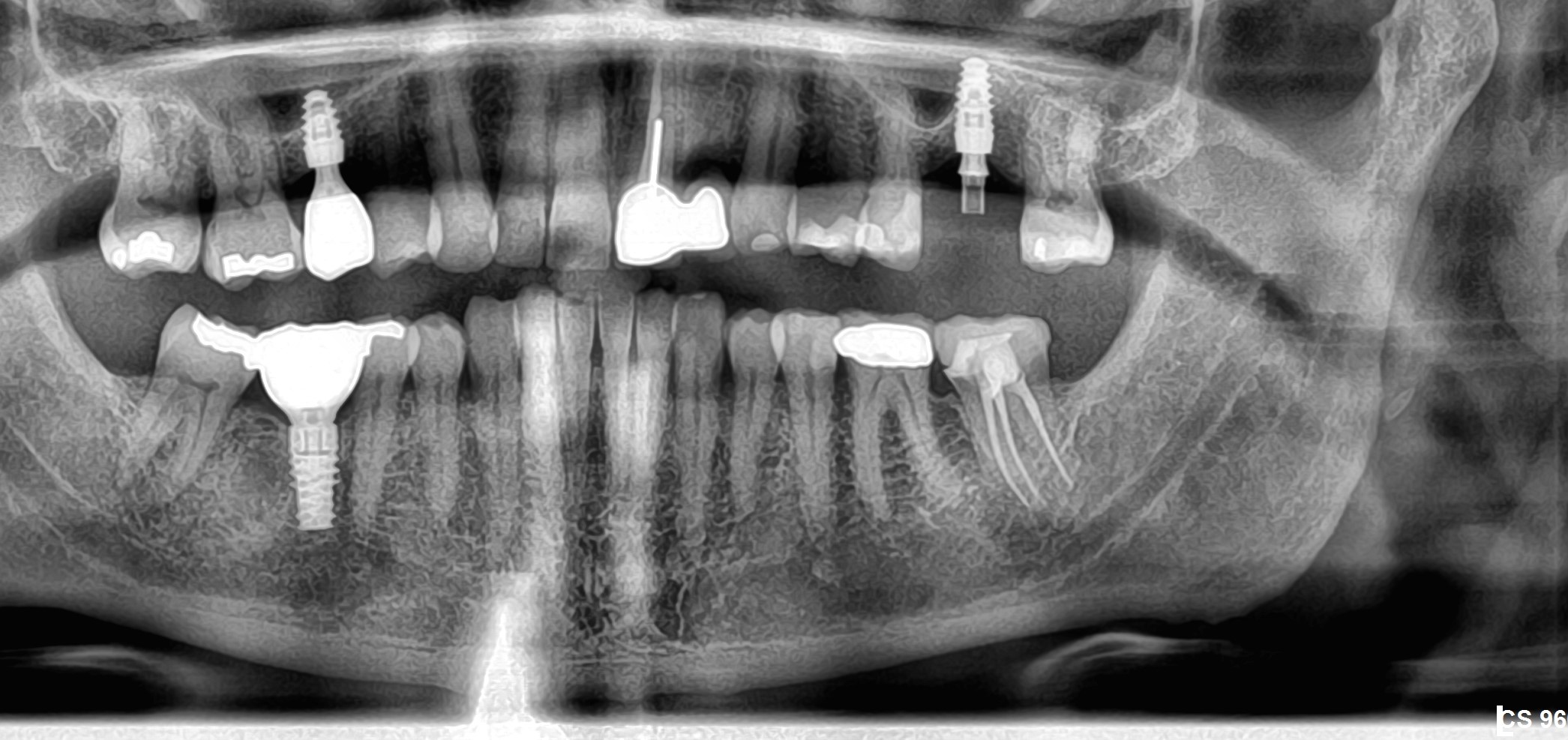
Dr. Krauser: A panoramic radiograph does not provide all available,
valuable information. But a CT scan can provide a 3-dimensional
representation where we can actually measure the vertical,
buccal-lingual, and mesiodistal dimensions of bone accurately. We can
also measure the density of the bone. Panoramic radiographs generally
do not provide that kind of information.
ON: How do you use that information?
Dr. Krauser: After incorporation of the data into planning software,
we can generate a 3-dimensional model of the patient. This can generate
a surgical guide/template for precision flapless placement of the
dental implants. Our trained laboratory teammates can generate a milled
titanium bar and prosthesis that will fit the implants after they have
been placed if they use the powerful Procera System software. Using
this information and available technology, we can assemble this data
and produce a restoration where we can provide a final prosthesis for
our patients at the same time as the surgery. This is now known as
“teeth in an hourâ€.
ON: Could you run through your protocol for “teeth in an hourâ€.
Dr. Krauser: First of all, we want the patient to understand exactly
what we intend to do. In order to accomplish this, we use the XCPT
patient presentation software program. With this software we can
demonstrate to the patient where we are going to place the implants and
how we are going to restore them. This can all be accomplished at the
initial patient visit using data from a digital image gathered in 2 or
3-dimensional image or xray.
ON: Does the XCPT also increase patient acceptance of the intended treatment plan?
Dr. Krauser: Definitely. And any staff member can be trained in a day to use the program with a patient.
ON: Please describe your placement protocol.
Dr. Krauser: After patient acceptance of our treatment concept,
several exacting pre-surgical prosthetic steps are performed. After
careful planning in 3 dimensions, we create a computer generated,
stereolithographic, surgical guide/template to place the dental implants. The
guide enables me to place each implant at the correct site, desired
angulations and to the desired depth. These parameters are very
important so the prosthesis can be inserted on the implants at the time
of placement. This all must be done with precision.
After placing the dental implants, the restorative teammate places the
prosthesis. It can be as simple as that. If I place the implants
correctly, and the laboratory technician creates the correct framework
and prosthesis, the final prosthesis will fit with precision and
comfort.
ON: How long does it take you to perform the procedure?
Dr. Krauser: About one hour, from start to finish.
ON: About how often does everything go the way you have planned?
Dr. Krauser: Most of the time we complete the case in an hour and
most of the time everything fits perfectly. Our tolerance for implant
placement is extremely accurate. Planning in 3-dimensions eliminates
complications like perforating through a cortical plate or anatomical
structure.
ON: In those rare instances where you need more treatment or adjusting of the prosthesis, what do you do?
Dr. Krauser: We have an operatory equipped with restorative
supplies. This is maintained for those times where the restorative
dentist needs to do more than simply insert the prosthesis. We move the
patient from the surgical suite into this operatory and the restorative
dentist adjusts the prosthesis.
ON: How closely do you work with your restoring dentist?
Dr. Krauser: We have to work as a team. We have to work together
closely on each case. Teamwork is essential. I meet regularly with my
restoring dentists. At times we have functioned as a study club in
order to teach each other updates in materials, techniques and
treatment solutions available for our patients.
ON: What are some of the benefits of the i-CAT cone beam system (Imaging Sciences) in particular?
Dr. Krauser: This in-office system occupies similar or less space
than a conventional panoramic radiograph machine. It can fit a small
space, basically in any office. With the i-CAT, you can scan only the
area you are interested in which reduces radiation exposure to the
patient. In fact, a full scan of both the maxilla and mandible with the
i-CAT requires 10% of the radiation the patient would normally receive
if you sent the patient out for a Medical CT scan, and takes less than
1 minute to take the scan, and another few minutes to get the data to
the CT computer terminal.. In my mind that is a great advantage of the
system. Instant data, and instant confidence in presentation.
ON: What is the approximate cost of the i-CAT?
Dr. Krauser: About 150 thousand dollars. A leasing program is also available and seems to pay for itself.
ON: Does Imaging Sciences provide adequate training and do they have a help-desk?
Dr. Krauser: The time required for training is minimal. Installation
and staff traing are done in two days. Their phone or on-line internet
help-desk is excellent and I am speaking from experience.


