Extensive Ridge Deficiency Reconstructing using Bone Graft Cement

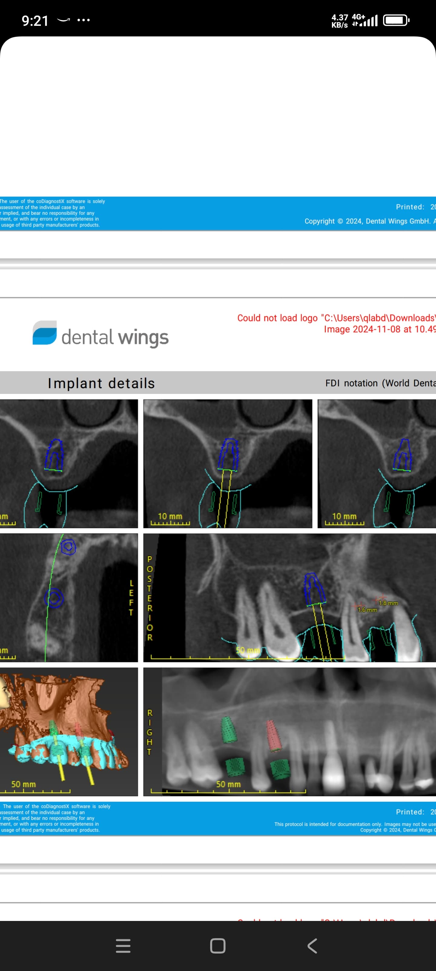
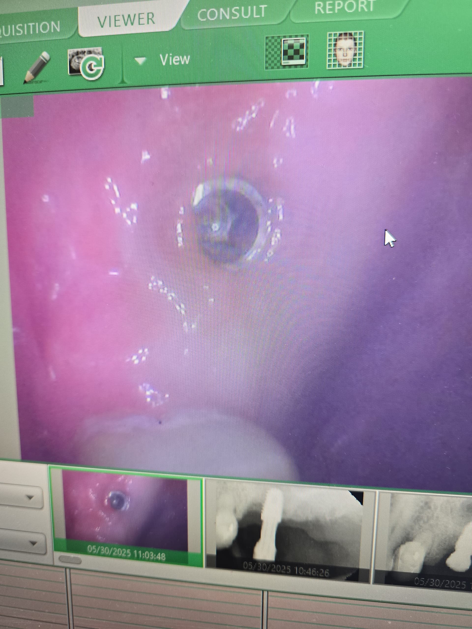
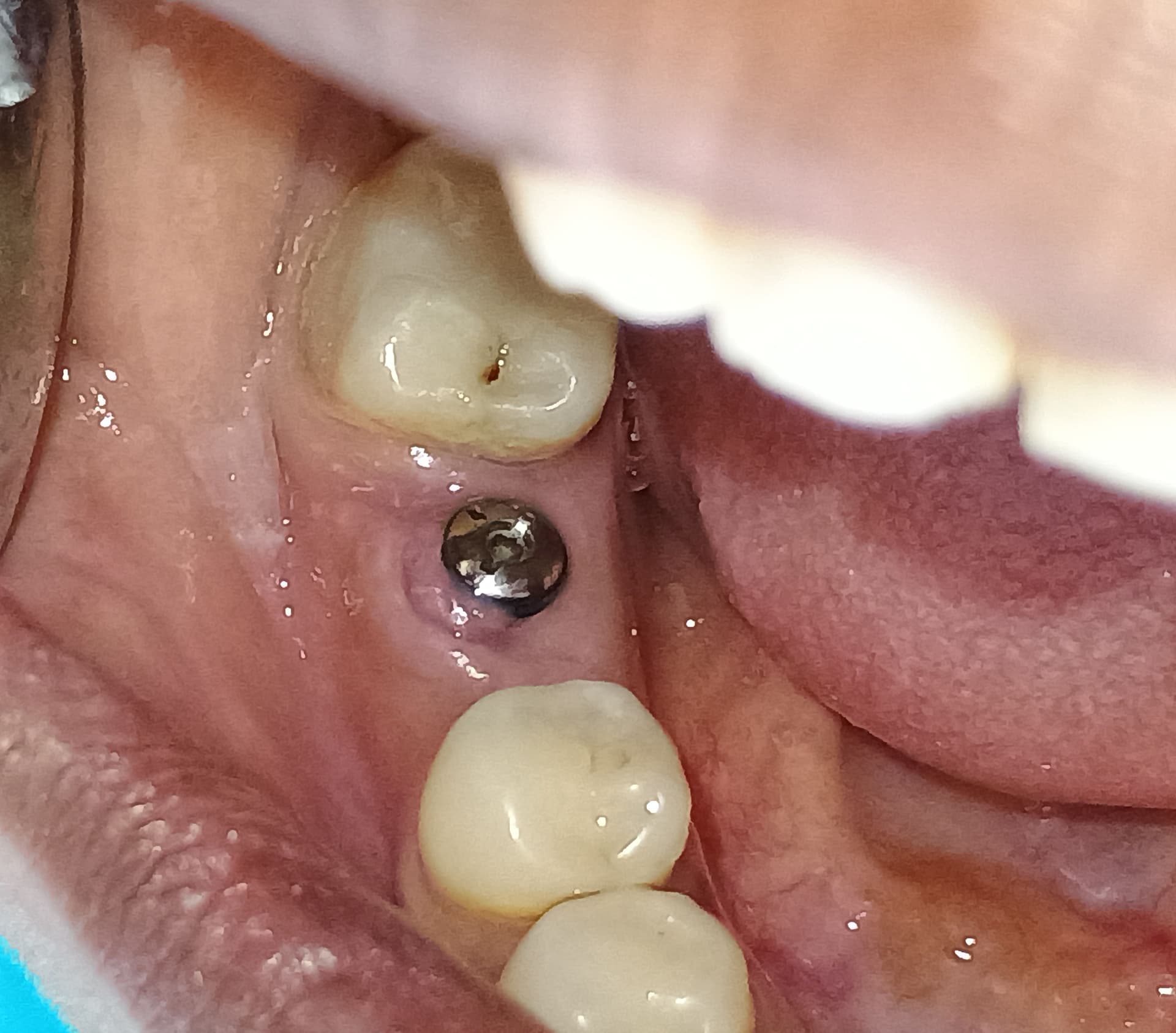
This case describes the rehabilitation process for the upper jaw. As a result of advanced periodontal disease, severe bone destruction is evident. It was thus necessary to laterally augment the narrow ridge in the area of the incisors with regeneration of the alveolar crest and bone restoration in the area of the molars following their removal .

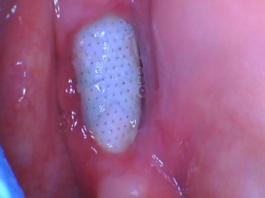
Following the extractions a composite cement graft material was used which contains biphasic calcium sulfate and HA ( Bond Apatite “ Augma Biomaterials LTD).

The surgical procedure of graft placement, using the bone graft cement, is easy and user friendly to the surgeon and minimally invasive for the patient. This is due to the fact that the full thickness mucoperiosteal flap is minimal, without the need of flap releasing dissection or periosteal incisions in order to achieve a tension free flap as is mandated with other bone substitutes and grafting techniques.


9 Comments on Extensive Ridge Deficiency Reconstructing using Bone Graft Cement

New comments are currently closed for this post.
Miguel Martinez
5/23/2018
Very nicely done. How long did you wait after bone graft? Did you use Versah’s osseodensification burs at time of placement? Post op X-rays please
Dr Yahav Amos
5/23/2018
3 months post none grafting implant was placed . Regular drills was used
nissim levy
5/25/2018
what kinde of temporary rehabilitation do you recomend
Dr.Amos Yahav
5/26/2018
In this case few compromised teeth were left for temporary no removeable aclilc bridge , and those teeth are extracted later on . It is important that your technichian will preform a cast metal bar or frame within the temporary bridge so it can resiste during the waiting time of healing .
Lukas
6/2/2018
Dear Dr. Amos Yahav, My name is Lukas, and I’m a dentist from Warsaw, Poland. I’m enthusiastic about this bone cement, but I have little experience working with it. I’ve used 4Matrix bone cement 3 times so far (this one is available here), and I’m still waiting for the final result. I have some questions about this product which I would like to ask you, as you are an expert in this field. 1) After implant placement, where there is a thin buccal bone plate, but the implant is fully surrounded by bone, and a bone cement is only used to thicken the alveolar ridge, is it possible to close the implant with healing cap instead of cover screw? Must primary closure be achieved then, or we can leave 2-3mm bone cement exposed? 2) If a buccal bone plate was missing, and after implant placement there were some threads exposed on buccal side, is it possible to augment them with bone cement? Would it be possible to use healing cap in this situation or cover screw would be necessary? And again, is primary closure a prerequisite in such case? 3) Is perforating a cortical plate before using bone cement is beneficial, or not? 4) If there is a bigger gap between the flaps after bone cement placement (more than 3mm) what is the best way to do? To release the periosteum a little bit? To place a collagen plug / membrane on it? To cover a gap with with PRF or perhaps with subepithelial connective tissue graft? I would be very grateful for your answers to these questions.
Dr.Amos Yahav
6/3/2018
Thank you Dr. Lukas for yours very good questions , Please find my answers below . Question 1: After implant placement, where there is a thin buccal bone plate, but the implant is fully surrounded by bone, and a bone cement is only used to thicken the alveolar ridge, is it possible to close the implant with healing cap instead of cover screw? Must primary closure be achieved then, or we can leave 2-3mm bone cement exposed? Answer 1: In this case you can use both cover screw or healing abatement .it is important to keep your flap under tension and not tension free as we used to with other bone grafts here its work differently . also during closure no membrane or PRF should be used above the cement it should be in direct contact with the periosteum .if you perform a vertical cut mostly one cut is enough it should be in a distance of one tooth mesial or distally up to your preference , the cut should be 2 maximum 3 mm into the mobile mucosa not more .You exposed the required grafted site, before graft placement take a forceps hold the corner of the flap and stretch the flap .It will give you the feeling how much you can stretch it for closure (because stretching is against our instincts that is something that we never did before ) so stretch it with no fear ,and as you will see 2 mm into the mobile will enable you to stretch 4 mm 3 mm will enable 6 mm together with 3 mm of exposure that you can leave exposed it will be sufficient for almost any grafting procedure that you would like to preform .but remember exposure should not be more than 3 mm otherwise the cement will wash out and you will lose volume . Now you can eject the cement directly to the site ,take a dry sterile gauze place above and press strongly for only 3 seconds ,the compression on the cement will compact it and the setting will take place immediately. Now take the buccal mesial corner of the flap stretch it and suture it to the mesial lingual corner ,then the distal and then in between them . If during closure and suturing the cement is breaking down a bit just take a dry gauze and press again for 3 seconds and continue suturing . Question 2: If a buccal bone plate was missing, and after implant placement there were some threads exposed on buccal side, is it possible to augment them with bone cement? Would it be possible to use healing cap in this situation or cover screw would be necessary? And again, is primary closure a prerequisite in such case? Answer 2: The answer here is similar the former one the only deferent is that the implant should be placed one mm below the crest. Question 3: Is perforating a cortical plate before using bone cement is beneficial, or not? Answer 3: Since we don’t use a membrane we do not block the periost and the cement is with direct contact with the periosteum in most cases there is no need for decortication .the only times that I decorticate is rarely in the lower jaw in cases that the bone looks extremely dry . Question 4: If there is a bigger gap between the flaps after bone cement placement (more than 3mm) what is the best way to do? To release the periosteum a little bit? To place a collagen plug / membrane on it? To cover a gap with with PRF or perhaps with subepithelial connective tissue graft?. Answer 4: as I wrote in A1,the vertical cut of 2-3 mm will provides you up to 6 mm of stretching ability with the 3 mm that you can leave exposed is up to 9 mm it is sufficient almost for everything and yet you don’t connect the flap to the mussels movements .if you need a little more you don’t dissect the flap you use your periosteal elevator and undermine a littlie bit in the base of the flap .do not use anything on the cement. The only time that we can leave a gup is when we have bony frame such as in socket grafting and than we must protect it with a simple collagen sponge that resorb in 7-12 days. The sponge is covering the cement but it should be secured to the soft tissue by the first stiches and then a cross suturing above. PRF is not good for that since it will resorb too fast Should you have any additional questions please don’t hesitate to contact me Best Amos
Lukas
6/3/2018
Thank you Dr. Yahav for your quick reply. I appreciate your advice very much. The only thing, that I'm not comfortable with, is to make vertical incisions. That is why I have an idea to elevate a full-thickness flap in larger area and to release the periosteum with horizontal incision deep at the base. Then I would gently dissect the flap, stretching it a little bit, to achieve expansion of the flap that way. I remember of course, that the flap must be sutured with tension, not tension free. Do you think, that this idea makes sense? I would also like to ask for your opinion about placing bone cement with tunnel technique? Is it worth trying? The last thing I would like to know, is how long does it take to see the cement transformed into bone in CBCT? I know, that after placement the material becomes radiolucent and it takes some time to see it again. Regards, Lukas
Dr. Amos Yahav
6/5/2018
With pleasure Dr. Lukas , It is not obligatory to perform a verticals cut you can do it by envelop technique as you suggested but do not make any incisions in the baseof the flap ' there is absolutely no need and it might compromise the stability of the flap . Make intra crestal incision that will be a bit longer than your grafted site (if there are teeth do it intra sulcular) and create a pocket by elevation of full thickness flap and undermined with the periosteal elevator not too much into the mobile mucosa it will give you sufficient flexibility to stretch the flap (do not make any dissection in the flap .(a nice tip to gain more flexabity is by undermining a bit mesialy and distaly it will dubbel the pouch size ) .now take a forceps and stretch the flap you can see you will get the feeling that it will gives you quite a lot free tissue to close .Hold the flap with you forceps aside to exposed the envelop and eject the cement inside .place a gauze above and exert pressure with the periostal elevator above the gauze for 3 seconds and push it down to the bottom ,remove ther gauze and make sure with the periostal elevator that the material went down into the bottom of the envelop ,if you need add additional layer of the cement and press again with a dry gauze .than suture the mesial corner after the distal after in between .3 mm exposure is completely fine not more .if in this stage you have some material access you can remove and continue with the suturing .that procedure can gives you easily 7-8 mm additional wide . The radiographic appearance –first day radio opaque 2-3 weeks it will mostly disappear and in 3 months it will appear radio opaque again .it will be less radio opaque than what we see with grafts that do not replaced into the patient own bone ,and it is completely normal since it is a new bone . Amos
loyalty
12/29/2018
N?w membes hav joined t?? ?roup and are plqnning special events t?roughout the town on Mar?h 5th f?om 5: 00 to 8:00P?. Think homemade hummus, feta, rice wrappe ?n grape leaves. ?t is lovely t? observe s? many couples ?t their tables holding hands.

Featured Products

OsteoGen Bone Grafting Plug
Combines bone graft with a collagen plug to yield the easiest and most affordable way to clinically deliver bone graft for socket preservation.
CevOss Bovine Bone Graft
Make the switch to a better xenograft! High volume of interconnected pores promotes new bone. Substantially equivalent to BioOss and NuOss.