Implant close to root: best way to avoid future complications?

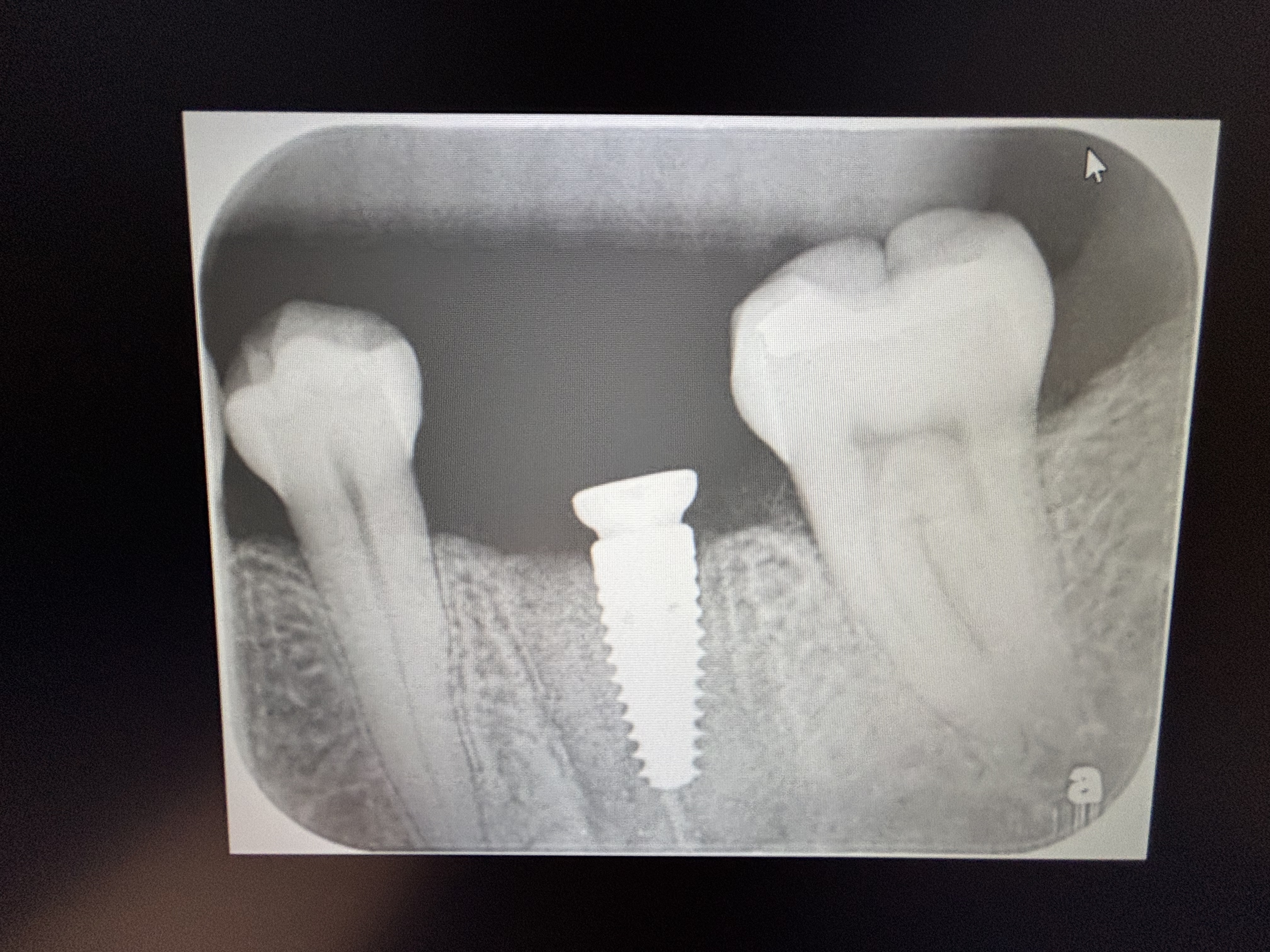
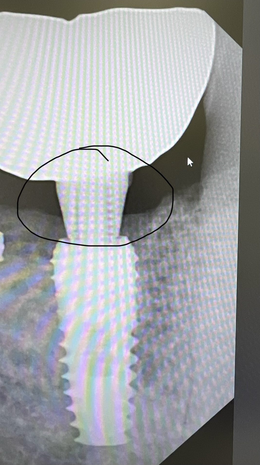
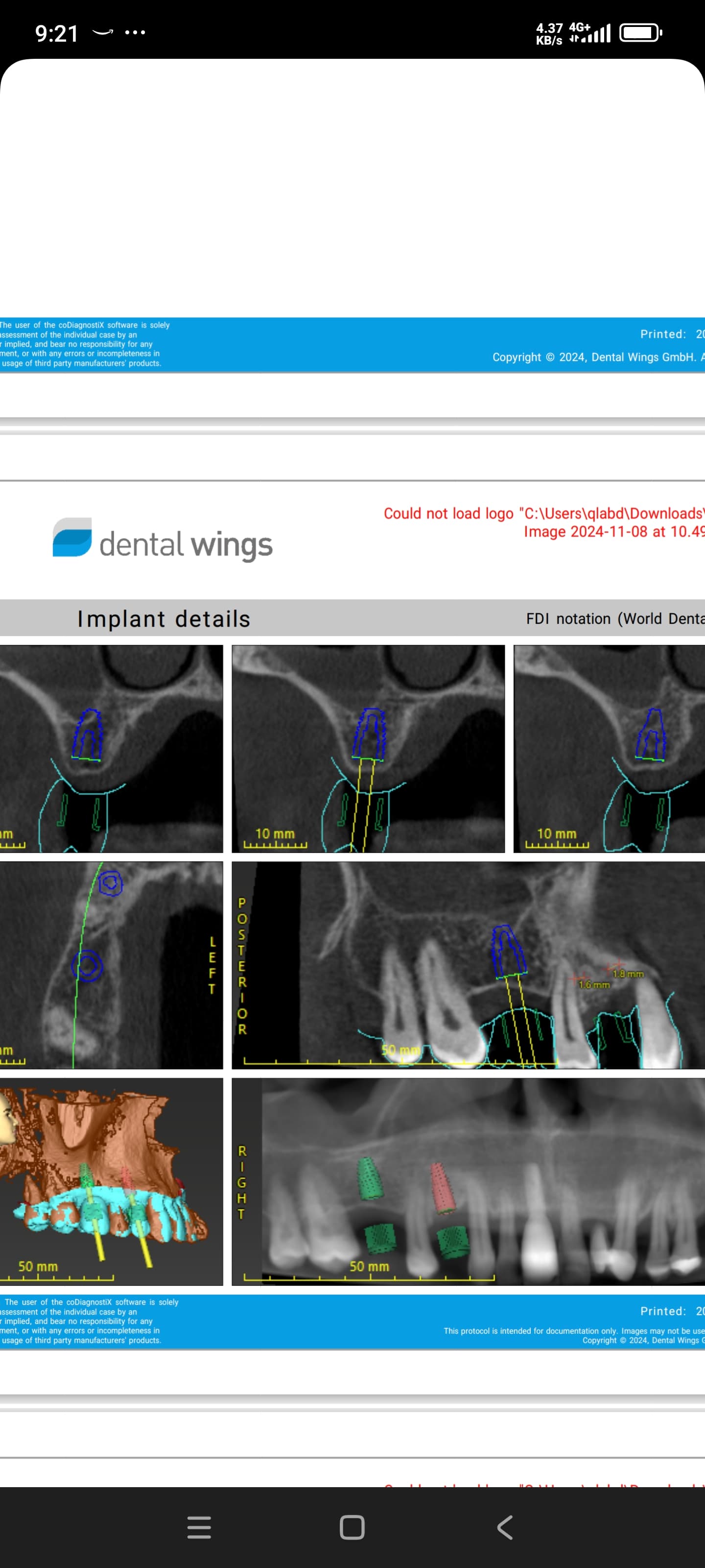
This patient presented for extractions, socket preservation and implant installation. Implants were installed in #10, 19, 20, 29 sites [maxillary left lateral incisor; mandibular left first and second premolars; mandibular right second premolar; 22, 36, 35, 46]. The implant in #10 site is rather close to the root of #9 [maxillary left central incisor; 21] and I am concerned for long term success and avoidance of complications. Would it be better to explant the implant in #10 site and reposition it more favorably? Would it be best to then do a block graft, wait 6 months for osseointegration and healing and then install a new implant? Or should I just observe the situation for now? How long should I wait before restoring it in the absence of symptoms?

(click images to enlarge)


Pre-implant panorexPre-implant panorex
Post-implant panorexPost-implant panorex
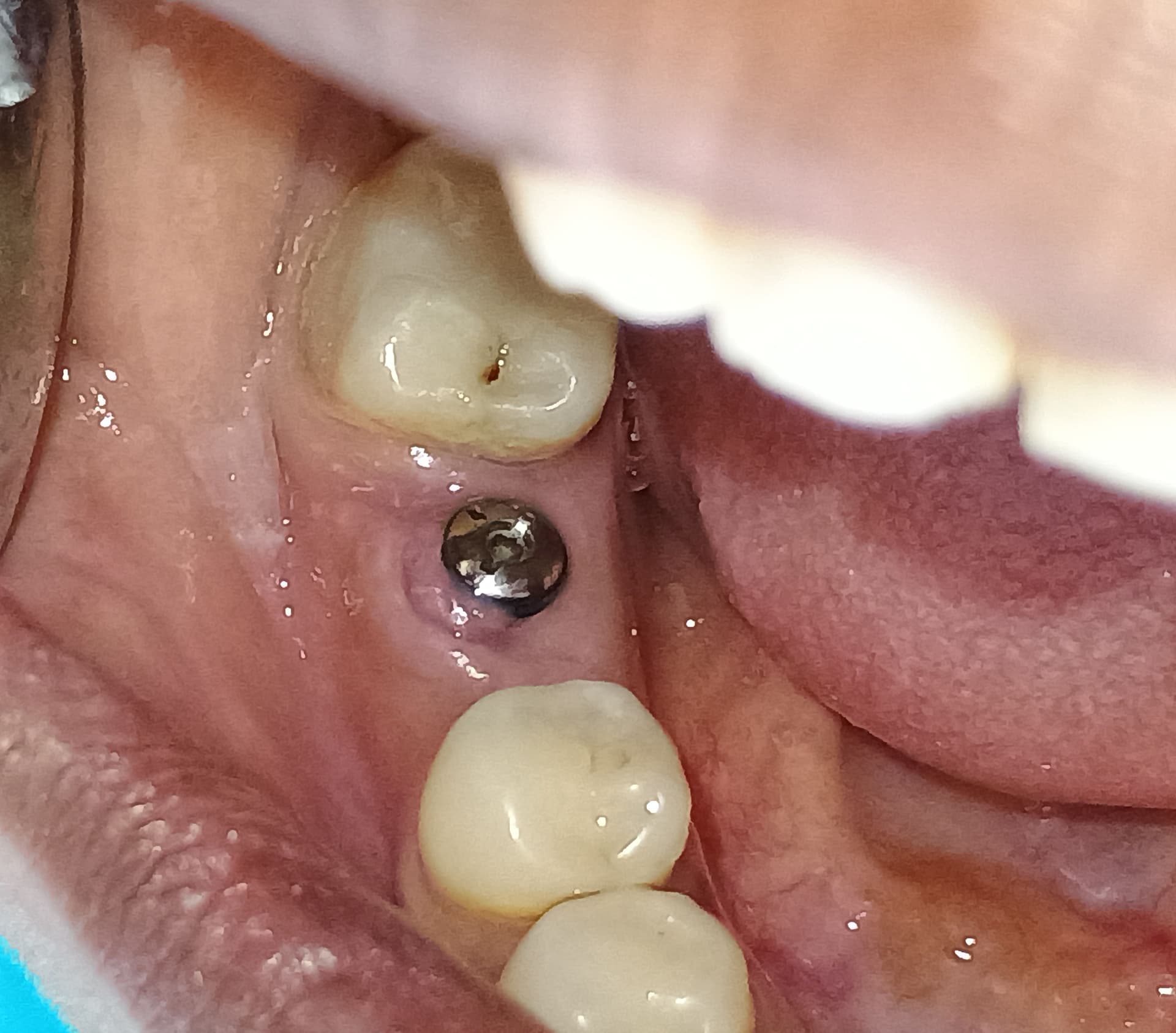
Single upper implantSingle upper implant
Single lower implant leftSingle lower implant left
Single lower implant rightSingle lower implant right

30 Comments on Implant close to root: best way to avoid future complications?

New comments are currently closed for this post.
CRS
7/15/2013
If it were me I would take it out, graft and line it up with the canine. All the other implants are perfect and it would bother me to see this oddly placed implant. Remember you will be replacing the molars next so you will be seeing a lot of this patient.I would suggest getting the perio status under control. A ct scan would truly show where the implant is, you are probably okay but why not have this one perfect also. The central root is oddly shaped. How does it look in relation to the other lateral? Is it symmetrical?
Jerome BHUNJUN
7/17/2013
Hi , Am new to this forum. I have had such a situation with TWO implants. My advice is as follows: First of all WARN the patient of possible complications. Secondly , wait and see , do x-rays evry 3 or six months and at the first sign of trouble remove the implant and replace+ graft . You never know and if all is ok you will be relieved that you did NOT remove the implant. If you have trouble, the patient will recognise the fact that you informed him beforehand. All the best
Michael Eurs
7/17/2013
Removal of this implant would require a trephine. This is like taking a core sample with the implant in the middle of it. Looks like there is no way to remove it without damage or devitalization of the adjacent tooth as there is not enough room. I would advise observation only.
mwjohnson dds, ms
7/16/2013
Is there any symptomology with the #10 implant? If not, there appears to be bone between the implant and adjacent tooth so there's no way I would remove this implant. Too much risk of damaging the adjacent tooth. It also could be somewhat superimposed on #9 since this PA is only a 2D film. If this implant is off angle a custom or cadcam abutment will realign the path of insertion to allow a dependable restoration.
DrO
7/16/2013
I would not expect there to be a problem as it is in the exo socket of the lateral. I would watch the central for any signs of distress and advise the patient it is a little closer than you like but do not expect it to be a problem. Removal and grafting at this point seems to offer more opportunity for damage and delay. I would prefer to wait a little while and see.
ST
7/16/2013
Although this fixture is indeed placed very close to the adjacent tooth, the PA indicates that you are clear of the root. CRS is correct to advise a CT scan (CBCT to be more precise) would correctly portray the true position of the fixture in relation to the adjacent structures. The first thing you are obligated to do is to inform the patient, then consent and proceed with CBCT. If the root is clear, personally, I would restore as normal. The patient should be informed of the possibility of implant failure and of necrosis of the adjacent tooth (#21). You should also offer the patient free remedial treatment in relation to this issue if problems ever arise. I believe that removal of this implant will almost definitely lead to necrosis of #21, and , this combination with an RCT nad potential bone graft, will be very invasive and unnecessary procedures; why put he patient through all this, would you do this if it where your mother, child, e.t.c? The implant and adjacent tooth can possibly have a favourable long term prognosis, the angulation is not perfect but acceptable, thus restorable. You also need to monitor this site with pa's and vitality test (for 21) every 6 months for 2-3 years, and yearly for at least another 3-4
Edoardo Calvi
7/16/2013
To CRS: Sorry but you said it right: "it would bother me ........". I am sure the patient would be fine with it, or at least he would bother more for your idea of having at least other 3-4 not necessary surgeries. It is a well done case there was not so much space but the collegue managed it pretty well, I do not see why this case is here. Thanks for reading, Edoardo Calvi
CRS
7/18/2013
Nope disagree that's why I would not do an immediate in this case, I would graft it and place the implant where it needs to be 1mm minimum from the central, not using the extraction site but the middle of the available bone. An implant is not a tooth no PDL and a papilla may not form. I'm assuming that this is a post op film prior to osteointegration so it is simple to remove just back it out. We all have our standards, that's mine. Hope to see how this works long term with restoration and hygiene. I have to fix these often within 5-8years, just my experience and opinion. Thank for reading.
Kaz
7/16/2013
If you do not have any symptoms or signs of issues, then I would leave it alone. I have seen worse cases with no issues as long as the cervical portion is not touching a tooth and you have 1.5mm space or more from the tooth.
Dr. Gerald Rudick
7/16/2013
I am very happy that you brought up this problem.......because it is not a problem at all, but an artifact in your panorex and periapical radiographs. Judging by the ideal placement of the three other implants you have placed in this patient, your spatial perception is excellent, and I am sure you did not impose on the adjacent natural tooth ( #21 or aka 9) when placing #22 implant or aka #10 implant. If you have nicked the #21, the patient would be suffering pain similar to an endodontic flareup...which you do not mention....so I am sure that tooth is fine. My best advice is to wait and see......do not intervene when intervention is not necessary. A few years ago, a woman was referred to my practice to place an implant in the #22 area, but in this case the root had been extracted some years ago and there was a buccal flattening of the available bone. I purposely placed the implant closer to #23 to be sure that I did not perforate the buccal plate in the area of #22. To be certain that the implant was well placed, I screwed on an abutment and photographed it, before placing the cover screw....as I wanted to wait four months before loading. I took a final periapical film as well as a final panorex, and was shocked to see that the radiographs indicated that I had impinged on the canine!!!! As the abutment and implant body appeared in the mouth,( and in my photographs) I was nowhere near the canine....I felt greatly relieved, and I had the photographs as proof of my proper placement.....I told the patient to come and see me in four months to uncover the implant......as well as calling her several times during the initial healing period, and she reported she was doing well, no pain, swelling, and the canine was absolutely asymptomatic. During the four month waiting period, the patient visited her periodontist for routine maintenance..... and when she told the periodontist that she had been to see me to place an implant.......he took a radiograph, and condemned my work saying the implant was in the wrong place, and immediately proceeded to trephine out the well integrated implant for no other reason but that his pride was hurt because "he did not get the job". The patient called me, very angry with me for " the misaligned implant, as she now was in the midst of extensive bone grafting, and going to go through more surgery to place another implant in its place".....because the periodontist said "the implant was in the wrong place". Had the periodontist, not allowed his insecurity take over his clinical judgement, I would have shown him the photographs to prove that two dimensional xrays are not always the most accurate, when "rounding a corner".... and suggest that the patient return to me to place a temporary crown that would be very esthetic with proper gingival contours........ but this is where politics interfered with the welfare of the patient. So do not be hard on yourself, time will tell....and I am giving odds that you did a great job. Gerald Rudick dds Montreal assoc.F. AAID ; F,D, M. ICOI
John Manuel, DDS
7/16/2013
You basically just placed the implant in the extraction socket, it appears. To avoid this is difficult on immediate or closely spaced extraction/implantation procedures. I'd guess you'd need a pretty sturdy guide sleeve and a long-healed site to avoid such a placement. If you've not perforated the inter proximal laminae durae, not much trouble is likely. John
Periodoc
7/16/2013
The implant is in the socket and isn't contacting the root, so I agree with the other posters...leave it in place. Get the periodontal breakdown under control and continue with your good work.
john townend
7/16/2013
I agree with Dr Rudick, this is largely an imaging artifact on the OPG. If the lateral incisor implant is placed with its coronal end in the socket but its apex palatal to the socket, i.e. tipped somewhat palatally, it will give this appearance of being angled over or towards the central incisor apex. The implant is not in an ideal position but if you took a 3 dimensional view you would find it is well clear of the central incisor. However, what's the point of subjecting your patient to unecessary radiation just to prove the point? Incidentally Dr Rudick I suggest you advise your periodontal colleague to invest in a NeoBiotech implant remover before trephining out any more of your implants. That is if you're still on speaking terms!
CRS
7/16/2013
Thank you for reading my comments, now here is my question isn't it prudent to leave a minimum of 1mm between an implant and a tooth? I find that grafting the extraction site when teeth roots are so close it is easier to use a smaller implant and line it up parallel vs taking the path of the original natural root. I think this looks odd since it had to be placed more palatal, I would just remove it since it is not osteo integrated, graft and place it straighter not so close to the central. I have had cases where the implant appeared superimposed and I know you don't treat X-rays, but the clinical appearance is key. If it is restorable and hygienic then it's fine . I routinely advise my patients if the extraction site is not perfect, I will graft. Now is the time to fix it and the only person who knows is the doc who placed the implant and who will ultimately restore it. The lateral incisor is very tricky. I agree with John Manuel and Gerald Rudnick also. Distal shifting of the periapical a la Clark's rule would determine if the placement is palatal or a CT scan( cbct) whatever your flavor is would also be of help. I Just like to be in as much control of the implant site vs relying on available bone or an extraction site.A clinical photo to make sure the implant is not jutting out buccally would also help, thanks for reading
Dr. Gerald Rudick
7/16/2013
JohnTownsend, thank you for the advice on the Neobiotech Implant Remover, I wonder if the same company makes a "bad attitude" remover from insecure dental collegues...that I know you know. Best regards Gerry Rudick
Don Rothenberg
7/16/2013
I would leave the implant where it is. You will cause greater damage if you try to take it out. If you were going to remove it ...it should have been done at the time of insertion, when you saw the post-op xray. Also using a shorter implant 8 or 6MM would have made this a "non problem". Why are you still using 12 and 14mm implants...it is just not necessary! We very rarely place an implant over 8mm...and if we do it is only 11mm long. I truely believe that using "long implants" ( 11mm and longer) will become a thing of the past,once we understand that ...implants are not teeth...and are effected by different principles.
Richard Hughes, DDS, FAAI
7/16/2013
If there are no symptoms, I usually do not worry. It appears superimposed. A CBCT may help one sleep better but first try PAs at different angles.
CRS
7/17/2013
So that one mm rule about adjacent teeth and implants is moot? I guess there are no do overs in dentistry!
S.Lin
7/16/2013
I would not worry about the adjacent tooth too much, you most likely will be fine. Looks like you follow the socket well and inclined the implant lingually, which is the correct thing to do, as the anterior maxilla concave inward at root apex. If such is the case your PAX or panoX will often appear superimposed. Bucal extrusion is often the risk for case like this. Sure, CBCT is the sure way to tell.
Anand chawla
7/17/2013
i agree with most of u. as the 21 is asymptomatic and the implant has already ossteointegrated, there is no need to remove the implant. if some complication is there in the future,u can go for root canal treatment with 21,if uhave already informed the patient about the possible complication. b/c even if u remove the implant now u will be damaging 21 for sure plus the probably unnecessary surgery. so in my opinion take a c.b.c.t. and keep the patient under observation for any symptoms with 21. all the best.
dr.pradeep
7/17/2013
i have placed 2 implants closed to the roots of adjacent tooth, both were fine, in this case i can see some bone i think you should not worry, wait and watch
CRS
7/17/2013
Please post the restored case would love to see how it turns out, esthetically and how the papilla develops Thanks
Robert Ngan
7/19/2013
In comparing the pre and postop panexs, part of the lamina dura of the 22 is clearly visible on the distal aspect(?) which suggests misalignment. It appears minor, I think the angulation is not an issue. If 21 is asymptomatic, I would leave it alone, still a good effort. The preop panorex shows signs of bone resorption distally. Is this really a good case for immediate placement or is it inviting peri-implantitis and possible loss of papillary form? Hopefully the aesthetics will be good.
Lucy Gold
7/21/2013
Thanks to all for good debate. Still seating on the fence. The POST pictures are fresh after implant. So by today the implants are about 10 days old. Probably there is still time to just screw it out without damage. Reading the debate I get the sense that those who would not remove would avoid it because of potential damage, not because the installation is good. Know that it is just 10 days now - would you take it out by screwing out (not coring), block graft it, and do a guided drilling 6 months from now and implanting a maybe 2.4 mm dia. Or what type and size would you use? For interested: the bottoms are Nobel Active, and the top is Nobel Replace.
CRS
7/21/2013
Dear Lucy, this is what I would do if it were me, just simply tell your patient you want to make the placement perfect, back it out it will be very easy ten days post op, place a particulate graft with primary closure. Wait 14-16 weeks, you have a beautiful provisional and just place the implant more parallel and closer to the canine. My patients are very accepting of my going the extra mile to fix this at the appropriate time. That way you don't need to go so far palatal and the implant will be lined up. When you replace the implant under drill it slightly in width and push vs cut the newly formed bone. You don't need a core, block graft or guide, just time, healing and sincerity. Remember these implants are self tapping and the last drill can be the implant itself. In the long run the case will be much easier to restore with a better esthetic result. Most of the posts did not consider that this is a recent placement, 6weeks being the rule of thumb for easy removal. I sense that you picked that up And if this placement is correctable then do it the implant size seems fine it is easier when there is not a socket to deal with . Best wishes.
Gregori Kurtzman, DDS, MA
7/23/2013
The best way to avoid this issue is by ignoring the root space after extraction and using the pilot drill parallel to the adjacent teeth then place either a pin or the pilot drill into the site and take a radiograph to verify the orientation with the adjacent teeth. then proceed or adjust trajectory accordingly. With this case right now the bone is very thin between 9 and the implant and at risk for loss after restoration of the implant over time. if the implant has been in less then 3 weeks you should be able to back it out and then graft the site and wait 4-6 weeks before attempting another implant but if its been in longer then 3 weeks you may see alot of crestal bone loss on the distal of 9 with removal graft or not.
DrO
7/24/2013
If the coronal portion of the implant was well positioned as I suspect it was, I can see no good reason for removal. A custom abutment can make up for any minor angulation issue and the close proximity to the adjacent tooth is no big deal. The pursuit of ideal is just that. We pursue it, actually achieving it in all circumstances is not practical. Leave well enough alone.
CRS
7/24/2013
Or you can just move your practice every five years, this may come back to haunt you.
rsdds
7/31/2013
i actually think this is a very nice case !!
CRS
8/7/2013
Yes it is, the funny looking lateral on the panorex is due to the immediate implant being placed more palatal (Clark's. rule) The other implants are perfect. The poster wanted to know if the lateral was too close since the implant had not osteointrgrated I advised placing it more in the middle of the alveolus for long term hygiene and allowing at least 1mm from a natural tooth as advised in most implant literature (Misch etc) along with the implant manufacturers. The implant slid along the extraction site. It will probably be ok but since one at easily intervene and place it more optimally vs hoping that it would have an uneventful course. I would have removed it after I saw the periapical, grafted and replaced it in 12-16weeks, not a big deal. If this implant is problematic in the future one could be faulted for violating the 1mm rule. That's just how I try to practice with ideal placement, patients seem to appreciate this. But it Is a beautiful case!

Featured Products

OsteoGen Bone Grafting Plug
Combines bone graft with a collagen plug to yield the easiest and most affordable way to clinically deliver bone graft for socket preservation.
CevOss Bovine Bone Graft
Make the switch to a better xenograft! High volume of interconnected pores promotes new bone. Substantially equivalent to BioOss and NuOss.