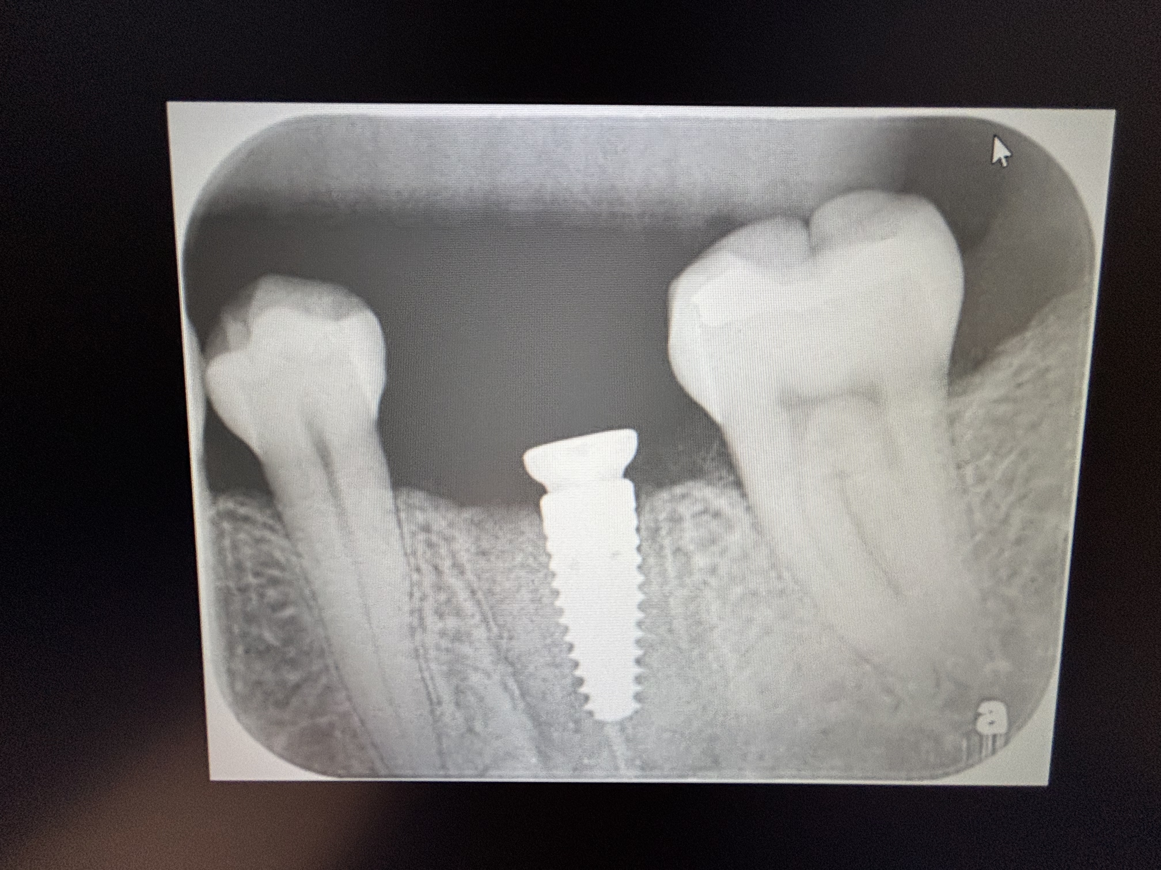
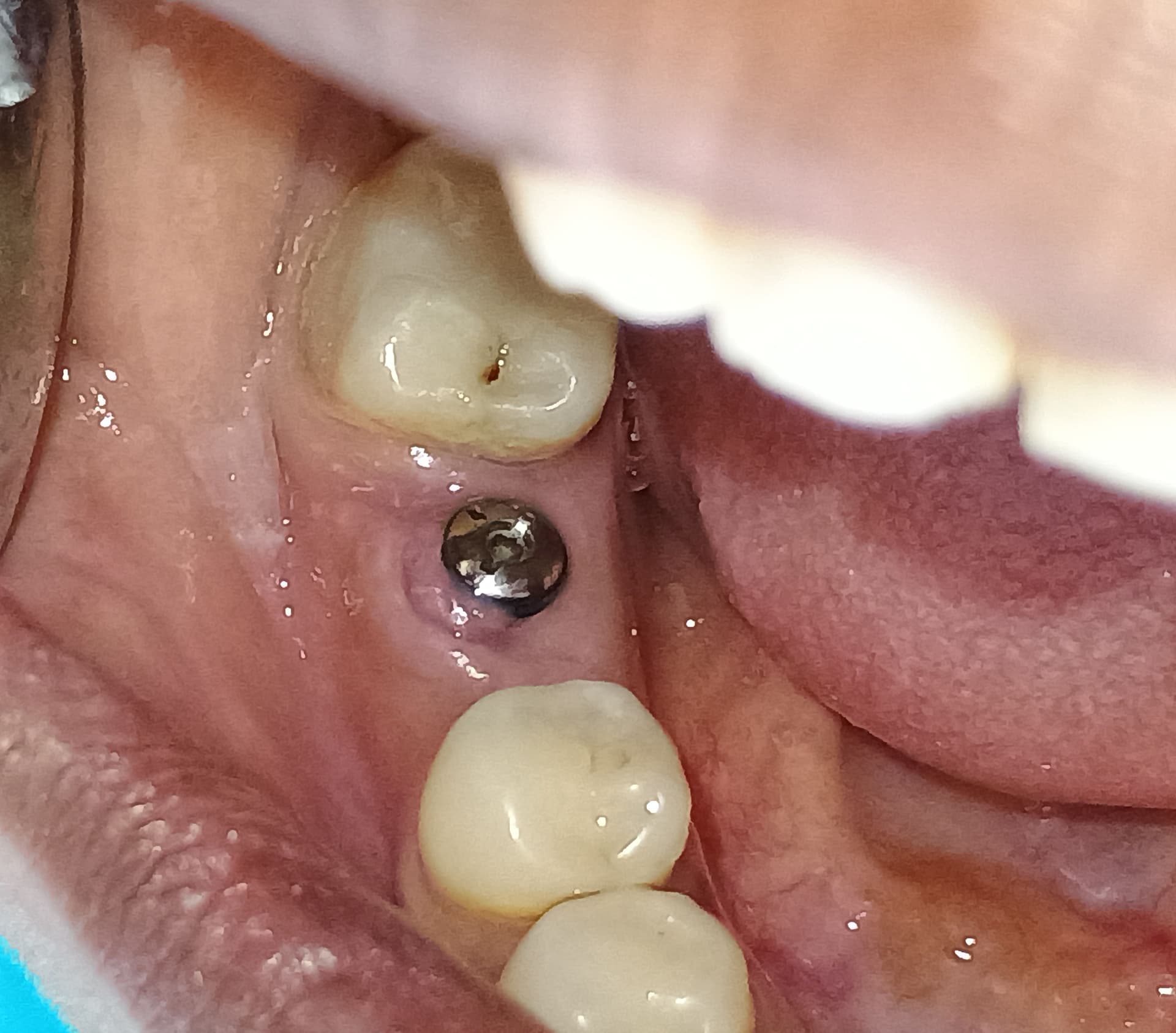
Implant missing for tooth 46 with 11mm space: thoughts?
I have a patient missing the mandibular right first molar and first premolar. The second premolar has tipped mesially. The mesiodistal space between the second molar and second premolar is 11mm. I would like to place one wide platform implant in the space. Will that be the best treatment plan?


42 Comments on Implant missing for tooth 46 with 11mm space: thoughts?
New comments are currently closed for this post.
greg steiner
2/8/2016
Place the implant. It does not look like this patient would be interested in ortho. From the radiograph you can see that the adjacent teeth are overloaded and the implant would stabilize the adjacent teeth and remove the excessive forces. Another issue is that you need to expect to find very pooly mineralized bone when you are drilling your osteotomy and you will need to be ready so you don't "fall in" to the manbdible. You should be prepared to treat the site if there in little to no mineralized tissue under the crest. Greg Steiner Steiner Biotechnology
Dr Gilani
2/9/2016
Hi Greg,
How do you treat a poorly mineralized spongiosa?
Joe Nolan
2/10/2016
Hi Greg, how can you foretell the lack of cortical bone ...from the relative traslucency of the bone perhaps?
In the event of softer bone, what would your best approach be, in terms of type/size of implant?
Best wishes
Joe Nolan
Dr Schiavone
2/8/2016
Yes, that's the best plan
DrT
2/9/2016
I agree but I would recommend ortho to patient...second molar has fused root so uprighting should not
be too difficult. If pt is investing in an implant he should at least be advised of what the optimal
treatment plan would be
jaime schutt
2/9/2016
habria que injertar a que se refiere sobre la mandibula cae y ver que tanto esel ancho vestibulo lingual
DrT
2/9/2016
Would you kindly post in English. Thank you
Peter Hunt
2/9/2016
A simple, fast and very effective solution would be to remove the mesially inclined second premolar and to place a second implant in the bone adjacent to the canine. The extraction site should be socket regenerated as the distal of the new implant might be partially within that socket.
Dr Gilani
2/9/2016
No need for wide platform implant. Use anything from 3.75 mm diameter and ca 10 mm long.
DrT
2/9/2016
Remove a virgin tooth with greater than 50% of its remaining bone support?? What are we coming
to????
Paul Newitt
2/9/2016
Agreed!
Albert St Germain
2/16/2016
Yes, I agree with the assessment by Dr Hunt.
Sometimes it's better to sacrifice a tooth to assure a more ideal outcome. We've done this when the patient has had a deficient mandible existing between #18 and 21 requiring extensive bone grafting prior to placing a bridge but adequate bone in the existing tooth sites to place implants and restore the edentulous space with an implant supported bridge. Thus the extraction of the molar and bicuspid and immediate implant placement followed by a bridge in four months.
Frankly, that's the first thing that came to mind when I saw the radiographs. However, It would be best to advise the patient the he will wind with a triangular food trap between the molar implant and the existing molar.
So are we back to uprighting or a crown on the molar?
Food for thought.
Jawdoc
2/19/2016
If that's the case, Why not just do a conventional bridge then? Preserves good teeth & saves u the trouble of implants. No?
Peter Hunt
2/9/2016
Well it's easy enough to upright it and bring it distally, especially if you have an implant in the molar region to provide anchorage. The distal molar can also be uprighted at the same time. We used to do that all the time many years ago when treating bite collapse cases. These days, it is is not so common because implants are being used more and more.
Dariush Radman
2/9/2016
It seems to me that the second premolars rotated 90 degree, if I am right, then the most predictable treatment plan would be first Orthodontic approach to correct the position of 29 ( to derotate it and move it to the position of 28 uprightly), up righting the 31 and then you will get enough space to place 2 implants and load them with proper occlusal contacts on implants and the adjacent natural teeth.This will render almost usual size of first molar and second premolar crowns on implants.
RC
2/9/2016
Thanks everyone for comments.
Patient is not interested in orthodontic treatment and would just like to get implants done.
The premolar has tilted mesially and has not rotated as such.
Would it be detrimental if an implant is placed in this space without orthodontic treatment of adjacent teeth.
Should I go for narrow or wider implant?
Thanks.
DrLSD
2/9/2016
Again, consider conventional Maryland Bridge or PFM 3 u fixed brig
Richard Hughes, DDS, FAAI
2/9/2016
If ortho is out of the question then extract the bicuspid and place a bi and molar implant.
Amir H. Mostofi
2/10/2016
Hi
When you ask the the question " Is it the best treatment plan?" I assume that you are looking for a comparison to other treatment option?
Clearly there are 2 different treatment options here:
1- Implant treatment
2- Fixed prosthetic (3 units bridge).
In the days of evidence based dentistry a dentist decisions should be based on reliable high quality clinical evidence and not just what we "feel" is the correct answer. This website is an Implant website and most of us prefer to have an implant placement rather than an conventional metal bonded ceramic bridge.
But contrary to our beliefs, and if you would like to have a academic look into this issue, the most recent data (systemic review of randomised clinical data) shows that the survival rate ( please note the survival rate is different from success rate) for implants is the same as end abutment fixed prosthetic. For further information and research look at systemic reviews in for example Cochrane Library. Or for example use a PICO treatment intervention comparison to find out about the reliability and longevity of different treatment interventions.
In addition the aesthetic outcome of an end-abutment bridge is more predictable and the patient does not need to undergo additional treatment (ortho.). The practical difficulty is to do the abutment parallel when preparing the teeth. The OPG is a two dimensional X-ray image and it is hard to do a correct judgement in this case, about the possibility of parallel abutment preparation when doing a bridge preparation on lower right side.
However in my opinion you need to consider both options.
Jawdoc
2/19/2016
I would have to respectfully disagree. It's a well known fact that conventional bridges do not last as long as implants.
RC
2/10/2016
I am sorry that I did not frame the question correctly.
I would rephrase it .
The patient is wishing to have missing area replaced by a dental implant and I wanted to know if single implant supported crown will be a good option in this case.
Thanks.
Bülent Zeytinoğlu
2/10/2016
Even if you have the skill to place an implant between the molar and the premolar without giving harm to the adjacent roots I think you will have difficulty in the prosthetic step besides there will be big triangular gaps at the mesial and distal of the crown so it may not be a bad idea to make a conventional bridge but taking care to the parellelism of the anchor teeth.
CRS
2/10/2016
Width of bone will determine implant diameter. Place implant with ideal crown dimension so that it will be in interproximal contact with molar to prevent more drift. Leave a cleansible space between premolar,it will be symmetrical to the other side. Have pt sign consent staying although not ideal this is treatment recommended to restore space since tooth drifted. Spell out risks benefits and alternative treatments, patients don't care about evidence based or spongy bone just what is going on in their mouths. I like to educate the patient, let them chose the most beneficial and least harmful option based on the circumstances. Remember this is elective. Be wary of trying to close the space with a wide crown on a narrow implant, two narrow implants may not have enough room. Get the calipers out and measure the model refer to the prosthetic section of the implant manual to determine best option for the implant. Remember that the prosthetics determines the placement, a cleansable space on the mescal may be a good compromise. Good luck hope I helped😊
ST
2/10/2016
Hi
have you considered placing 1 "standard" sized fixture, say 4.5x 11.5, distal and close to molar, and create 2 small premolars with a mesial cantilever, or large molar with mesial extension (hence cantilever again)? Mesial cantilevers, as opposed to distal, are well documented and quite successful. This way you avoid
* large triangles
* poor implant positioning
* need for wide diameter fixture and risks of
* poor aesthetics
Your upper 1st molar #16 is tipped distally and would not create heavy occlusal forces on mesial aspect=protected occlusion
Not a perfect outcome, but not a bad one either; Just A thought
Another option is to place a TAD (ortho implant) to upright lr7, you can pass this on to pt as implant t.t and not ortho, then place 2 small fixtures, say 3.5's?
Dr Bob
2/10/2016
Yes a single implant in this space will work OK. Not ideal but OK. Place the largest diameter that will fit and still allow for approximately 2mm of bone or more all around it. Use a rather narrow facial - lingual table to minimize forces on the crown. Try to keep the forces in line with the vertical long axis of the implant, and keep lateral forces also to a minimum.
RC
2/10/2016
Thanks everyone for your valuable comments.
This is going to my first case of dental implants, so does it mean that it is going to be a complex case and I should refer the case to another dentist ?
Thanks.
Dr Gilani
2/11/2016
RC,
Unfortunately, most of the above comments are too hypothetical and fear mongering. Go ahead and drill a hole and torque in you implant in the right angle and length as you won't find simpler case than this.
Best wishes
peterFairbairn
2/11/2016
Yes Dr Gilani , world is never a perfect place , go ahead RC . Only word of advice check the ridge width as CRS suggested . The slight shadow on the x-ray may suggest modelling of the bone and hence may not have the width for a wide platform ......
Peter
Alex Zavyalov
2/11/2016
If the author were in the patient’s shoes he would definitely consider a molar-fixed-cantilever treatment plan. That's why I partially agree with DrLSD because of the defect size. This Forum discusses a possibility of an implant treatment as a better choice for any patient but this should not become the only possible option.
greg steiner
2/11/2016
Dr. Gilani
I treat poorly mineralized bone by stimulating osteogenesis during implant integration. First prepare your implant osteotomy and then fill the osteotomy with OsteoIntegration then place the implant that disperses the material into the bone. When you go to load the implant you will have normal to superior mineralizaton. OsteoIntegratiopn was developed for this purpose in addition to treating "spinners". Greg Steiner Steiner Biotechnology
greg steiner
2/11/2016
Joe Nolan
The best indicator for poorly mineralized bone is when you have a thickened crest over bone of reduced radio-opacity as you see in this radiograph. The thickened crest is a result of endosteal bone formation under the crest in response to poor structural support provided by the underlying cancellous bone. Rather than "falling into" soft bone it is best to diagnosis it first and tell the patient how you plan to treat it so they understand the process and of course the charges for treatment. Greg Steiner Steiner Biotechnology
joe nolan
2/12/2016
Many thanks ,Greg
What is your opinion about underpreparing the osteotomy, or using wider blade implant ?
greg steiner
2/15/2016
Joe Nolan
The only time I under-prepare an osteotomy is when I am placing an implant in 1 to 2 mm of crestal bone in combination with a sinus augmentation because I don't want the implant pushed into the sinus. When I am putting an implant into poorly mineralized bone I know the bone has poor vitality and underpreparing the osteotomy will not improve the degree of integration. I treat the bone to improve it's vitality by stimulating osteoprogenitor cells and osteoblasts to regenerate the vitality of the bone that will improve integration and eventually support the implant. Greg Steiner Steiner Biotechnology
greg steiner
2/11/2016
Amir H. Mostofi
Yes bridges having the same survival rate as implants is contrary to our beliefs. Did the studies look at survival rates in general throughout the dentition or did they focus on survival rates for bridges and implants when replacing mandibular posterior teeth? My understanding is that it is the movement of the mandible under a fixed bridge that is responsible for their reduced survival rate. If the studies looked at bridges and implants throughout the dentition then the studies are of no value. If they looked specifically at comparing bridges and implants in the mandible posterior dentition then I have something to learn. Please clarify. Thanks Greg Steiner Steiner Biotechnology
Amir H. Mostofi
2/16/2016
Greg Steiner,
Yes there are plenty of good systemic reviews available. Broadly speaking the evidence today shows the survival rates for Bridges and single tooth implants are the same. They take both technical and biological complications into the account. For example have a look at the review below:
http://www.ncbi.nlm.nih.gov/pubmed/17594374
If you can not access the site then you can get the PDF format here:
http://www.ykdent.com.tw/pdf/sam.pdf
As I mentioned above, for an specific treatment or intervention, the best way forward is to do a PICO search. I hope it helps.
greg steiner
2/18/2016
Hello Amir
Thank you for the article. It was a well done review but I could not see where they looked at restorations in various locations of the mouth. I am commonly removing failed bridges in the posterior mandible and placing implants. I seldom do this in other areas of the mouth. Do you know of any research that looked at survival rates of various restorations in the posterior mandible? I strongly advise against fixed bridges in the posterior mandible and I would very much like to know if I am misinforming my patients and referring doctors. Thank you for you help. Greg Steiner Steiner Biotechnology
Juan Rumeu
2/15/2016
Dear colleague: This is a pretty straight forward case. Firstly a CBCT is needed to evaluate whether wide o regular platform has to be placed and chances are based on the full pano Rx that you will need a GBR procedure. Good luck. Cheers.
Jalil Sadr
2/17/2016
Hi, how long patient has been like this. No upper teeth eruption. if from occlusion point of view is ok, after CBCTand its evaluation, it seems you could insert two narrarow Implant ( 1mm+ 3.1 + 3mm + 3.1+ 1mm = #11mm), with minimally reshaping mesial surface of 2nd molar, even one buccally and other one more lingually. As we see roots of both teeth with little mesially tilted are Parallel togather. Insertion should follow the same direction which is hard to handle.
Joe Nolan
2/18/2016
Fixed full coverage bridges and crowns are designed to fail, whereas a biomimetic approach will give a 20 year implant more than a run for the money...an onlay will allow flexion, ever notice how onlay retained bridges rarely ever show caries?
KDS
2/28/2016
In my opinion you should place the implant, it last longer than the bridge. Good luck.
Stefan Gollwitzer
3/6/2016
Hi,
place an implant as long and as big in diameter as possible. Secure the abutment, and then the crown with concave / convex contact to approx. teeth, do not overload contact to antagonist teeth and everybody (Patient/Dentist) will be happy for a long period of lifetime. regards Stefan
Dr. Dennis Nimchuk
4/18/2016
From the radiograph I see a crestal dip which means that there is ample vertical height to develop a decent emergence profile without making a lollypop on a stick. The mesio-distal dimension is a normal 11 mm. If CBCT imaging allows it place a wide implant 5mm or greater. If not, then graft, or accept an implant diameter in the range of 4mm and produce a custom abutment that will develop the best emergence. This case does not seem so complicated.


