Implant placed adjacent to non-infected previous failed site: will it survive?

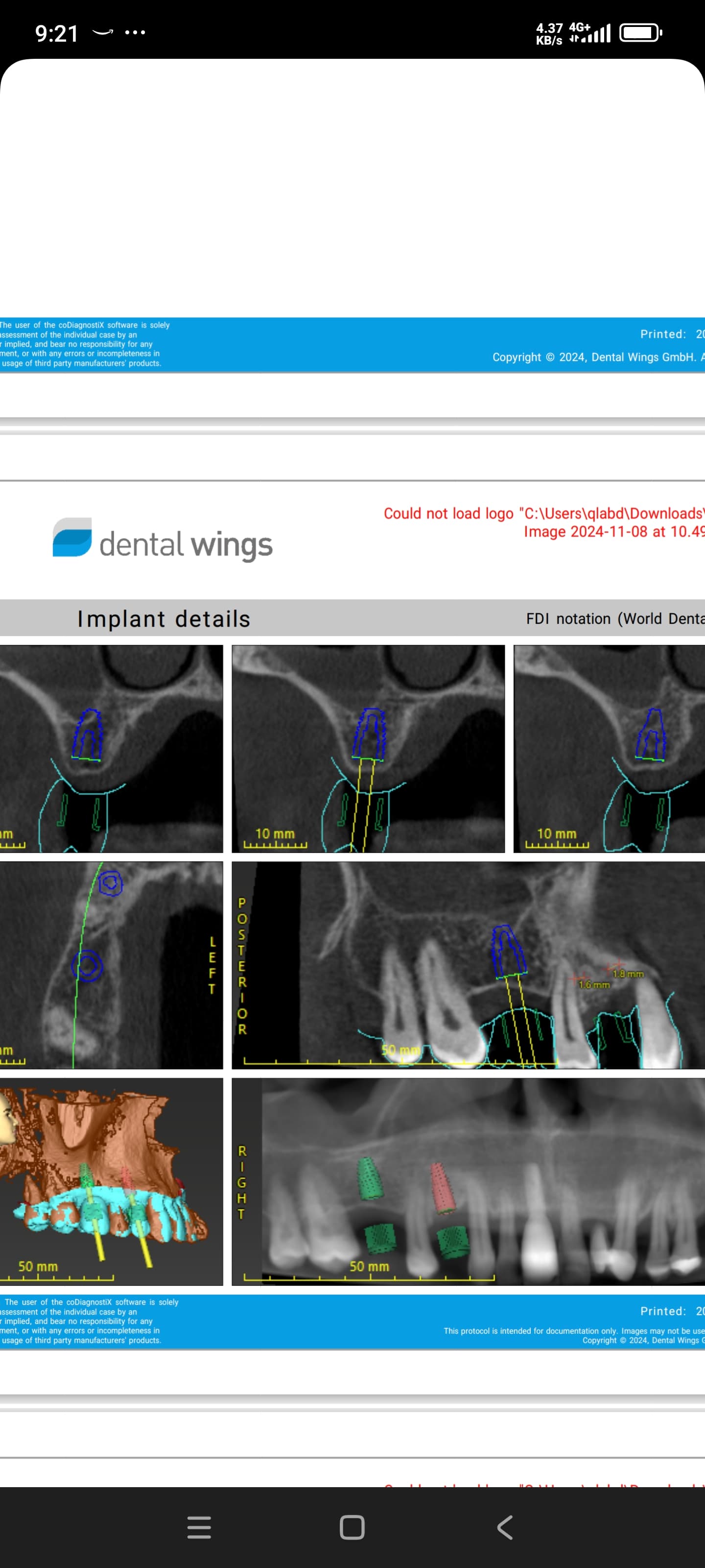
I have a new patient who had 6 implants placed in her maxilla 5 years back by another dentist. The patient said out of 3 implants placed on left side in #9, 10 and 14 sites [maxillary left cental incisor, lateral incisor and first molar; 21, 22, 26]. 2 implants failed by second stage surgery in #9 and 14 sites and were not replaced. Only the implant placed in #10 site survived. The 3 implants in similar position on right side #3, 7, 8 [maxillary right first molar, lateral incisor and central incisor; 16, 12, 11] osseointegrated. The previous dentist provided a palateless fixed-retrievable (hyrid) denture with UCLA abutments on the remaining 4 implants and was not retrieved once for cleaning. The denture broke and patient came to my clinic. I retreived the denture and found that lone standing maxillary left implant in #10 site had failed and took it out. There was no pain or infection. Unfavorable occlusal loads seems to be reason for failure. I treatment planned 3 new implants on left side to support a fixed partial denture. I placed implants in #13 [maxillary left second premolar; 25] and 14 sites and planned to place an implant in #11 [maxillary left canine; 23] site as well. At the surgical installation visiti for #11, the buccal bone was thin and perforation of buccal plate happened, so I had to give up placing an implant on that site. I then placed the third implant in #9 site [maxillary left central incisor; 21] . Due to proximity to incisive canal, I had to be cautious in the surgical installation. In doing so the implant was touching the previous failed site( failed implant was removed 20 days prior) and the implant did not achieve primary stability. This implant was Osstem TSII. Will the implant survive or what should be done?


iopa of implant touching failed siteiopa of implant touching failed site

19 Comments on Implant placed adjacent to non-infected previous failed site: will it survive?

New comments are currently closed for this post.
Peter Fairbairn
5/5/2013
There are a number of worrying issues here , the lack of primary , the clear lack of curretage of the previous granulous site and finally it appears some graft material has been place ...possibly with HA ( Allo or Xeno ) which may complicate matters . So you may be lucky but failure may occur , I would not have followed this protocol . Good luck. Peter
CRS
5/5/2013
I would have treated this case with grafting if the infected, failed site with curettage, laser sterilization and grafting with prgf,bone and Teflon membrane. I would plan preop with A CT to determine the implant placement for the planned prosthesis. I would have my prosthodontist opine on the prosthesis design making sure there is enough inter arch clearance and also use the successful implants in the design. This case does not appear to me to have a flight plan. The short answer based on the X-ray with in implant placed in poor bone is that it will fail. A complex case with a lot of factors to consider, Inagree with Peter. Thanks for reading.
ttmillerjr
5/6/2013
Okay, So let's talk about ct's. What kinds of cases "need" a ct? More complex cases, correct? I think ct imaging is becoming the standard of care (in many areas) for all but the most straight forward cases. Here you have a case with a history of some failed implants, a case where the denture design may not be appropriate for the number of implants present, a case needing multiple new implants, etc. This case would easily pass muster for a ct. I think all would say you didn't need a ct to realize you didn't have anywhere to put #9, but a ct may have made this more obvious. It looks like maybe locators are being employed, so you need a fair amount of parallelism, which looks like it may be a problem here (looking at the implant distal to 9). So without planning appropriately, I don't know that you have helped her much. 9 will soon be gone, and the only other implant we can see looks like it is at an unfavorable angle. Remember, implants are planned based on the final restoration in mind. We all make mistakes, so learn from this experience and spend more time planning.
Sam Jain
5/7/2013
Dear Dental Colleague Do not sweat. Let us think through it. First my sincere advice to u would be to buy a CT today! You can SEE and you won't have to guess the z dimension. Now with this case if u let this implant heal and integrate, it will always remain sick and would have to come out due to part of implant body being sorrounded by soft tissue. So this decides that u would remove the implant right away. Now what can be/should be done really depends on your surgical skill level. You can thoroughly debride and graft /block graft the area at the same surgery but if Px wears a stay plate it will kill the graft. If this Px was in my office and had been promised immediate loading using three old implants on the right and three new on the left, I would remove this implant, debride, and place 3.7 x16 implant anchoring in the tons of apical bone and what we do is harvest bony cylinders (called huggers)from ramus using double trephine method, to suit the 3.7 body. Place one hugger on the facial of implant and one on the distal exposed surfaces and we ALWAYS use bmp soaked sponges( for these kind of difficult scenerios) placed along these boney cylinders and covered by prf membrane. OD abutment screwed to 30 N , titanium cylinder screwed to 20N and the flap sutures tightly at the neck of implant. If the Px was promised immediate loading, the Px gets the immediate teeth. Sorry I am not able to give u some practical easy solution. May be u can use 16mm mini implant for fixed temporary and as well use it for tenting for GBR. You can call me at 510 574 0496. I can help if you can show me CTslices. Take care buddy. Sam Jain DMD Center for Implant Dentistry
Loyd Dowd, DDS
5/7/2013
What cases need a CT? All of them in my opinion and in the opinion of every personal injury attorney around. That's why I bought my own CT machine about a year ago. I know it's a large investment but since I can take CTs at the drop of a hat I'm doing a lot more implant, surgical, and even endo cases. My gross production has gone up 40% in the last year from treatment that I would have not seen the need for or would have been leery of doing blind using 2D x-rays.
Adam
5/7/2013
Based on history and the radiograph, the implant will fail: no primary stability, apparent radiolucency around the implant in the 2D xray - which could be many things but it is not quality bone. If you do not get primary stability, do the necessary grafting and come back to place the implant. Violating the incisive canal is not as big a concern as you may think. Oh, while planning with a CT is an inarguable asset, most of the success rates that we quote today to our patients were established well before the use of CTs became common. Good luck with the case.
CRS
5/7/2013
The bone looks very poor on the periapical and nothing was done to augment it. Standard of care is not determined by CT scan but by the jury deciding what a reasonable practioner would do. And often you may be held to the standard of an oral surgeon. The history given on this case concerns me, there may be a retrograde periimplantitis and cutting the new flutes in the bone may stir up the embeded bacteria. I only mentioned the CT for the overall plan of the fixed prosthesis and plan for implant placement it is pretty obvious that the periapical is sufficient in this case. Also placing bone cylinder huggers soakedin BMP won't get rid of the bacteria, hence I suggest laser sterilization. I am very happy for those pracitioners who are increasing their bottom line with their cone beams but the amount of radiation must be considered and used judiciously. Thanks for reading
Dr M C
5/7/2013
Hi Doc, there is absolutely no problem in placing implants in a failed site. Site needs to heal for 6months in maxilla and 3 months in mandible or there needs to be sufficient bone for a bigger diameter implant. While doing osteotomy if the bone is soft then undersize your osteotomy and place implant with great primary stability. Have done cases like this with great success. As you have noted this is not an infected site. As for the present implant you have placed , if there was good enough primary stability its going to work, just leave it for 6-8 months with a review every 15 days to check if there is any concern.
Dr M C
5/7/2013
Hi Doc if it is recent placement then remove the implant and place a larger length so as to get primary stability from the apical half of the implant. I would take a 2.3 drill and place a 3.7 tapered screw vent implant from zimmer. It is HA MTX. and fantastic primary stability.
Sam Jain DMD
5/8/2013
Removing abscesses teeth and scrubbing the socket with gauge soaked in IV clinda/ metro for 20 min, and placing implants with grafting is the work horse of my practice....every day business.....once you clean the area properly the area is not infected anymore. And I 100% believe in it. Placing implant in thoroughly disinfected site has never ever become an issue. If a px comes with a tooth in the mouth, they almost always get a flap less extraction and implant and a screw retained temporary also if it is in the esthetic zone. And now with advent of Bmp, there has been a paradigm shift in the way we think about implant treatment possibilities. Bmp has left behind any other biologics that we have known.....it is a giant leap.... You guys have to look at the work of Dr Marx, a very charismatic omf from Miami. You will be impressed by his passion, voice, and style. He has been my inspiration. CRS keeps talking about his laser disinfection, there must be some thing special ..... I have not personally seen any convincing work/cases in this field. So I do not have much knowledge about this ..... Can it heal a peri-implantitis?..... I have heard a lot of Dr Stuart Froum's lectures on growing bone on exposed implant threads, but he never talks about laser for disinfection. So I never payed any attention. Good night friends. Sam Jain, DMD Center for Implant Dentistry cases.
CRS
5/8/2013
It is a newer application. This bone is not optimum here. I am a charter member f the tissue engineering society and have followed Bob Marx for many years he developed the Bisphosp protocol but I personally stopped using BMP since it has been linked to cancer and the company is in trouble. There are other safer methods to grow bone. You probably get lucky with the second implant placement since you are mechanically n chemically removing the bacteria present . I see value in disinfecting the bone with laser energy this is an application that I feel will be beneficial especially in occult chronic infection as we see in localized osteomyelitis and perimplantitis. It is good however that you are following Marx he also was a pioneer in bone grafting for head and neck cancer, his techniques are widely used today if you were an OMS you would know this since I is part of our curriculum. He is partial to PRP but I like PRGF better. And yes grasshopper there is something very special about laser disinfection I'm just ahead of the curve on this and nice enough to share this pearl.
Peter Fairbairn
5/8/2013
Hi CRS wise words on BMP , fortunately banned for use here in Europe . Side effects and poor initial bone are the main issues , the body wants to heal , after 10 years of developing materials ( Alloplasts ) I feel there is a major breakthrough in host regeneration and it will introduce a more frienly more effecient way to acheive better results . Regards Peter
Sam Jain DMD
5/9/2013
Hello Dr CRS I also know his work on bisphophonate INDUCED onj and also his work on hyperbaric oxygen on radiation treated head and neck cancer-----exploiting the power of Bmp-prp-mfdba( "taco salad" is dr Marx' s term) to grow back the resected area instead of using fibula harvest etc. I am very cautious in the use of Bmp and really enjoying what it has been doing for me. After reading your long posts I keep wondering what I really learned. Nice to know you are ahead of the curve. Sam Jain, dD D
CRS
5/10/2013
For some reason my posts seen to generate a reaction for you vs an exchange of information if you don't learn anything then perhaps don't read them. I don't get the defensive attitude, it is very simple I have a different opinion from yours. I do find some conflicting ideas in your treatment plans which you seem to immediate start to defend when another idea is presented. Sorry we cannot seem to agree and perhaps you may have stated the issue you keep wondering what you've learned. In future I will try not to educate you and let your treatment plans speak for themselves. I still wonder what you find inappropriate for GPs? We all have dds degrees and different experiences.thanks fr reading all feedback is helpful
Sam Jain
5/9/2013
Hello CRS I realize you are an oral surgeon but I would refrain from using inappropriate language for the GPs. Sam Jain, DMD Center for Implant Dentistry
CRS
5/9/2013
What do you find inappropriate?
Sam Jain
5/9/2013
I realize you are an oral surgeon but I would refrain from using inappropriate language for the GPs. Sam Jain, DMD Center for Implant Dentistry
E
6/25/2013
What is the settings for YSGG for implant decontamination before bone grafting. At the clinic, there are Waterlase YSGG and also 810nm waterlase soft tissue laser and periowave, any suggestions as to which machine gives best results for decontamination and what settings if either waterlase YSGG or 810nm is used? Thankyou.
CRS
6/26/2013
Waterlase will decontaminate the implant surface without harming it but it won't kill pigmented bacteria since it only sees water and hydroxyapatite. The 810 diode will only cook the tissue, like the crust on cream brûlée. The wavelength is key 1064 to kill p.gingivitis and have depth of penetration in the privileged sites, I am developing a protocol for this. Thanks

Featured Products

OsteoGen Bone Grafting Plug
Combines bone graft with a collagen plug to yield the easiest and most affordable way to clinically deliver bone graft for socket preservation.
CevOss Bovine Bone Graft
Make the switch to a better xenograft! High volume of interconnected pores promotes new bone. Substantially equivalent to BioOss and NuOss.