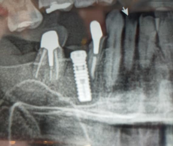
Is the Implant Fixture Too Close to a Natural Tooth?

Dr. G. asks:

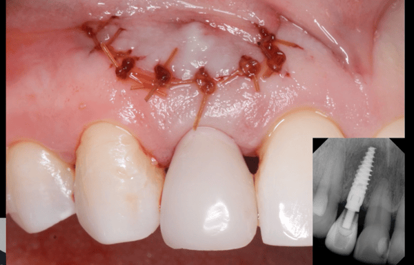
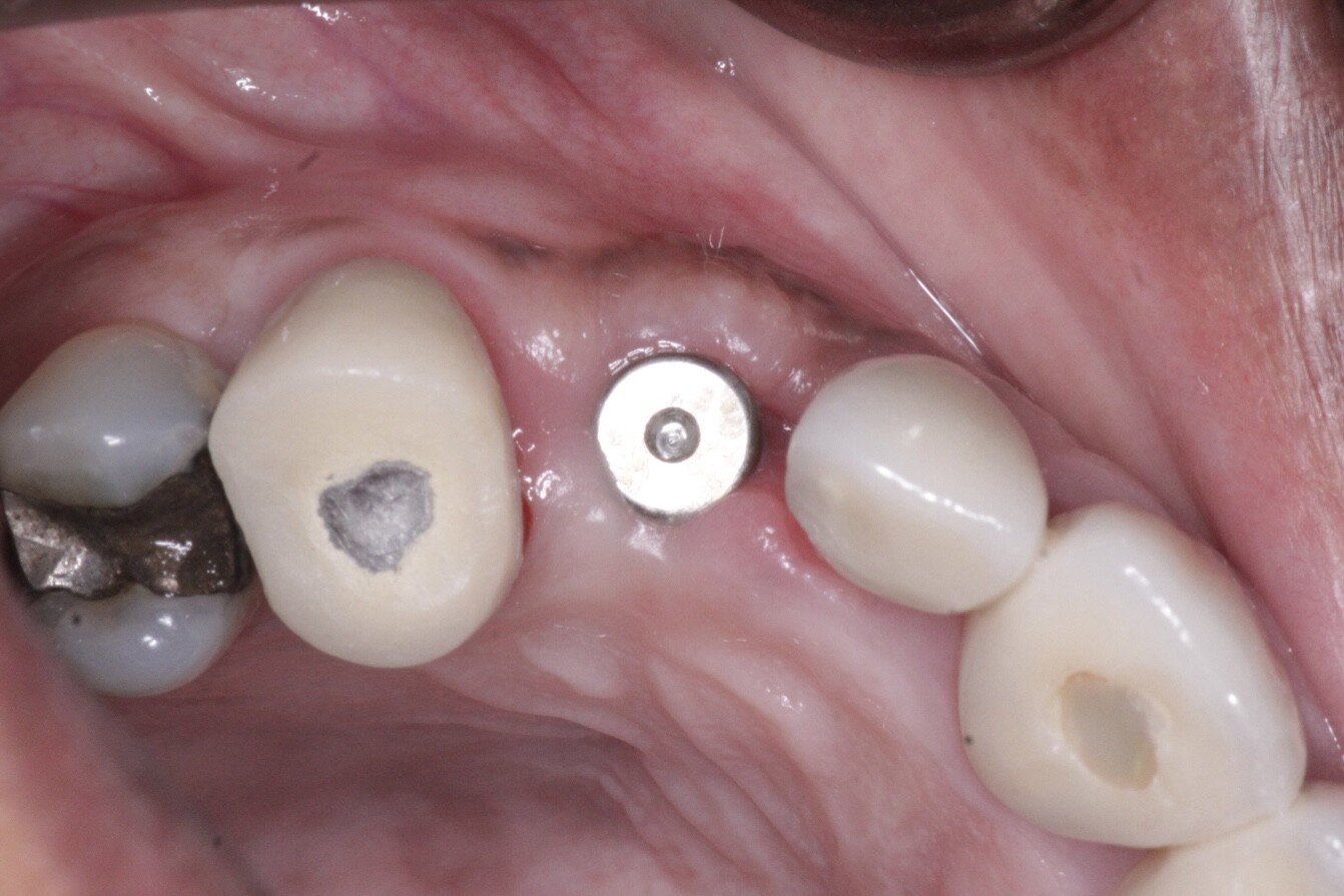
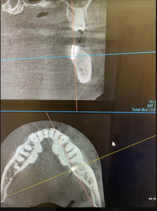
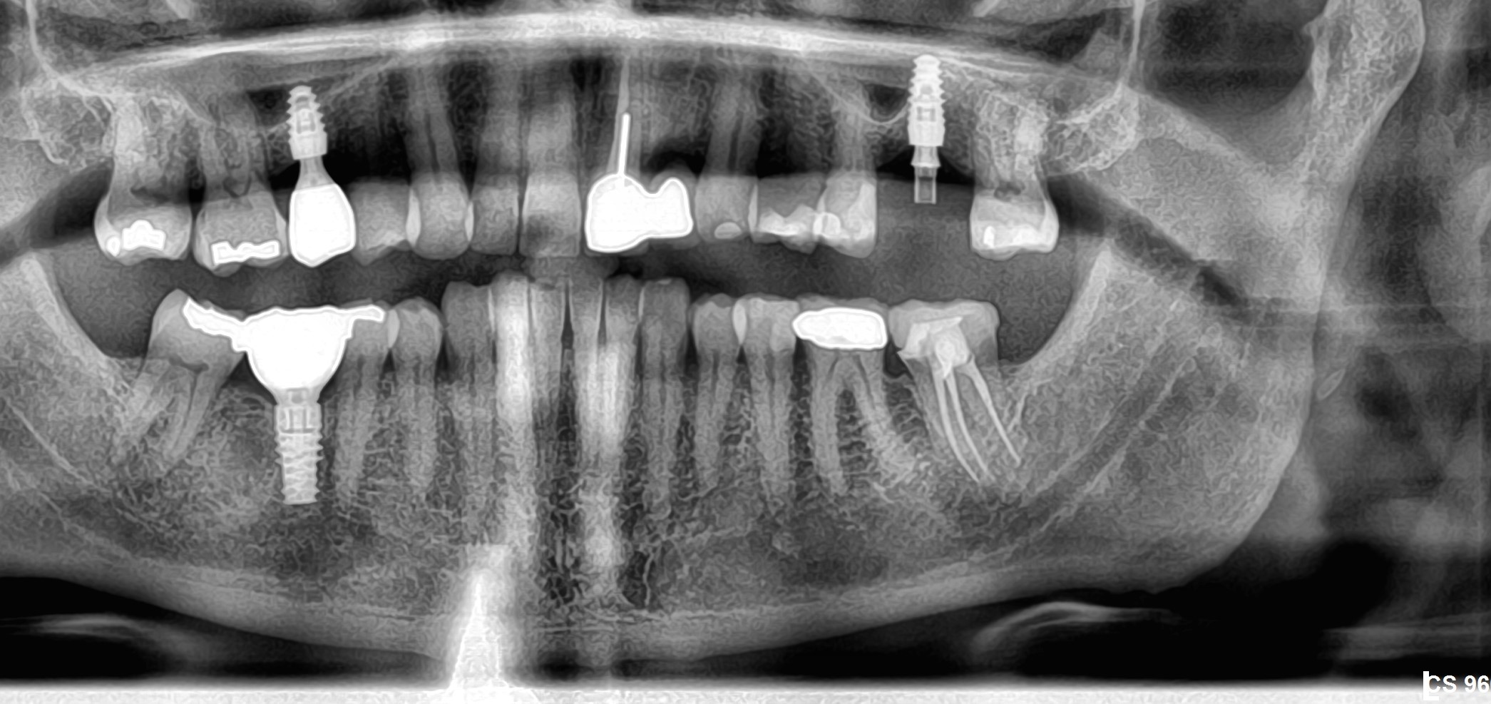
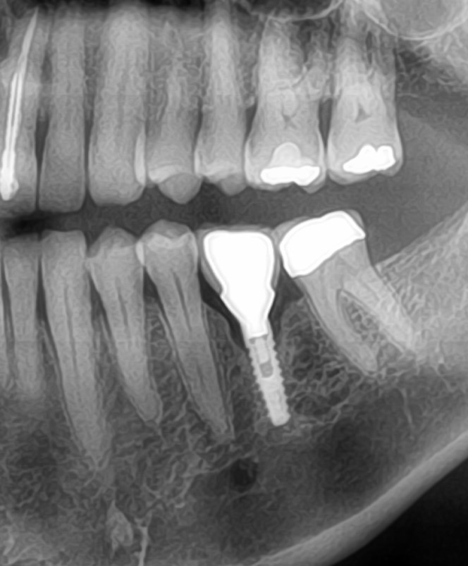
Please see photo below. I placed 2 implant fixtures in the maxillary anterior zone to support a 3 unit fixed partial denture. The implants were placed in #7 and #9 sites [maxillary right lateral incisor and maxillary left central incisor; 12, 21]. #8 will be the pontic [maxillary right central incisor; 11]. I am concerned that the implant fixture adjacent to #11 [maxillary left canine; 13] is too close and may have damaged the root. I feel there may be enough bone separating the implant from the root to prevent permanent damage. Do you think that I should advise the patient at this time that #11 may need root canal treatment? How long should I wait to monitor the signs and symptoms before coming to a final diagnosis and treatment plan?

24 Comments on Is the Implant Fixture Too Close to a Natural Tooth?

New comments are currently closed for this post.
Fahim Changizi
2/7/2011
please providing an other priapical Xray of the site with XCP(PARALLELING TECHNIC), this is for confirmation of your guess about collision of fixtures by next tooth. if you see collision , don't worry up to facing any problem of natural tooth. usually after two weeks if your patient has any questionable complain of next tooth you may be need to treat it . of course if you haven't any problem by the tooth after osseointegration putting crowns of fixtures.
Dr. Martin
2/7/2011
You should also have in mind that your x-ray is only 2D and usually the maxillary canines (rootstructure) are positioned very facially (anteriorly)whereby implantpositions in the maxillary incisorregions are much more palatal placed, due to following the bone angulation and avoid fenestration. So it is primarily the marginal area of the periimplant bone, which is of concern, and it looks from your xray (maybe at little overprojection) that it is OK, not ideal, but ok. Resorption of interapproximal bone with loss of papilla, can be a consequence, then hopefully the lipline/smileline is low positioned :-)
Dr .T
2/8/2011
Looks fine. Probably more distance there than you think. Still advise the patient of potential risk but don't lose any sleep over it.
Pankaj Narkhede, DDS; MDS
2/8/2011
It looks OK. If the canine is not rotated in the mouth notice the pulp chamber. it looks wide, therefore the angle is from the distal of the canine. Even after that you still see a space between the tooth & the implant. Follow the comment by Dr. T :-)
brian
2/8/2011
I think it looks OK, if you are concerned, get a CBCT and check the palatal position of the implant as mentioned previously. Vitality issues will appear after ~3 months or so, and bear in mind that the innervation and vascularity comes from distal to mesial (as I am sure you are aware).
cpgunner
2/8/2011
You will be fine. You can make any implant too close with the right angulation on the cone with plain film radiographs. These teeth should have no issues!
K s
2/8/2011
I think there will be no problem in future , just monitor the case dr Saini , good luck.
Gregori M. Kurtzman, DDS
2/8/2011
Dont jump the conclusion that Endo is needed. There appears to be space between the implant and natural tooth. Monitor the tooth and if it becomes symptomatic then do the endo. What may be more of a factor leading to endo is the crown prep. Also check the tooth to the left on the mesial root at the cervical may be something going on cant tell for sure in this rad
Willliam Ingram V DMD, MA
2/8/2011
Canine may need endo, but it is probably not the implant's "fault". Canine was way compromised prior to implant placement?? Very little coronal tooth structure remains but no apparent 2D lesion? As others have mentioned CBCT is a great asset here. If you don't have one do a search or ask your Kodak or other manufacturer's rep if any of your friends/collegues has a CBCT locally. I do CBCTs at no to low cost as a courtesy to local GPs and specialists as well as a learning experience for myself. IMHO Bilbo
Ali Hossein Mesgarzadeh
2/9/2011
Dear Dr.G You are rigth your implant is too close to the maxillary canin ,you may have some problem in position and fabricating of prosthetic crown and bridge.But don't worry it may happen for all clinician in all field of surgery.I think It will solve by using angulated abutment in this area, moreover a consultation with an honest prosthodontist will be useful.As some of other clinician recommended cone beam CT is useful.But if you don't want to force your patient for high price of cone beam CT ,it is a good idea to have a vitality test and if maxillary canin dosen't have any vitality response follow it 2 - 3 week later,with the same non-vitality results performing of a root canal therapy will be a good treatment of choise.Finaly A persian(iranian)proverb said" Unwriten composition don't have any mistakes". Good luck Dr.Ali Hossein Mesgarzadeh DDS - OMFS - MS Tabriz / Iran
MEU
2/9/2011
I think that tooth 13(maxillary right canine) should be fine. If anything happens to this tooth it may be related to previous dental work. You mention that you are planning a fixed implant supported bridge using the implants at the level of 12 and 21 as abutments and 11 being the pontic. Based on the provided radiograph, it appears to me that the implants were placed too close together and that perhaps the space could be tight for three teeth. I base my observation by looking at the bone distal of implant 21 where I am unable to see the mesial of tooth 22. A panoramic radiogrph may provide a better look at this. Hope it helps
Dr. Samir Nayyar
2/9/2011
Hello Load the implant & go 4 endo in future if & whenever required. Jus Chill.............
Faisal Moeen
2/9/2011
Seems like this peri apical x-ray got a little bent while the image was being captured assuming the cone was properly placed, hence the implants appear a bit stretched and that could essentially effect everything that we might be seeing here, including the inherent problem of this image being 2D. An OPG would have been comparatively more accurate. If not, I think that the implant being close to the canine is not your major concern. The end result needs to look good, which is why the patient walked into your clinic on the first placed. It seems like you might be ending up placing a teeth which might be smaller than the patients natural teeth and also shifting the midline a little bit as well. You might need to root treat the canine which is snug next to the implant or even both canines with the objective of providing your patient with a good looking, harmonious 6 unit bridge. Its all about the end result!
Natwar
2/9/2011
I believe in these cases I find 2 surgical stents method is really helpful. First one for location and second for angulation so that no worries later.
Dr Sanjay Jamdade
2/9/2011
Is that an Arrow implant from Alpha bio or a Bio Horizon implant? The reduced distance between the implant and the tooth may not be as bad as it seems. The canine is the tooth around which the arch turns backwards. Ideal film/sensor placement is difficult. As well to place Xray tube and shoot lateral incisor and first premolar zone without superimpositon of canine root is also difficult. This is a zone where you may never get a true picture no matter what you try. In all probability this is a parallex error. I agree with most of the above comments. Try to shoot at a better angle. Try an OPG, A CTscan. Do a vitality tests often. It's too early to panic. Ask me how I know! Been there done that!
Shirley
2/9/2011
Doctors, Please correct me if I'm wrong ... Looking at root morphologies, that "canine" is much too short compared to the adjacent tooth to be considered a canine. Besides, what comes after tooth #9 if not tooth #10? Furthermore, is that not the mid-palatine suture I see in-between the implants? Sirs, we are dealing with MEDICO-LEGAL documents, the need for CARE and ACCURACY in our entries is of utmost importance for the benefit of everyone concerned. I hope you catch my drift. Evidently your designated implant #7 is on #8 space, it explains why the space available is too narrow. At any rate, if one is proficient in directing the central beam, one can get a better angulation and idea about the space in question. What you're concerned about is --- will your bone support be able to tolerate the kinetic energy imposed by occlusal loading? It is a 50/50 configuration which is dependent on many factors... you may want to try a provisional restoration before making tha final decision. Given the arrangement of your implants, you may end-up cantelevering #7, which is okey. Warmest regards...
Ik
2/9/2011
Angulation of canines and laterals are different in Bucco-lingua aspect so U are most probably fine. Take PA after your pilot drill next time for orientation . Panoramic Xray is a good idea to see another Image. Good Luck
Guy Carnazza
2/9/2011
I would monitor the patient during the healing phase and test periodically. An easy way to avoid this problem is to wax up the case and then fabricate a surgical guide from there.
john townend
2/9/2011
So far so good. I agree with just about everybody else. The implant is too close to the adjacent tooth but is very unlikely to have devitalised it. You'll probably get away with it. Don't waste money on fancy xrays. They may (or indeed, may not) allow you to sleep better at night, but won't make a jot of difference to the point of emergence or angulation of the implant. Just pray you don't get problems with peri-implantitis (which, of course, can affect even the most perfectly positioned implant)in due course and find yourself having to trephine the implant out. This will certainly b++++r the adjacent tooth and send your patient on his way to a lawyer. I know - I've just been trying to defend a colleague who placed an implant in an almost identical position. Implantology is the surgery of millimetres I'm afraid.
Dr. R.
2/11/2011
Yes, too close but as said before. Stuff happens. Documentation and advising patient of situation is imperative. Ensure proper consents are used; as someone already said, 'Been there, done that!' Follow everyone's sage advice. You may want to get a tomo for the record. (at your cost)
Dr.ganesh shetty
3/13/2011
I think the implant will work fine.dont fret over it.
Dr. Dan
3/21/2011
Dear Dr. The xray is two dimensional and doesn't accurately portray its closeness. A 3D conebeam will tell you more accurately, but not completely as accurate as laying a flap and doing a physical measurement (which I do not recommend doing). Even these 2D xrays look ok to me. You are not hitting the roots and my only suggestion is to inform the patient and to keep on eye on the natural tooth during regular maintenance check ups that the patient should be having anyway after implant placement. Good luck
Carl
3/24/2011
Dr. Sanjay, these implants appear to be Legacy 3 from Implant Direct. Considering their location, they appear to be their small diamter tapered 3.2 implant.
Dr Sanjay Jamdade
5/6/2011
Thanks Carl! The radiographic difference between brands is getting lesser and lesser!

Featured Products

OsteoGen Bone Grafting Plug
Combines bone graft with a collagen plug to yield the easiest and most affordable way to clinically deliver bone graft for socket preservation.
CevOss Bovine Bone Graft
Make the switch to a better xenograft! High volume of interconnected pores promotes new bone. Substantially equivalent to BioOss and NuOss.