Patient with Hemangioma: Thoughts on implant treatment?
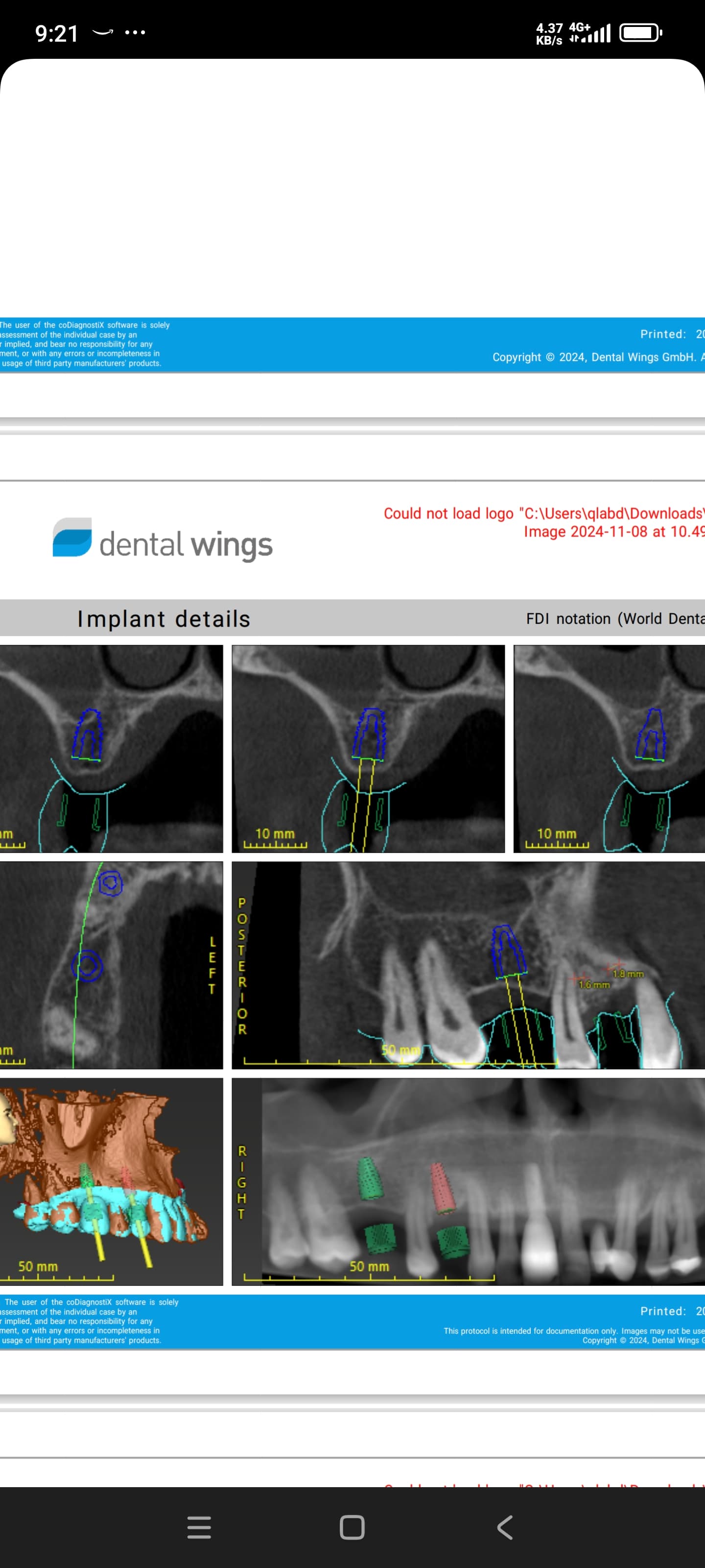
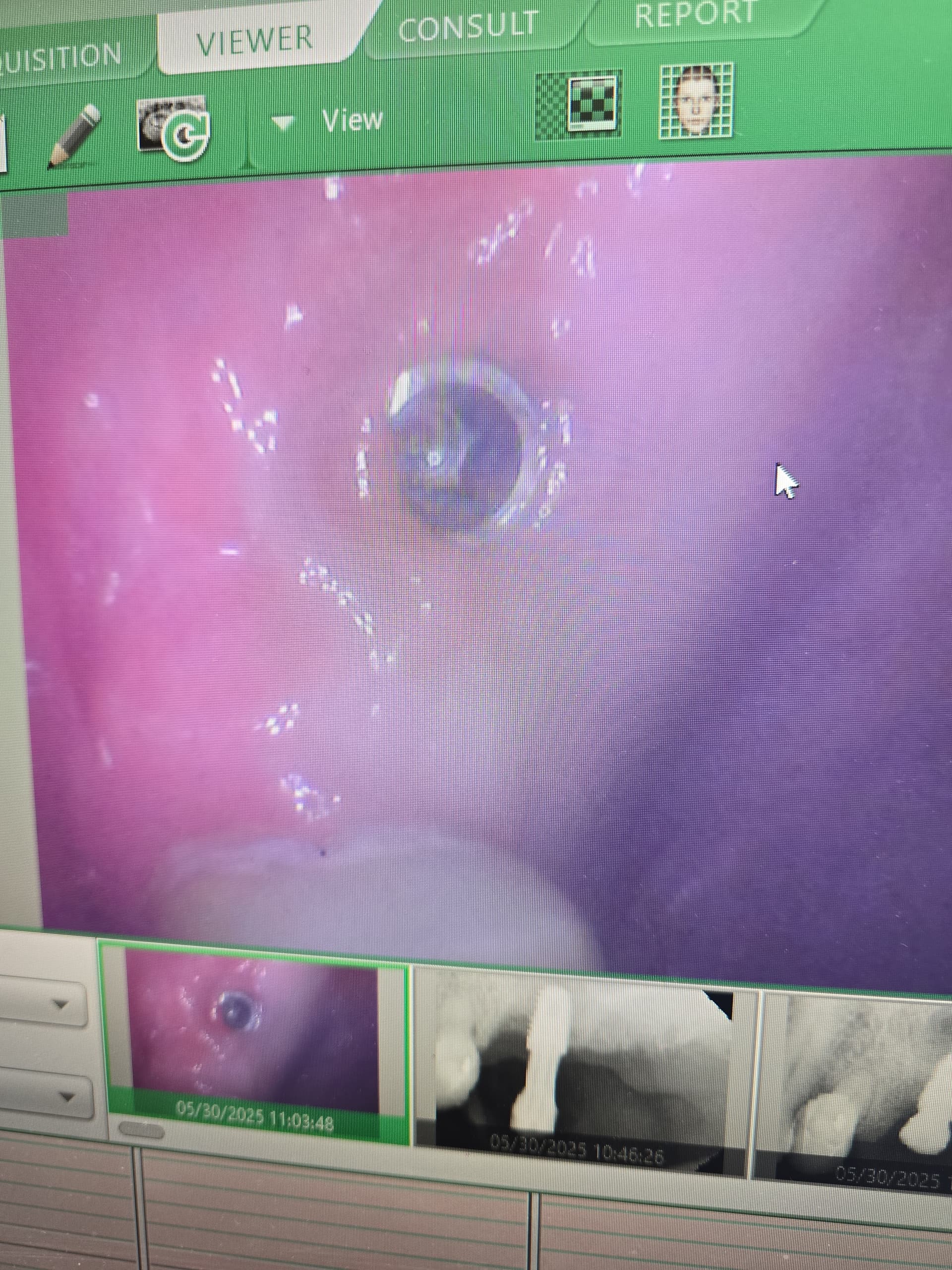
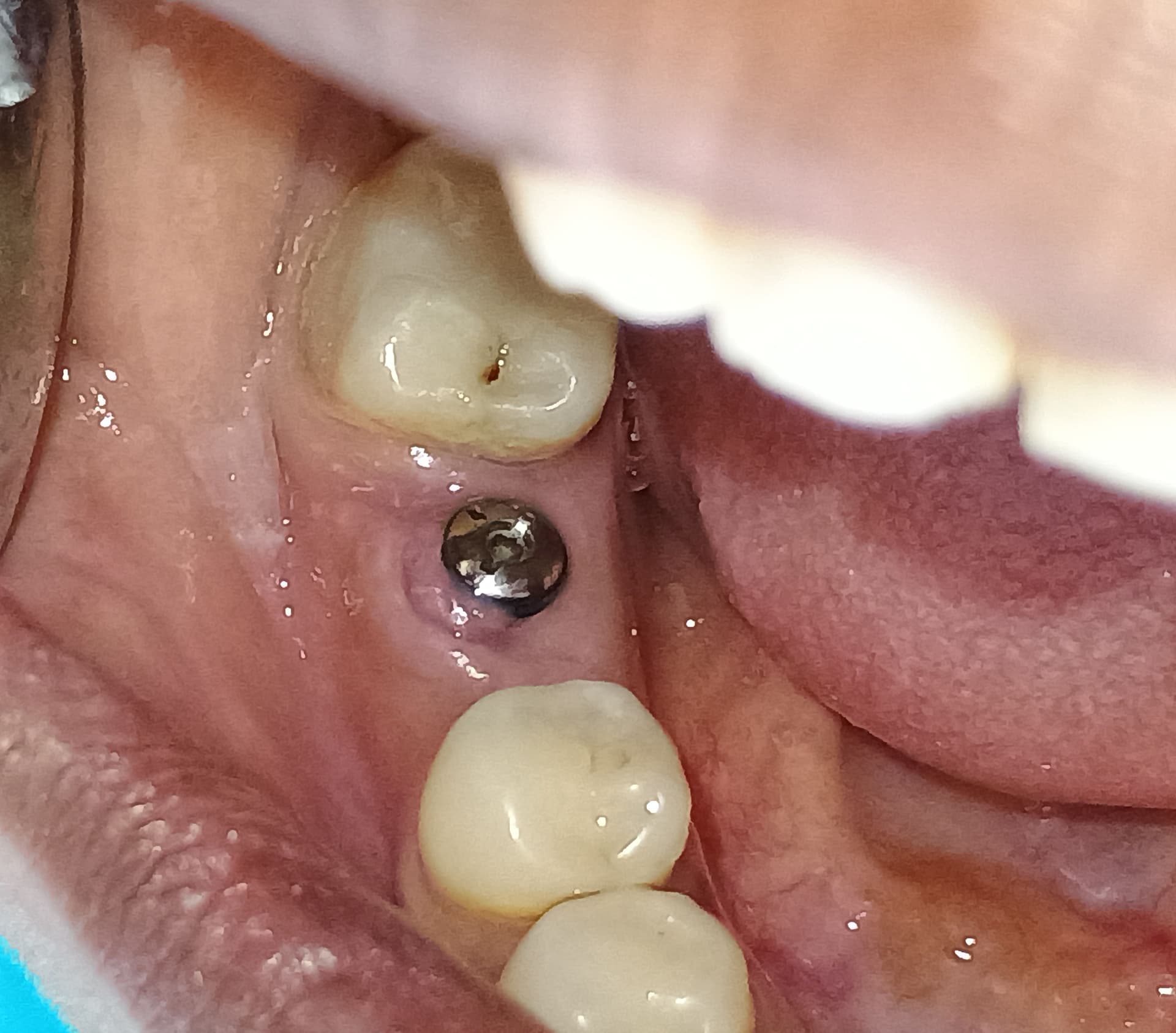
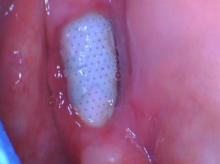
I have a patient who presents with a hemangioma of the facial region. This is the first such patient I have treated. I want to ask several questions for those who have experience treating patients with hemangiomas. I have treatment planned my patient for implants in the premolar and molar regions. Hemangioma is present on the right side of the patients face, continues intraorally at the gingiva and reaches the midline of the palate. The underlying bone is adequate on both sides. The patient’s present molars are going to be extracted. Has anyone had a similar case before? What is the possible reaction of the soft tissues? Is it dangerous to place implants in this patient?


21 Comments on Patient with Hemangioma: Thoughts on implant treatment?
New comments are currently closed for this post.
Mark Bornfeld DDS
4/24/2017
Disclaimer: I have never placed an implant in a hemangioma. Having said that, the chief concern is uncontrollable bleeding, depending on the volume of blood pumping through the lesion. Additionally, it it sometimes difficult to delineate the extent or the hemodynamics of a hemangioma without special imaging-- usually MR with IV contrast. This is a particular concern with large lesions, or those associated with angiomatous syndromes (e.g., Sturge-Weber). The lesion could extend to the bony areas under consideration for implant placement.
In short, you should consider sending your patient to a vascular surgeon for proper evaluation and mapping of the lesion, and place implants only in those areas of soft or hard tissue where the lesion has been demonstrated to be absent.
Mark Bornfeld DDS
4/24/2017
P.S. Naturally, it would be a good idea to do the necessary imaging prior to the planed extractions.
ap
4/24/2017
This is not a hemangioma. You are looking at a capillary malformation aka port wine stain.
Osbert Usher
4/24/2017
First thought that came to mind. I do believe the same as Ap.
Danev
4/25/2017
It is a Hemangioma. The pink color is because of the laser treatment in the past. Unfortunately the latter wasn't successful enough.
Thanks for the comments!
Andris Bigestans
4/25/2017
With a small flap only to open a space for placement there is no problem , and as someone mentioned this is a blood vessels malformation not a hemangioma.Local situation is good enough.
Mark Bornfeld DDS
4/25/2017
Hemangiomas may be capillary, cavernous, or compound; they may be peripheral, central, or both. Don't get too hung up on semantics; the nomenclature of vascular malformations hemangiomas, port wine stains, etc., has been a moving target. To suggest that surgery is safe simply by looking at a few photos is presumptuous at best, and catastrophic at worst. More formal evaluation of vascular lesions is always warranted when contemplating surgery anywhere near them.
Dr .Adian Nistor
4/25/2017
This is probably Sturge -Weber syndrome Type 2 . Facial angioma (port wine stain) with a possibility of glaucoma developing( not any evidence of brain involvement). Symptoms can show at any time beyond the initial diagnosis of the facial angioma.
I do not recommend to insert implants for replacing the premolars due to osseus possible involvement.I recommend facial angiography and the revue of diagnostic.
rsdds
4/25/2017
play it safe do a 3 unit bridge... that's my recommendation
rsdds
4/25/2017
thank you for posting this case
Steven A. Guttenberg
4/26/2017
1. Did the patient have a bleeding problem when the teeth were extracted?
2. Does the osseous architecture appear normal or abnormal?
dj
4/26/2017
Most important questions, Bravo. Also, does the patient have a history of bleeding problems in the area considering obvious past treatment? Consult patient's physician and go from there. Consider flapless options with minimal osteotomy.
Danev
4/26/2017
There no evidence of abnormal bleeding after extraction.
Mark Bornfeld
4/26/2017
As the broker said, "past performance is no guarantee of future results". Whether or not there is profuse bleeding following extraction may hinge on whether the roots of any particular tooth are within the lesion. You may have just been lucky with those previous extractions.
Alok Bisht
4/26/2017
Since there was no complaint after extraction I believe placing the implant would be non eventful.
Danev
4/26/2017
I want to insert an Ortopantomografy into the post,but I don't know how to edit it.
OsseoNews
4/26/2017
You can post another photo either by Post another Case (link at top of page) and use the same title and contact information of your last post and we will add photos to this case. Or more simply, log into your account with OsseoNews.com and come back to this page and you will a button to upload a photo for your case when adding a new comment. Contact us, if you have any problems. Thanks.
Steven A. Guttenberg
4/26/2017
If there was no unusual bleeding concurrent with the extractions, there will likely not be any with implant placement. Just take a close look at the bone morphology to see if it is similar or the same to other areas of the mouth that are not affected by the vascular anomaly.
Danev
4/28/2017
My opinion coincides with opinion of Dr. Mark Bornfeld .The question was whether you have experience with a similar case.The rest is conjecture.The risk is unjustified.
Thanks everyone for the opinions.
Dr. Shet
4/30/2017
Primary concern for this patient is to evaluate the extent of the lesion with CBCT or MRI. MRI is more applicable to check the extent. This cavernous type some times restrict in the soft and is not contraindicated for Implant surgery. According to your statement it seems involve the alveolar bone as well. If it is found with bony involvement than very difficult to control bleeding during the surgery. Moreover excessive bleeding can impair the healing process due to more iron deposition that leads to osseointegration failure.
RLFaler
5/3/2017
Dr Shet is correct. An MRI will show if the site is involved. Implant should be very possible.


