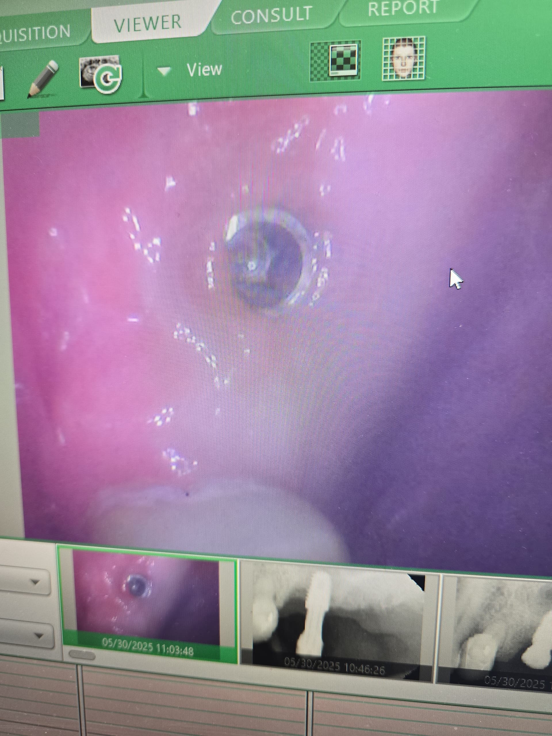
Patient with history of draining sinus tract: treatment plan?
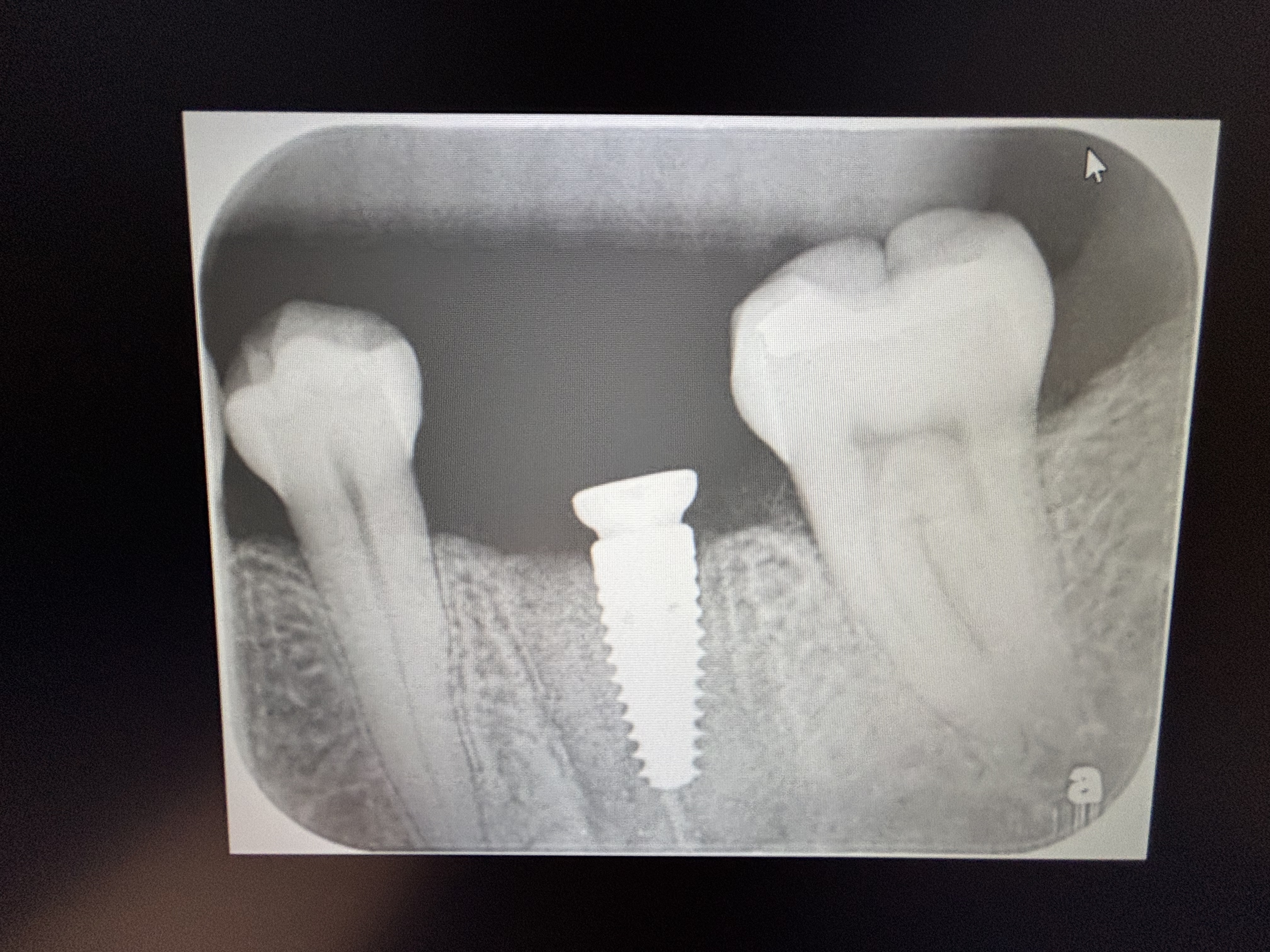
I have a 45 year old female patient with a history of a draining sinus tract for the last year from tooth #9 [maxillary left central incisor;21]. It was non-vital and discolored. The extraction was done 1 month prior. The patient is asymptomatic without any signs of soft tissue swelling or purulence. The periapical radiograph shows a periapical pathosis with bone loss. I plan to debride the extraction socket and install the implant fixture slightly to the palatal and to do a bone graft with PRF. Is this a treatment plan with a high expectation for success? Do you have any other recommendations on how to proceed?

16 Comments on Patient with history of draining sinus tract: treatment plan?
New comments are currently closed for this post.
CRS
2/23/2015
Honest answer, good possibility of failure in th esthetic zone. This is a challenge even for a specialist. There is a chronic localized OM present which may spead to your implant.
bijander jain
2/23/2015
I am looking for treatment planning in above case.
Steve
2/23/2015
I agree with CRS.
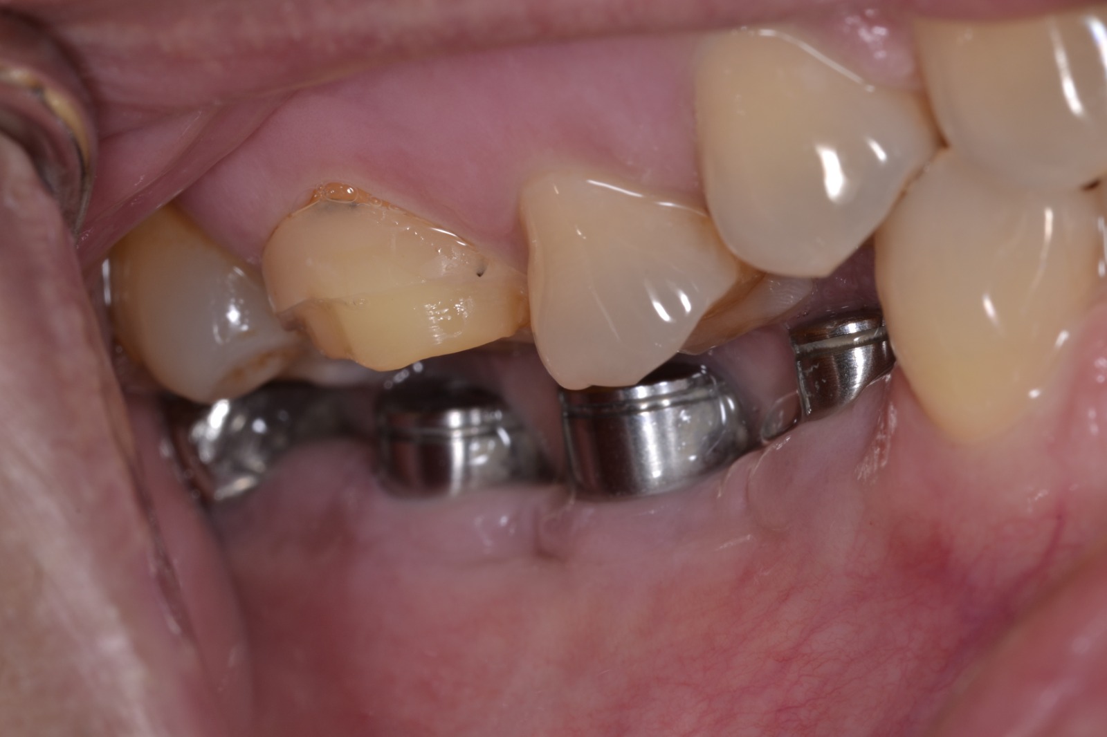
I wouldn't worry about the implant's osseointegration, but the aesthetic outcome is very challenging in these cases. Do you have any photos of the area to share with us?
CRS
2/24/2015
Just curious why you did not disenfect and graft at extraction to maintain the space and treat the chronic infection? What is key here is how the area is cleaned up, grafted and regeneration of the labial plate for esthetics and implant longevity. Good news is the adjacent teeth will help with the papilla.
Dr Daneshgar
2/24/2015
To be in safer side I think it is better to open it again debride the extraction socket, Graft and wait for at least 3 months then proceed with an Implant.
All the best.
CRS
2/24/2015
Disagree, lose space and create more scar tissue with additional surgery. Waiting does nothing to disenfect the site, the bacteria will hang around. Extract, disenfect, graft with sonic weld or Teflon. I see this often I have to go back in and fix it have less of a base to work with. I would use the time wisely by allowing the graft to heal then place the implant.
Leonard Smith DDS, DICOI,
2/24/2015
Please consult with an experienced surgical implant dentist. You are starting on the hardest of cases. Maxilla, esthetic zone. First: start with the final crown in mind. What does the patient expect? Is she high lip line, high profile, beauty oriented? Will you have inter-dental papillas? Will you have a zenith that matches #8; is it time to approach patient to do more teeth, look better, laser zentiths, that way you can move the outcome to your favor more easily. You need to plan for less, if she doesn't like possible outcomes, refer her off. The only way anyone can answer you is by viewing a CBCT scan, there is no way, bone sounding, pa xray, etc, to know the 3D anatomy of the now healed socket. Did you extract, that was the time to bone sound in 3D, how much facial plate was there, what is the labial bone width and where is the labial crest. Are you going to prepare bone for RAP? Autogolous fibrous membranes will not hold a labial advancement. Do you have bone tacks, membrane selection that will hold space? What implant selection: current thought is for smaller diameters in anterior maxilla. Counsel patient on need for possible follow up CT grafting or second stage bone grafting at time of implant placement.. Will you charge her for additional soft tissue and /or bone grafting? I am just giving you a few things that have to be known before you proceed. This is not a case to get experience. If the treatment has any shortfalls and the patient comes at you, you will be glad you heard all of this. The reality of implant dentistry is not all riches and success. PS: if this was an ideal socket for an implant, you would have either put in the implant at extraction, or done ridge preservation or GTR at that time. Of course this is only my opinion and only passing on another way to look at things. Good Luck
Dr Bob
2/25/2015
A fixed bridge with connective tissue graft, if needed, would be my first choice in this case. I see a very difficult time ahead to establish good contours of soft tissue as well as length and shape of the implant crown. If the patient insists on an implant and refuses a fixed bridge I would refer the case. I fear that after completing the implant placement and crown the resulting aesthetics would leave much to be desired and would likely then have no way to be improved upon. The remaining infection must be cleaned up in any case.
Asif
2/25/2015
Hi
I am not sure if what is happening with the lateral incisor apical aspect Perhaps a cbct may help in excluding an underlying larger pathology. We have all come across those. That could explain the draining sinus.
Regards
A
Dr KKS
2/25/2015
I do not understand to why sinus drainage related to this tooth as anatomically this tooth is away from the sinus.
3D scan would be very useful.
I would consider treatment and exclusion of any OroMaxilloFacial pathological condition before placing implant/s and other dentistry work.
CRS
2/25/2015
I believe it is a sinus or fistulous tract from which the infection drains to outside the bone, not the maxillary sinus or floor of the nose.
Sam Jain
2/26/2015
No meaningful diagnosis w/o CT
No meaningful advice w/o CT
CRS
2/26/2015
Okay, one month is not enough time for a socket to heal. On a X-ray there is not enough calcification to show up until four months. The bone is still remodeling and probably bring lost since it was not grafted. You don't need a cone beam to tell that. There is a fistulous tract from the long standing infection which needs to be cleaned up. This is a straight forward case with decent interdental bone so you will be able to keep pappilas if the site is adequately disinfected and grafted hard and soft tissue. If you are not comfortable doing thus then refer it otherwise you may be chasing a failing implant in th esthetic zone. It Is the biology of the area. You have already burned a few bridges in just extracting and delaying treatment. There are some good comments here heed them.
Docoms
3/3/2015
A "history "of sinus tract sounds like it was in the past and resolved with the extraction ? In the same way that history of MI doesn't mean patient is having an MI at the moment. Kinda hard to tell with the way the written presentation given.
I'm gonna assume there is no draining fistula currently.
Why do you think that the radiograph demonstrates "pathosis"? Given the description of events, it would seem more consistent with healing osteoid bone that has not fully mineralized as suggested by CRS.
At this point, its probably best to allow 4-6 months for healing then see what you have to work with in terms of defect. If you have the experience you can graft it now to minimize further bone loss but it looks like there was a fairly good size abscess that is filling in nicely and disrupting it now will just re-start the clock on healing.
Dr J
3/10/2015
Consider pulp testing the left lateral incisor for vitality. Treat this PRN. If there is a persistant sinus tract then consider culturing the pus, or empiracally treating with clindamycin 150 mg qid if suspected OM. Dry up the infection and then see what you have left there and plan accordingly. This could range from site reconstruction with block grafting, or possibly a barrier/ PRP and pariiculate, or a conventional 3 unit bridge.
Thanks for posting the case.
linda Rebstock
3/13/2018
Tooth#14... Broke the cap on the first implant. Had to have it pulled. Long story short, he placed a second implant. When he pushed the cap on it broke through the membrane. I then had an ENT doctor place bone graft material in the area. That has mixed with my sinus and gone down my throat and in my stomach and GI tract. It says it can't be dissolved and is causing problems. Have you ever heard of this and can you recommend anything?


