Possible palatal perforation with bicon implant: recommendations?

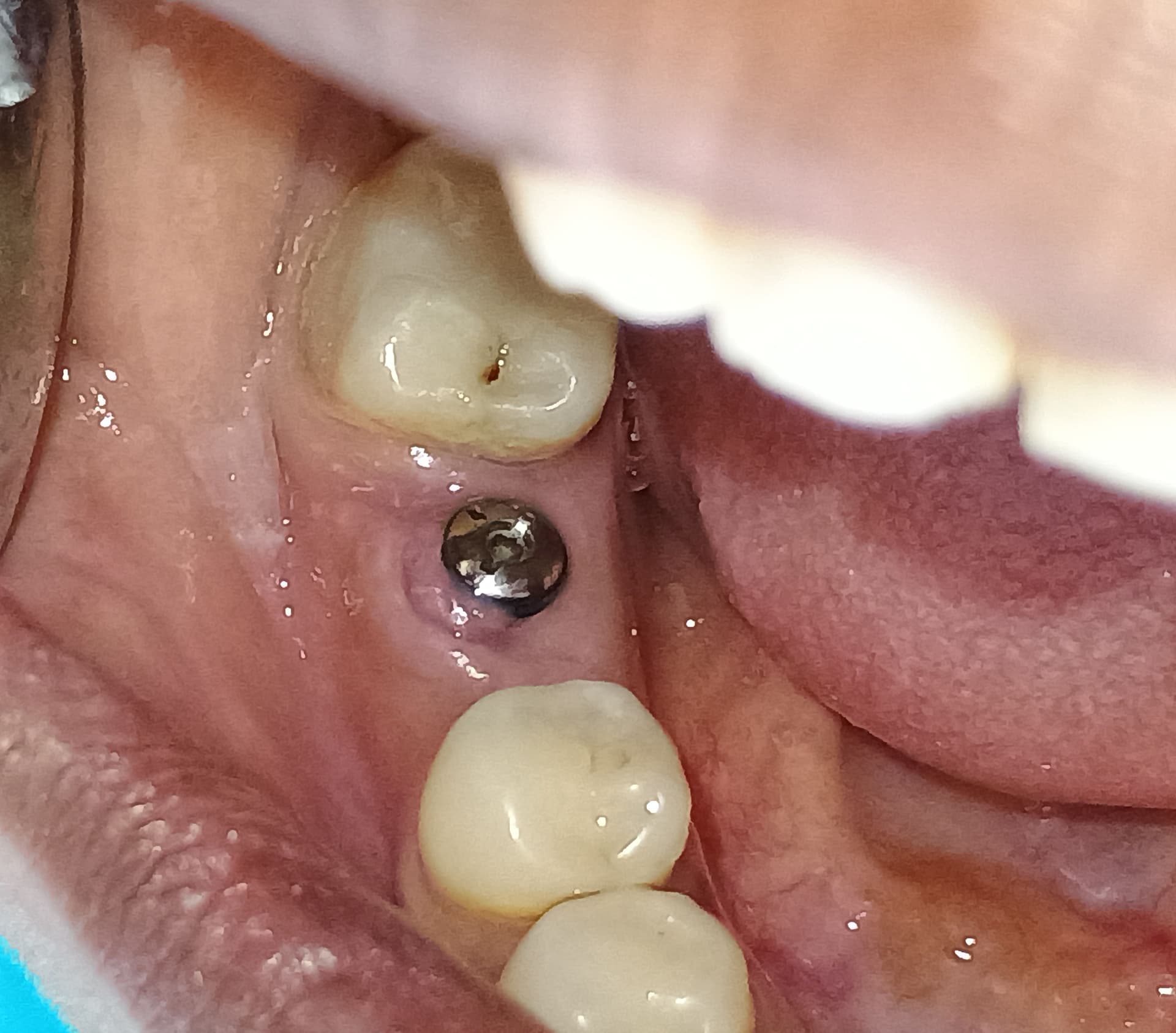
I had a seemingly straightforward case of missing maxillary left premolars, for which I placed two Bicon implants just over four months ago.

As can be seen on the x-rays, there was a weird “dip” in the bone in the area of 25. This did not bother me at the time, as this part of the ridge was very narrow to begin with, but gets wider more apically, so I simply used the base of the dip as my depth gauge. This measured 15mm from the base of the dip to the sinus floor, plenty of space.
I tried to line the 24 up with the 23 and the 25 with the 26.

Directional pins were placed (one is angled, as I only had one 180 degree one at hand) depth and position looked right.

I trephined up to a 4.5mm final drill. I harvested less bone from site 25 than site 24. At the time I assumed the difference was due to a difference in bone density.

I placed the implants (both 8mm in length) with the Bicon mallet. Before doing so I scraped the osteotomy site and didn’t feel any perforations. Sutured and patient returned for follow up 2 days later. Patient wears a denture over the submerged implants and hasn’t experienced any undue discomfort.

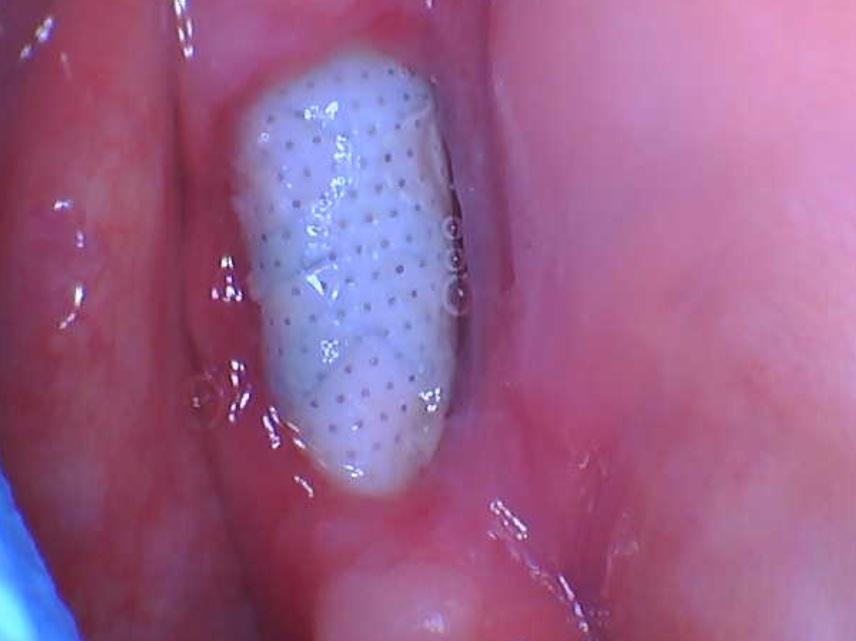
Four months later, the patient is happy, but there is plenty bone loss visible on x-ray around 24. Upon palpitation of the palate, the patient felt “something” when I pushed my finger in the area of the 25 apex.

My concern: I think I may have misdirected the drill, aiming too much towards the palatal, and with placement tapped the tip through the cortex. This could easily be verified via conebeam, but I would have to refer the patient for one and even if the outcome is as I fear, I have no clue as of how to proceed from there.

My questions:
– If the tip of the implant is indeed exposed through the palatal cortex, is it a significant problem?
– If it is not 360 degrees integrated, will loading dislodge it and push it into the sinus?
-Will the possible failure of the 25 influence the 24 which also experienced bone loss?
-Will loading the 25, even if it is partially integrated lead to ulceration and eventual exposure through the palatal mucosa?
-If the cone beam shows that my fears are realized, but in the absence of symptoms and with a seemingly stable implant, can I proceed with restoration and hope for the best? And restart when/if it fails in time?
-And if the implant fails, or the overall recommendation is to remove and restart, is there a way to remove a Bicon (fins, not screws) without a trephine?


8 Comments on Possible palatal perforation with bicon implant: recommendations?

New comments are currently closed for this post.
David
9/10/2018
Do you know how to perform a lateral windo sinus lift?
Don Callan
9/10/2018
If bone has been perforated, it would be best to remove the implant., most if not all will fail. You may graft the site or, let it heal and reinsert the implant after complete healing.
Mahmoud
9/10/2018
If your implant is integrated, you can do a flap in palatal and just cut the apex of implant that perforate the palatal bone.
Dr Dale Gerke, BDS, BScDe
9/10/2018
I would suggest that you are trying to find a solution before you have identified the problem. As the saying goes, "don't put the cart before the horse". I would recommend you get a cone beam done, identify if something is wrong and then design a way to fix any issue. Speculation is fine, but really you need to narrow down what is going on (if anything) and move on from there. I would point out that if you had done a CAD/CAM work up and used a resultant surgical guide, this discussion would probably not be happening. Implantology is much easier if your treatment plan proceeds that way.
bigjulie
9/10/2018
"a seemingly straightforward case" can be a trap in your thinking and readiness because those simple ones can suddenly become complex. Using a mallet is dangerous so close to the sinus: implants can be like bullets or ICBM's ... the old Branemarks had a mounting shaft so if the implant lost thread holding in soft bone, one could grab the large mount before the implant shot into the sinus or elsewhere. An implant system should be chosen on the basis of "if all goes wrong, how can I have options?" A threaded implant has a reversal option albeit a friction mount is difficult to re-engage.
David Levitt
9/11/2018
You stated the implant is integrated. There is more than adequate research showing an implant penetrating a millimeter or two into the sinus will not cause a problem. The problem then becomes possible perforation of the palatal cortical plate. Once again not an issue if the patient is asymptomatic on the palate. If the patient has symptoms (pain, fistula) flap the palate, remove the protruding portion of the implant, place some graft material, and close it up. Even without the graft it should be fine as the source of the symptoms would be inflammation of the soft tissue from the rough surface of the implant. See "Apicoecotmy of an Implant" Journal of Oral Implantology 1998 D. Levitt
Z
9/11/2018
I think you're imagining a problem. Likely the transitional denture caused the bone loss. It can happen even though the implant is submerged. I would guess that if you put pressure on the palatal of 25 again you wouldn't get a response. Cbct best idea to confirm though.
Dr. Mahmud
9/15/2018
I would like to inquire why a short 8mm Bicon implant was used when 15 mm of available space was available from the " base of the dip to the sinus floor" as you mentioned. My understanding is that Bicon implants should be used as an alternative to situations where bone quantity is limited. Do you routinely use Bicon implants for all your cases? Also the comments made about the use of a custom surgical guide are appropriate. The main disadvantage is the added cost of utilizing a surgical guide which makes the entire implant fee significantly more for the patient.

Featured Products

OsteoGen Bone Grafting Plug
Combines bone graft with a collagen plug to yield the easiest and most affordable way to clinically deliver bone graft for socket preservation.
CevOss Bovine Bone Graft
Make the switch to a better xenograft! High volume of interconnected pores promotes new bone. Substantially equivalent to BioOss and NuOss.