Radiolucency or just Over Drilling?
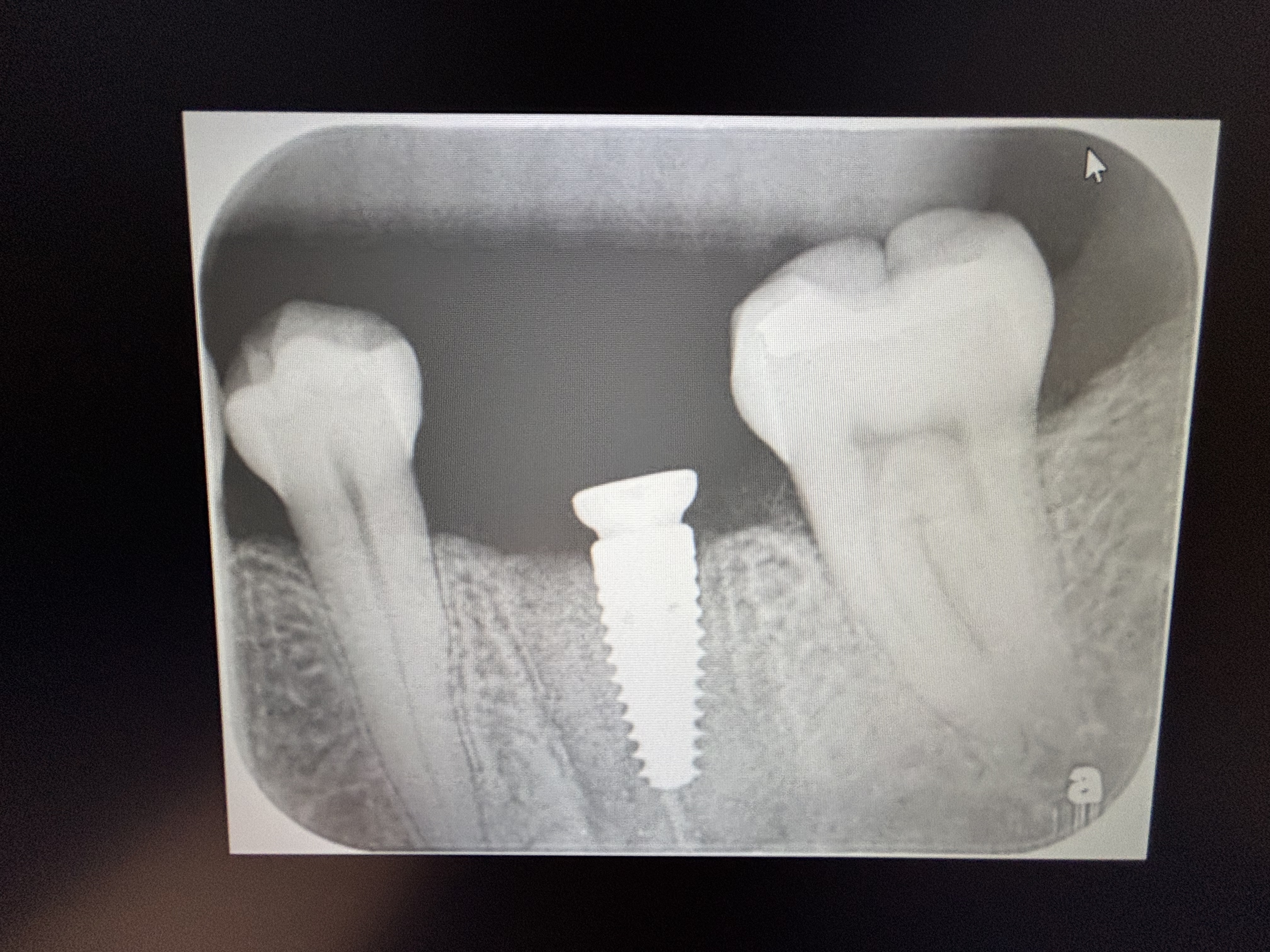
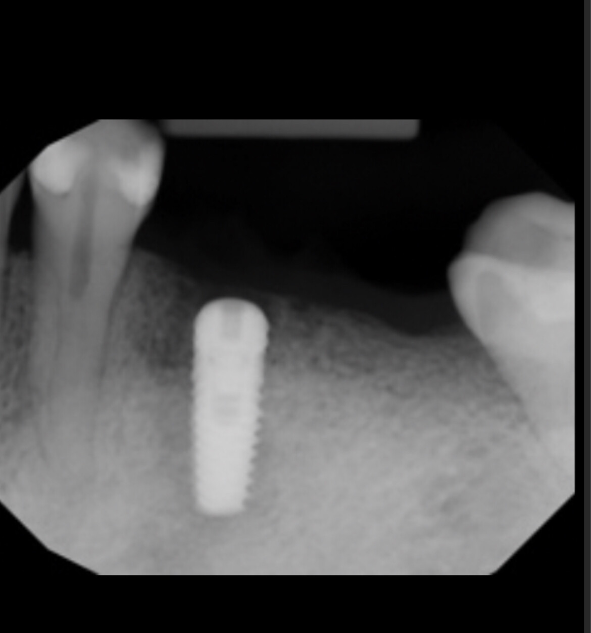
This implant was installed in extraction site of #19 [mandibular left first molar; 36] 2 weeks ago. Patient is completely asymptomatic and palpation is normal. I believe I did some over-drilling and may have perforated the lingual cortical plate. However I can see an intact cortical plate below the implant. I am concerned with what seems to be a radiolucency below the implant or is it just due to over drilling? what to do?
![]20140305_145938-1](https://osseonews.nyc3.cdn.digitaloceanspaces.com/wp-content/uploads/2014/03/20140305_145938-1-e1394489961699.jpg)
27 Comments on Radiolucency or just Over Drilling?
New comments are currently closed for this post.
marc
3/11/2014
You sleep well and let it heal.
Do you always take post-op CBCT ?
Does it follow ALARA principle ?
Do you use surgical guides ?
CRS
3/11/2014
Over drilling you were very lucky. I would have extracted grafted and placed the implant in the middle of the alveolus with good crestal bone. In my opinion I feel the alignment is too far lingual, I don't like the extraction site to dictate where I place the implant. Why was the tooth lost may I ask? Agree with Marc's comments above with the high tech gadgets, a stent need to be used and the placement should be better.
gumdoc7
3/11/2014
tough crowd
you did perforate
it will probably be more than ok
if it is not too mesial or distal, very restorable
pre-op scan and guide where necessary can never hurt
Gregori Kurtzman, DDS, MA
3/11/2014
Appears some over drilling may have happened and standard radiographs give false sense of bone so taking a rad with instrument in place part way thru wont help much I would suggest keeping a finger on that area when drilling so you have some tactile feel if the drill is getting close
in this case is will heal keep an eye on pt if somethings going to flare up it will happen within a few days of surgery
Birgitta W.
3/11/2014
As an oral and maxillofacial radiologist, I would need to see the scan to see if you over-drilled. One view is not enough to make a correct assessment. Did you take a pre-op scan? Was there pathology in the pre-op scan in this region? Did you clinically feel that you over drilled? Bruising lingually? Clinical signs when placing the implant to indicate over-drilling? It looks like artifact to me as there is still cortical bone directly apical to the inferior aspect of the implant on the buccal side. In your pre-op scan was your lingual cortex thicker in this region? If you are interested an official consult with report please visit my practice website at beamreaders.com
Interesting case thanks for sharing,
Birgitta W.
DrG
3/11/2014
Darn right we are a tough crowd. That's what separates the amateurs from the professionals. With that said, let's be honest, your drill went from dense cortical bone to "whoops" I'm through. That prompted the post op cbct. Reminds me of the most terrifying thing I've ever seen in implant surgery. 22 years ago I watched an OS resident perf the Lingual plate in the same spot . It was a 1 in a million shot, he severed the lingual artery, it retracted right back into the patients neck/carotid. The patient was whisked off to the OR and a vascular surgeon saved the patients life. Thank god it was in a major teaching hospital otherwise that person would have died.
Short answer to your question, the implant will integrate.
The correct question is do you understand exactally what you almost did?
CRS
3/11/2014
I sure hope so, the implant is still poorly placed, will integrate that way and be a long term issue since the forces will not be down the long axis and parallel with the cortical plates. Remember a natural tooth has two roots an implant is a single cylinder. The anatomical structure you entered is the lingual shelf, had you been in the middle of marrow space the cortical plate would not had been a problem and the drilling would have been in homogeneous bone and you would not have had to push so hard perforating the plate. Seeing more of these types of cases suddenly in the literature, wonder why?
Birgitta W.
3/11/2014
What an interesting case thank you for being brave enough to share online.
If it is two weeks and your patient is asymptomatic, I am sure he/she is fine and will heal. Did you see clinical signs of perforation? Bruising, ecchymosis etc?
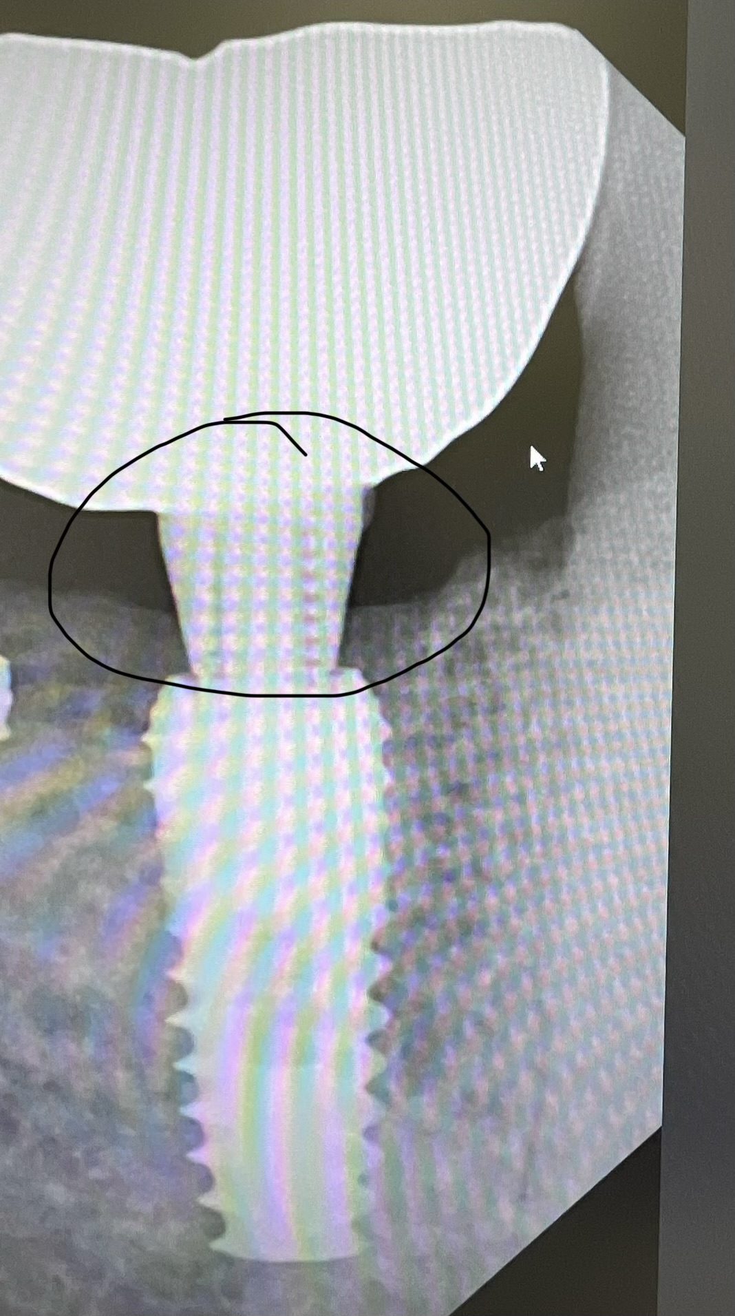
IT IS CRITICAL THAT ONE NOT MAKE AN OPINION OR A DIAGNOSIS FROM ONE ANGLE/VIEW ONE SLICE.
The AXIAL VIEW would be more accurate to observe a perforation (interruption in the lingual cortical plate) not the coronal view you have or cross-sectional slice you have provided. As well as viewing the pre-op scan if possible.
I THINK the people who have stated you have perforated are very brave as I would not feel comfortable making that statement without actually viewing the entire scan in all three planes viewing as well as knowing the resolution of the scan and what slice thickness your picture/image is above. The cortical plate is very thin but there is no perforation observed. There is still cortical bone directly apical on the buccal side directly inferior to the implant; if you did perforate it would have had to be with one of the initial drills smaller drills. Also your contrast is too high for viewing in this image. You can tell as your implant looks very white.
As for ALARA a CBCT scan is equivalent to being outside for about 3 days in the sun. The radiation amount is piddle compared to a Medical Head CT scan about 180 days. The new machines scans are less than a some Pano machines. If you felt that it was clinically warranted it will not harm an adult patient whose cells are fully differentiated. I feel that you would have not taken this scan without a good reason. Good advice about always checking you osteotomy sites with a probe after each drill. There is very critical anatomy which could cause your patient to bleed out but it would have occurred on the same day. Remember the lingual artery comes directly of off the external carotid so hitting this would be a very bad day in surgery. (second branch of external carotid)
If I was you and you had further concern I would send it to an Oral Maxillofacial Radiologist who can evaluate your before and after scan and tell you for sure if you perforated.
There are services online were you can upload your scan.
Some people actually lingualize there implants to engage the cortical plate.
Another thing the term radioluceny is not for CBCT only 2D images this would be described as a low density area with or without lingual cortical plate thinning and/or perforation. Here from one slice appears thin. It is important that you understand this as this helps to better understand what you are observing. Low density could mean marrow space, so the shade of gray you are observing is critical.
All the best, thanks for sharing.
CRS
3/12/2014
Looks like a perf to me. Sometimes it is best to be honest. But the person who placed the implant should be able to feel the "give" in the bone when perforating although a consultant can be helpful, it is the operating surgeon in the moment, who makes the call. If they can't tell then something is amiss.
ryoung
3/12/2014
Had similar incident, was placing immediate implant several years ago into the #26 spot. Had sublingual hematoma, sent to ED asap and ended up with an emergency tracheotomy. This was early days of the CBCT. I sent for CBCT to check after the fact and the implant had perforated the lingual cortex by about 1.5 mm. Definitely to need to be careful when placing these implants, and observe close for any sign of problems before discharging from the office.
CRS
3/21/2014
The best CBCT still needs to be clinically correlated there are many variables and artifacts in the technique and a radiographic interpretation is only as good as the clinical application. I would be careful giving this kind of advice since it is speculative.
Richard Hughes, DDS, FAAI
3/11/2014
Yes, you did perforate the lingual plate! It will heal uneventfully and the implant will integrate. Dr G. brings up a good point about the artery. This could happen to any of us but always be mindful of where you are in the mouth. Know and respect the anatomy. Also get into the habit of probing your osteotomy site after each bur.
dr nehal
3/12/2014
Marc-what is ALARA principle?.
i thinnk it will heal. so but in case of undercuts i prefer one size shorter implant so i remain safe. also tapered implant will sometimes save you from complications.
peter Fairbairn
3/12/2014
When drilling in the posterior mandible as anywhere when resistance to the drill is increased STOP and reassess your direction and angle .... drilling through a cortical plate feels significantly different to cancelous bone.
Peter
Tuss
3/12/2014
I think there was a case back in the day when someone perforated in this location and there was a life threatening bleed. Crestal bone profile on the implant does not look to good and angulation can be corrected if you are going cemented, screw retained might be complicated. I think you are lucky you did not hit an artery
Edoardo Calvi
3/12/2014
I think it is impossible to determine if the implant is too tilted without a pin inside the osteotomy in relation to the opposing tooth. since the you did not perforate the artery the implant will be fine. The finger tip is a good one.
Dr. Bill Woods
3/14/2014
Great comments. Yes being brave and honest are good things, and sometimes education hits with both barrels. I'm just fearful of the lingual anatomy of that area altogether. It's A healthy fear, whether it's with a scalpel or a drill. I almost never cut over there on wisdom teeth, either. Period. Preop measurements with a calibrated radiograph(5mm ball- I know "old school") or a CBCT would have sized the safe length of the implant osteotomy on the front end. The CBCT being the best. About ALARA. A perf throws all that out the window. All this discussion makes for a better clinician. We have all run into situations that makes the practice of dentistry the practice of dentistry. Me personally, I would have removed the implant, grafted and waited for another day. Sx guide the next time around. Two comments from someone WAY more experienced than me and past president of the AAID. One, " I'm always looking for my blood supply for this implant right after my initial incision." He didn't mean the lingual artery. Secondly, "Surgeons whistle by the graveyard way too soon"! Think about it. What is happening now may not involve any symptoms on the front end. Could this end up being a late surgical failure with a nightmare attached? I don't want one like that. I'll just try and stay away from all that anatomy back there. Some implants can be placed safely but it does take some thought process to determine how to go forward and when to punt and call another play on the next set of downs. Thanks for all the comments. They make me a better clinician. Dentistry is not a perfect science and patients teach us that every day. Bill
DrG
3/14/2014
Bill,
Great comments, almost poetic!
So many lessons to learn, so little time.
ben
3/14/2014
I always like bold comments CRS make. Here u went over the top. How on earth u manage to imagine prosthetic envelope . There is no information abt adjacent or opoosing teeth 9r occlusion. U can even imagination can even see axial and no axial forces. If we all do a ct of our placements will certainly find placement like that in our collection. Apart from lingual plate perforation rest is not a disaster. Over critisim doesnt help . We trying yo have a constructive forum.
CRS
3/15/2014
Dear Ben thanks for the compliment, and I agree that there is not much information regarding occlusion etc. My point is that using an extraction site usually a mesial or distal root site is not necessarily the optimal placement. You are placing an anklylosed titanium cylinder without a PDL and setting up a cantilever in the crown. This is just the shape of the implant we have to work with. Placing an implant in this case thru the lingual plate using a tooth extraction site as a guide is not something I would do but would prefer to line it up ideally in the center of the alveolus and in occlusion with the opposing dentition that's all I'm saying. I find it interesting how when I speak plainly with good intent and honesty it is considered bold but so be it. It is always gratifying to see my original comment repeated in the post thread so I guess the message is getting thru. Thank you again sorry for the bruised egos and I'm not perfect just stating an opinion. I'm actually a very nice person who cares passionately about the restoring doc and the patient.
Marc
3/16/2014
My comment was, if you are to take a CBCT, take it before. And if you took a CBCT , make a surgical guide in relation to the prosthetic plan. Simple,Predictable, Safe and as a bonus you save on the sleeping pills.The worst, I think ,about this case is that some doctors just dont have a CBCT machine and do the best they can, but in this case as demonstrated by the post-op CBCT image, clearly,he had a CBCT machine at hand, so then the question becomes :but why did you not used it before the surgery and if you did use it why did you not make a guide ? If I was a judge that would be my reasonning. If I was a patient that would be my reasonning.Because I am a dentist, that is my reasonning.
Tuss
3/16/2014
if we look back at the posting, the Dr's asking "what to do?" Hitting him with the first posting as a series of questions doesn't really help. The lingial plate is 90% more likely to be intact as his slice looks to be thru the long axis of his implant so more likely he has not perforated considering his final implant drill may have been a +4mm one. Talking "judge" again doesn't really help the guy. Yes it might be a restorative issue but he can angle correct up to 30 degrees with a cast-to or an angled multi unit type abutment to put the occlusal surface in the right place. The best advice is to wait then see if the "space" fills in
r
3/16/2014
Thank you all for your valuable comments.
I have a question; how did some of you jump to conclude that the it is malposed, from a single cbct slice???? Or is there some wizards here......with all my respect, a cbct was done pre op and that's why I placed the implant here, in fact it is in almost a perfect position for the future prosthesis. My question is.....Does this radiolucency represent an evidence of infection?
Thank u all again
Cliff Leachman
3/19/2014
You did that on purpose?
CRS
3/20/2014
That's a perf, periapical radiolucencies don't look like that and typically spread laterally thru the marrow not thru dense cortical plate. Bicortical engagement is not necessary with an osteointrgrated implant it is not a Steinmann pin treating a fracture. Based on your comments there is trouble in the future if this path is followed.
DrT
3/18/2014
If you took the cbct pre operatively then why didn't you angulate the implant
appropriately to avoid the submandibular concavity? The B-Li positioning is
definitely good.
DR C
5/31/2016
If any of us say "its never happened to me" we are either lying or haven't done enough to be commenting. The older I get and the more implants I do I now always get a CBCT in ALL cases and a guide in the VAST MAJORITY of cases. This way if something does go wrong at least we did everything possible to preempt such an outcome. It does not appear that you have a through and through perforation into the soft tissue but does appear awfully close! I would observe it at this point in time and watch for filling in of the radiolucent area.


