Radiolucent Lesions: How Should I Proceed with Implant Treatment?
Dr. I. asks:
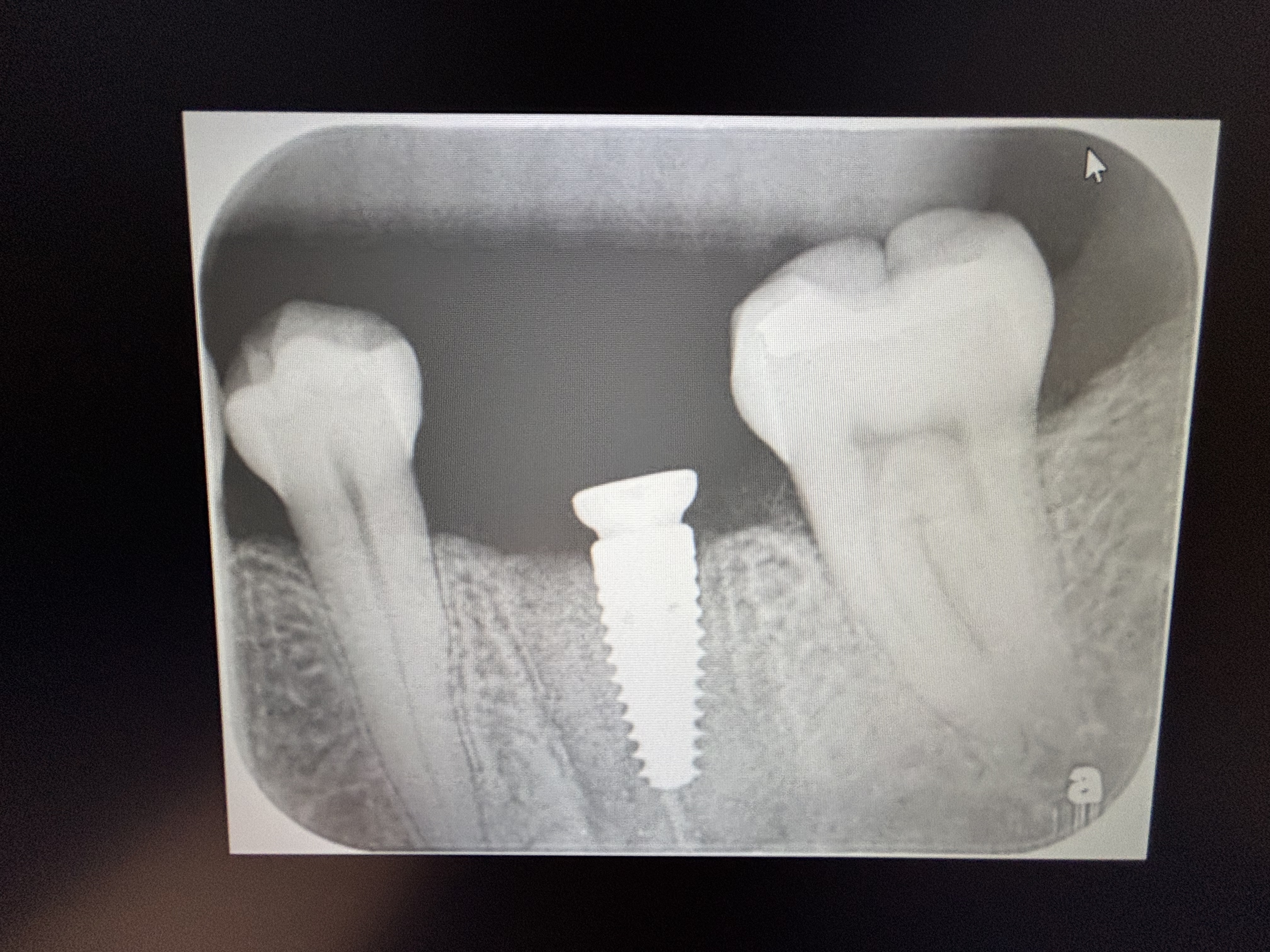
I have treatment planned this patient for extraction of #30 [mandibular right first molar;46] and #25 [mandibular left central incisor; 31] and replacement with dental implants. How would you recommend I proceed? Both teeth have radiolucent lesions around their apices which appear to be endodontic infections. Should I extract, curette aggressively and install the implant at the time of extraction? If so, should I prescribe an antibiotic? Or should I extract, curette, graft and wait for the graft to heal and then later install the implants?

22 Comments on Radiolucent Lesions: How Should I Proceed with Implant Treatment?
New comments are currently closed for this post.
Dr.B
11/6/2011
This would be my preferred treatment plan:
1. Extract and curette
2. After four weeks re-assess the ridges for possible guided bone regeneration
3. Implant placement (at least three months after extraction), otherwise six months if bone was grafted.
Guy Carnazza DMD
11/7/2011
CT scan would be more helpful in treatment planning the case. Extracting and grafting allows for more predictability. Ct scan will give you a better understanding of available bone and aid in decision making.
dr.med. dr. dent Alessand
11/7/2011
Along many years i have worked on a project of a protocol that goes very well :
a) extraction and courettage with a ball cutter o drill, under coverage of penicillin or cephalosporine from the day after.
b) fill up in the socket with cristalline cephalosporine and after few minutes with cristalline hidrocortisone for any minutes with a temporary tampon.
c) insert after five or ten minutes, an appropriate one piece implant eventually going deeply in the socket with a lanceolate drill.
d) suture with an U ring like tobacco bag, after having positioned an appropriate bone graft.
e) ten days waiting for suture and four months to proove the implant with a temporary crown.
All this said very schematically-
Carlos Boudet, DDS
11/7/2011
One thing about forums like this is that you will get as many answers as doctors responding.
I would extract and carefully debride and detoxify the sockets. Dr Alessandro Romano uses his protocol, I use the laser.
I graft with beta tricalcium phosphate and cover with PRF (also cover all my grafting with antibiotics Amox. 500mg q8h for one week). I defer taking the conebeam CT until 4-5 months have elapsed so I know what I have before placing the implants.
This period for bone remodeling will allow you to make your molar osteotomy more ideally.
Good luck!
Dr.B
11/7/2011
I agree w Carlos. There are many ways to "skin the cat". Do what you r most comfortable with. What matters is the end result. There are two reasons why I may not immediately graft here. One is the periapical lesion. The second and most important in my view is lack of primary closure. By waiting a few weeks I can guarantee primary closure. Call me crazy but I'd rather play it safe and take my time.
rsdds
11/8/2011
extract curretage and make an assesment of socket. is it intact? is it missing the buccal wall? how many millimeters to the IAN ? do i have a lingual undercut? i take a cbct before ext and before implant placement... you can graft or not or you can immidiate place and graft at the same time , to me every implant case is different !! good luck
John Manuel, DDS
11/8/2011
Using antibiotics in the graft can alter pH, and resorption rate of graft materials. Also can damage the bone.
Sterile water will kill more bacteria than bone cells without altering graft performance. Antibiotic per-med will have some success in reducing bacterial load at surgery w/o significant alteration of graft.
So, remove roots. Clean sockets by curretage. Place short implant centered in interradicular area and set low. Close with Collaplug. open and place abutment in 90 days. Then finish with crown.
John Manuel, DDS
11/8/2011
Important note: After curretage, irrigate socket with strile water and let is sit for 5-10 minutes before placing implant and blood/Synyhograft/bone mixture.
Mohamed
11/8/2011
evaluate after the extraction,if you see pus oozing and you have missing the buccal wall then postpone the bone grafting and /or implant placement
you can wait 6 to 8 weeks and start placing the implant and bone graft with resorbable membrane if the buccal plate is missing
if no pus is comming and you get a nice clean socket after removal of the lesion then you can place the implant or do grafting according to the clinical senario
it is so easy
in the molar area primary stability is important if you want to place the implant immediatly or delayed immediate
Richard Hughes, DDS, FAAI
11/8/2011
This is warmed over leftovers! Yet it is real life. I would extract the two teeth, detox, decorticate and graft the molar with a membrane. I would do the same for the incisor, but depending on the condition of the facial plate will determine if to place an implant at the time of extraction or give it tincture of time.
Richard Hughes, DDS, FAAI
11/8/2011
PS: as per my last entry. As per the molar site, I would place the implant after thengraft turns over. It can be very easy to bag the IAN, if you try to place the implant at extractin Tim, not to mntion the polymicrobic infection. It is rare that these are not polymicrobic infections! It is safer to be conservative in this case.
Pankaj Narkhede DDS MDS,
11/8/2011
Extract, currette graft the molar first.
For anterior extract curette well, may be loss of buccal plate
Check scan. Possibility of implant placement & graft.
gary omfs
11/9/2011
The most predictable and economic way is to go step by step. Patients accept this, if you explain them why. To Richard: I' ve had that exactly in one of my first implant cases; she was a sales manager of a major drug company, visiting lots of collegues every day! I wanted to impress while this immediate implantation was a hot topic many years ago. The septum was very tough so I had to put some pressure on the (probably blunt) drill. Suddenly it sunk 4 mm into the bone, severing her IAN. Worst case scenario. Unfortunately we learn most from our mistakes. BTW I think an apical infection is not an issue if you curette well, contrary to periodontal disease.
Tyler
11/9/2011
Please tell me you're looking at the other teeth... #2 and #15 come to mind right off the bat....
Mohamed
11/9/2011
you have enough height to place the implant in the molar area immediately.the inferior alveolar nerve is far
you are not going to place 18mm implant. remember that wider implant will help to get stability as well
it is not a way to push you to immediately implant but whenever the the clinical scenario permit why not?
Trace the inferior alveolar nerve to find that you have enough height
good luck
Richard Hughes, DDS, FAAI
11/9/2011
Gary Omfs, I did that once and ever since, I graft and revisit at a later case. This way it's safe and easy! Your candor is welcomed.
ttmillerjr
11/10/2011
Hi Dr. I,
So we know you haven't done a lot of grafting or implant work. Take my advice and do things the most predictable way in the beginning, as time goes on you can start combining steps. Bone grafting is more difficult than placing the implants. Getting passive primary closure over an extraction site is a learned skill. Soft tissue manipulation is what causes most of the pain and inflammation. The least painful way for the patient and the most predictable way for you is:
Extract the teeth and thoroughly cleanse the sockets. Let the tissue heal over the socket (7 weeks). Now go in and graft, primary closure is much easier now. Now just wait 4-6 months and place your implants. You will learn more than you think by following this approach and you will have happy patients and predictable outcomes. After you get really comfortable with this approach you will start to develop your preferences.
Just a few more tips; The older the patient the more of their own bone you should use. Use a bone scrapper, studies have shown that this bone collected with the scrappers works as well as bone collected from the Iliac Crest. Mix this bone with Dynablast putty, the putty is firm and hardens a bit when warmed to body temperature, doesn't migrate easily like particulate. Bone generally comes out better with membranes, yes you can do it without and there are arguments to be made, but if you follow these techniques you will have much success. As time goes, again, you will develop your preferences.
Good luck and have fun!
Richard Hughes, DDS, FAAI
11/10/2011
Dr I, Dr. Miller gave you excellent advice. You also have to decorticate the bone inside the defect. If not you most likely will have formed a honey cortical bridge without any deeper bone formation, since the blood supply is just from the periosteum. Duns last is fine. I like Osteogen with or without prp/prf. Later on when you have developed flap releasing techniques, membrane techniques, PRP/PRF and suturing techniques, you will be cooking with gas. We all start as beginners! You will make mistakes, we all have!
Richard Hughes, DDS, FAAI
11/10/2011
Tyler, good eyes. #2,3,4,5 & #15 need conservative treatment
Gregori M. Kurtzman, DDS,
11/10/2011
You wont get primary stability in that site and with the lesion present its seeded bacteria away from the failing tooth. Best to extract currette and graft then allow to heal for 3 months before placing an implant as this will give a more predictable result.
Richard Hughes, DDS, FAAI
11/11/2011
Correction:I ment to state "a boney cortical bridge"
Dr. Punjabi
11/12/2011
Hello Doctors!
I read lots of good advice we all can use in our implant practice. But dearest Drs. Hughes and Kurtzman: what does all the titles behind your name mean and why do you use them?
Just a curious colleague asking.
All the best,
Dr. P


