Soft tissue dehiscence 3 weeks after implant placement: thoughts?

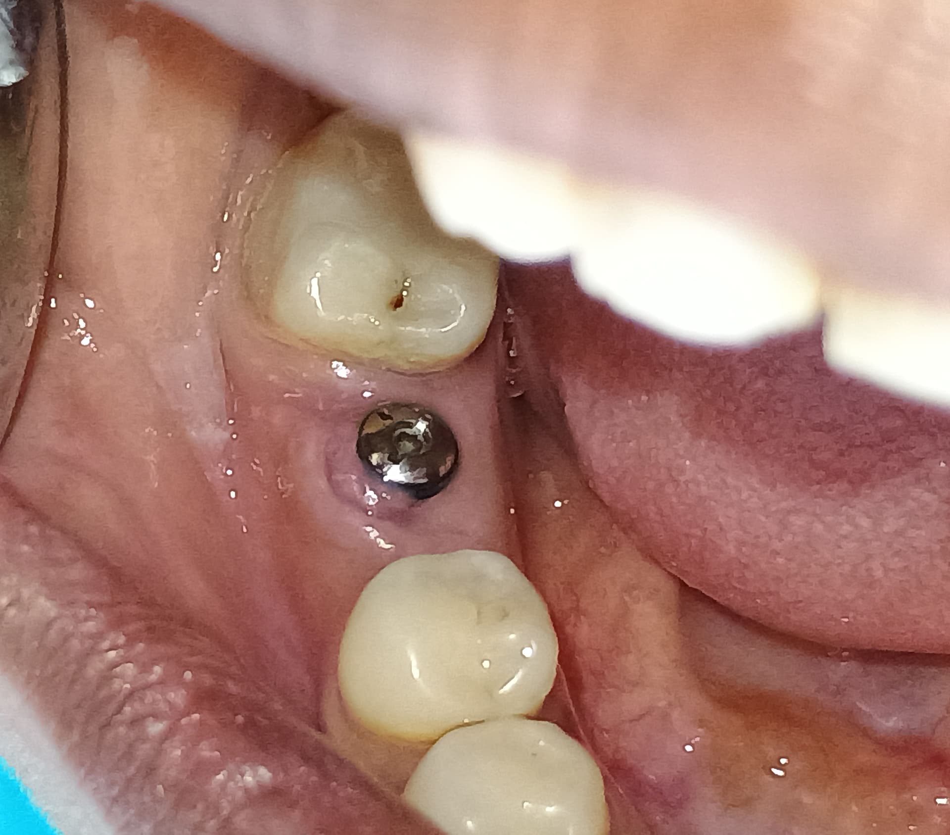
I had placed 2 Nobel Parallel CC implants in the 4th quadrant in the premolar and molar region [lower right]. It was a straightforward case, I thought, and I also placed 2 implants in the 3rd quadrant [lower left]. The patient experienced severe pain for a week and came back to me with a soft tissue dehiscence. As the patient was medically fit, I advised the patient to leave it for 2 to 3 weeks and hopefully it would granulate. I put her on chlorhexidine rinses and hyaluronic acid application. She has come back today but unfortunately it hasn’t healed yet at 3 to 4 weeks after surgery. The other side has healed fine. I am thinking of removing this exposed part of the bone and covering with PRF. Thoughts? Any other ideas are welcome.


![]soft tissue dehiscence](https://osseonews.nyc3.cdn.digitaloceanspaces.com/wp-content/uploads/2018/10/8168-24-20181011_162724-bbf45279dacb-e1539261928620.jpg)

29 Comments on Soft tissue dehiscence 3 weeks after implant placement: thoughts?

New comments are currently closed for this post.
Dr. Moe
10/11/2018
Hi, Seems like bone was overheated during osteotomy, leading to necrosis, especially if you are saying that bone is exposed. X-ray would help too. I would possibly remove, graft, DPTFE membrane. Let it heal, it's obviously not going to work as is due to either bone issue or bacterial infiltration, leading to failure. My $0.02
Dr. Moe
10/11/2018
Sorry, meant to say Titanium reinforced DPTFE membrane.
Sam
10/11/2018
These Titanium Reinforced PTFE Membranes are available at DDSGadget.com. They are textured on both surfaces.
A
10/11/2018
I was thinking of just removing the necrotic bone,making sticky bone and then placing prf at the site of bone and soft tissue dehiscence and leaving the implant as it is.What are your thoughts on that?
Timothy Carter
10/11/2018
All you need to do is rough the exposed bone with a high speed diamond bur to make it bleed. This will remove the outer necrotic layer and the soft tissue will close right over it. No big deal!!
A
10/11/2018
Thank u for the answer
Dok
10/11/2018
This relatively small area of infected/necrotic bone should be filed away. Release and advance the adjacent soft tissue ( full thickness ) to cover the defect and suture ( with stability and primary closure ). Wait and see what happens. You will know within a couple of weeks. If the lesion reappears, take out the implant and do what Dr. Moe suggests.
A
10/11/2018
Thanks for the answer.Lot of good suggestions. I'm now in a better position to take this further
Dan S
10/11/2018
More information is needed on this as there can be multiple reasons for this type of appearance. Is the patient on bisphosphonates? How about steroids? Diabetes? A number of medical conditions can result in poor bone healing. Did you get a CBCT after the implant was placed? Perhaps the implant is partly outside the lingual plate. It could be overheating of bone, but also many other things. Try to figure out what is causing this, then decide on a course of action.
Alejandro Berg
10/11/2018
I would check with a cbct of the area, you may have had created a green stick fracture of the lingual wall due to an anatomic variation of the patient. that would explain the pain. If so, you will most likely loose the implant and have to graft prior toor when replacing the fixture.
A
10/11/2018
Patient is not on bis neither steroids.Havent taken postop cbct though I was considering it.Thank you for the reply everyone
E Katch
10/11/2018
Looks like the mylohyoid ridge bone is exposed . It will heal but a few weeks and painful course. Did you inject a lot of local lingually ? Did u tear your flap ? Typically this doesn’t need to be exposed during implant placement.
A
10/11/2018
Hi.yes I did inject lingually though I didn't tear the flap.Thanks for the reply
CRS
10/11/2018
Not sure why you would use hyaluronic acid in the mouth probably exacerbating the situation. Exposed bone is dead bone especially after 2-3 weeks. Never place sutures or a membrane in this type of situation, the dead bone will sequester off no need to grind it. No that's not the mylohyoid ridge. My advice is that even these implants integrate there will be a bony defect that will be an issue later, remove them. May want to learn how to manage surgical complications, it is part of placing implants along with surgical principles of tissue management.
A
10/11/2018
Thanks for the answer.Thouhh I have placed and restored lot of implants faces this situation the 1st time.Still learning I guess
Adil albaghdadi
10/11/2018
First of all you need to know if the patient is wearing her old partial. as you place the implant the ridge is expanding and by relieving the tissue side of the partial will solve the problem. Second check the implant weather it is infected or not if it’s not integrated and infected that will cause pain and the cover screw exposure. Try with a probe to check any mobility and update us. Hope that will help.
A
10/11/2018
Ok.im planning to open this and see what's happening.If nothing else then I'll remove the dead done,and close.She isn't wearing partial.Thanks for the reply
CRS
10/15/2018
good thought
Drnitzin
10/12/2018
This to seems to be a operative issues as mentioned too much of vasoconstriction or tip necrosis due to non keratenised mucosal suture bite or double incision. Pain is just because of the exposed bone. Why not just wait and watch. Prescribe a local a Anaesthesic based ointment for a while may be it can take up to 21days
A
10/12/2018
Superb comment.thank u
Dr Saad Yasin
10/12/2018
This is due to soft tissue infection and trauma by the lingual flsnge of the partial denture and bone infection and or necrosis due to excesive generation during drilling and implant placement because the cortex is D1dense bone
Dr Saad Yasin
10/12/2018
This is due to soft tissue infection and trauma by the lingual flange of the partial denture and bone infection and or necrosis due to excesive heat generation during drilling and implant placement because the cortex is D1dense bone
PAAUH
10/13/2018
just to add to above points, make an acrylic plate with extending ligual flange to the affected area,then place some antibiotic and sedative inside the plate and give to pt ,just another way to deal exposed bone in lingual area
A
10/13/2018
Thanks for a good tip
KSD
10/14/2018
I'd have to agree with CRS - mostly. This type of situation is seen not all that uncommonly after difficult extractions in the lower molar area. What you're looking at is dead bone that has not sequestrated yet. Most surgeons, myself included, have learned that the best course of treatment is time. New tissue will often grow underneath the piece of dead bone and it will slough off or become loose enough to remove with tweezers or curette. Usually this takes 3-4 weeks. And yes, it produces pain out of proportion to what you would think, often radiating to the throat and ear. The depth of bone injury is difficult to predict but in my experience is limited to the lingual cortex or portion thereof. Depending upon where your implants were placed in the bucco lingual dimension, this may or may not affect them. Attempting to repair the thin and friable mucosa is fraught with difficulty and in many instances has led to a wider area of tissue loss and bone sloughing. This is one of the few instances where I will keep a patient on a longer course of narcotic pain relievers if they are really miserable and having limited or no success with NSAIDs. As soon as the piece sloughs and you can remove it, they will experience complete pain relief and the soft tissue will heal by secondary intention. Best of luck with it.
KSD
10/14/2018
Forgot to mention. Soft diet with avoidance of hard foods such as toast, croutons or other similar foods that may further abrade the area is important as is confining chewing to the contralateral side during the 3-4 weeks. Local measures such as moist heat externally may help also but it is usually pain relievers that will help the most
A
10/14/2018
Thank u for such a nice comment.I think this is what I'll be doing
Greg Kammeyer, DDS, MS, D
11/13/2018
I agree with DOK. I certainly think that getting some L-PRF will help heal the area.
Dr Bill Woods
12/10/2018
Im of the school that you should remove the implant. Repair the area fir future placement. Dead bone next to an implant isn’t going to repair itself and leave a future surgery for you. Better starting off fresh later on than fumbling around a potential and possibly lengthy trial and error cure. Conserve the operative site. JM2C.

Featured Products

OsteoGen Bone Grafting Plug
Combines bone graft with a collagen plug to yield the easiest and most affordable way to clinically deliver bone graft for socket preservation.
CevOss Bovine Bone Graft
Make the switch to a better xenograft! High volume of interconnected pores promotes new bone. Substantially equivalent to BioOss and NuOss.