What is the best technique to graft a canine?

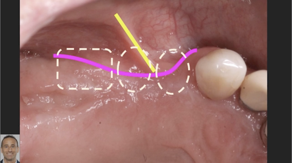
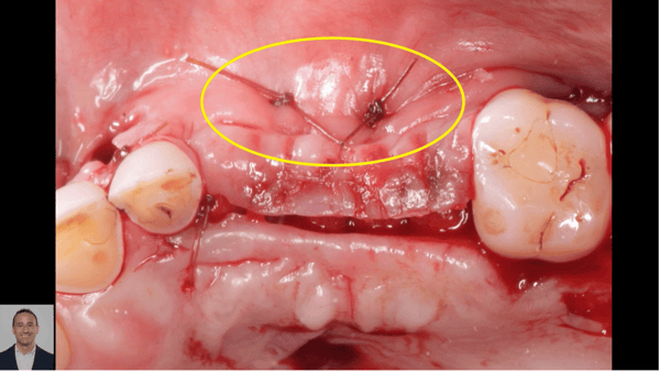
In this series of two videos, Dr. Simon talks about a case where there is a recession for an upper canine. He discusses the two main techniques, CTG (connective tissue graft) or Tunneling Graft, used to address gingival recession. In the 2nd video, Dr. Simon specifically addresses the incision outline for the CTG.

Part I

**Part II**
### About Ziv Simon, DMD, MSc

Dr. Simon practice is limited to periodontics and dental implant in Beverly Hills, CA. He obtained his dental degree and Bachelor of Medical Sciences degree from Tel Aviv University, where he held a teaching position in the Department of prosthodontics. He received his periodontal graduate degree from the University of Toronto with a Master of Science degree in Periodontology. He is a Diplomate of the American Board of Periodontology and a Fellow of the Royal College of Dentists of Canada in the specialty of periodontics. Dr. Simon is the president of the Beverly Hills Academy of Dentistry and the founder of the Multidisciplinary Dental Study Group of Beverly Hills. Dr. Simon teaches at the University of Southern California and lectures nationally and internationally on esthetic soft tissue procedures, computer guided implant surgery and tissue reconstruction. Dr. Simon is the creator of SurgicalMaster TM , a surgical training program for dentists.

3 Comments on What is the best technique to graft a canine?

New comments are currently closed for this post.
steven kollander
3/18/2016
best technique hands down is Pinhole Surgical Technique. Not a difficult procedure and much less morbidity for the patient.
Dominik Nagy
3/18/2016
Dr. Simon, I agree with you, the best way to cover the cervical part of the canine is CTG with tunnel technique! But my main question: What was the reason, to appear the cervical part of the canine, and happened buccal bone reduction with soft tissue reduction? If you treat just the symptom of the disease and not the cause of the disease, the long term succes will be bad. May be the too high traumatic force in laterotrusion movment cause this symptom? The patient has a thin buccal bone wall, with high dental axis inclination in the upper frontal part! The patient is dentaly compensated and may have Class I normo-occlusion but may has sceletal differences. The parabolic curve of the maxilla in the level of the dental apical is more thin like the parabolic curve in the level of the dental cervical. It's determinate the buccal wall reduction of the canine, and frontal teeth as we can see the video. The questions before the treatment: - was any orthodontic treatment before? -the Num 23 has the same symptom? -under laterotrusion movement help the premolars to derive the movement or just the canine get the full force? So I think to cover this Miller Class I recession is possible but to treat is very difficult. Yours sincerelly
Ziv Simon
3/21/2016
These are very good points and important to consider and assess before performing any surgery. I totally agree with you. I don't have the answer to all of these questions. As mentioned in the videos, I'm acting as a consultant to another doctor. My goal with the video series was to demonstrate in general how I evaluate cases and the treatment options. There is much more that goes into the analysis as you indicated in your comments. Regards,

Featured Products

OsteoGen Bone Grafting Plug
Combines bone graft with a collagen plug to yield the easiest and most affordable way to clinically deliver bone graft for socket preservation.
CevOss Bovine Bone Graft
Make the switch to a better xenograft! High volume of interconnected pores promotes new bone. Substantially equivalent to BioOss and NuOss.