Treatment of Large Bone Defect from Root Fracture
Last Updated: Sep 25, 2017
In this case, a large bone defect, as a consequence of fractured roots of the first molar, was augmented by Bond Apatite bone graft cement.The molar was extracted and a complete debridement of the granulation tissue was performed, followed by bone grafting with Bond Apatite bone graft cement. During the augmentation procedure, the flap was minimally reflected and minimally released. Thereafter, the cement was activated within its syringe and ejected directly into the site. After cement placement, a firm pressure was applied on a dry sterile gauze above the cement for 3 seconds to induce its hardening in place. The flap was placed directly above the cement without a membrane and was maximally closed with moderate tension.
![]](https://osseonews.nyc3.cdn.digitaloceanspaces.com/wp-content/uploads/2017/09/85de0d4c5b89dd86d904a7a58d7796a8.jpg)Pre op radiographic appearance

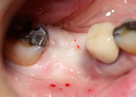
![]](https://osseonews.nyc3.cdn.digitaloceanspaces.com/wp-content/uploads/2017/09/4db18828aad0925a5a0b48068ea7ff35.jpg)Soft tissue appearance before reentry
![]](https://osseonews.nyc3.cdn.digitaloceanspaces.com/wp-content/uploads/2017/09/c6da194495a844062db48f6ee00ede6a.jpg)Newly formed bone can be seen
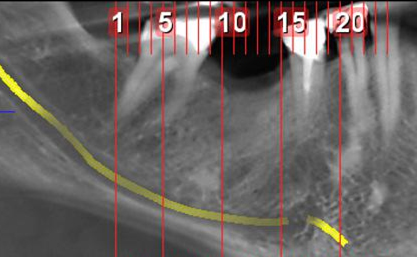
![]](https://osseonews.nyc3.cdn.digitaloceanspaces.com/wp-content/uploads/2017/09/e874d000ce00d95e61cd53d3557e7e6b.jpg)CBCT image 14 weeks post op
![]](https://osseonews.nyc3.cdn.digitaloceanspaces.com/wp-content/uploads/2017/09/119010048d8f9f9442b5215c359ecfe9.jpg)CBCT image 14 weeks post op
Pre)
Soft)
Newly)
CBCT)
CBCT)
5 Comments on Treatment of Large Bone Defect from Root Fracture
Dr. Amos Yahav
09/26/2017
Rainier Urdaneta
09/26/2017
dr. Amos yahav
09/26/2017
Rainier urdaneta
09/27/2017
Featured Products
Classic 50/50 Mix
Promotes osteoconduction
Provides structural integrity
Convenient Syringe!
50/50 Cortical/Cancellous
Available in 3 sizes.
Eliminate hassle of mixing particulate grafts
Sold in packs of 5 or packs of 10.
Proven safe, and clinically effective
Resorbable collagen membrane derived from purified porcine pericardium
Fast hydration and excellent tensile strength
Good adaptation to various defects
Excellent tear function and duration
100% allograft
Eliminates mixing hassle
Moldable after hydration
Peter Fairbairn
09/26/2017