Use of Surgical Guides
Dr. Tsanis asks us:
I have a couple of questions regarding the use of surgical guides in dental implantology.
To start with do you routinely use surgical templates when dealing with edentulous patients?
What is your opinion of computer guided surgical guides, as their costs seem to be increasing?
Finally, in terms of practical applications, I have an edentulous patient that needs 6 or 7
dental implants and I want to construct a conventionally made surgical guide (not computer guided). What concerned me was how I will be able to fit that template in the mouth after I have raised the flaps, given the interference? Any usefull tips or advice?
9 Comments on Use of Surgical Guides
New comments are currently closed for this post.
Anon
5/30/2006
Shouldn't you consider a bone borne surgical guide created by cone beam CT? Surely your conventional surgical guide will not fit upon the jaw after the flaps are raised. Your other alternative is to use a tissue borne guide, and do a flapless implant procedure, but again you need CT guided placement. I suppose you could use your conventional stent, drill through the mucosa to mark your implant placement, then reflect your flaps to see if that would be in the right place. If there is insufficient bone then modify your placement. Good Luck.
anton j voitik mdt
5/30/2006
We routinely develop exclusionary drill guides for a surface dentistry approach, that is, planning based on the pre-surgical diagnostic wax up, and I would be happy to demo such a case view per e-mail for you.
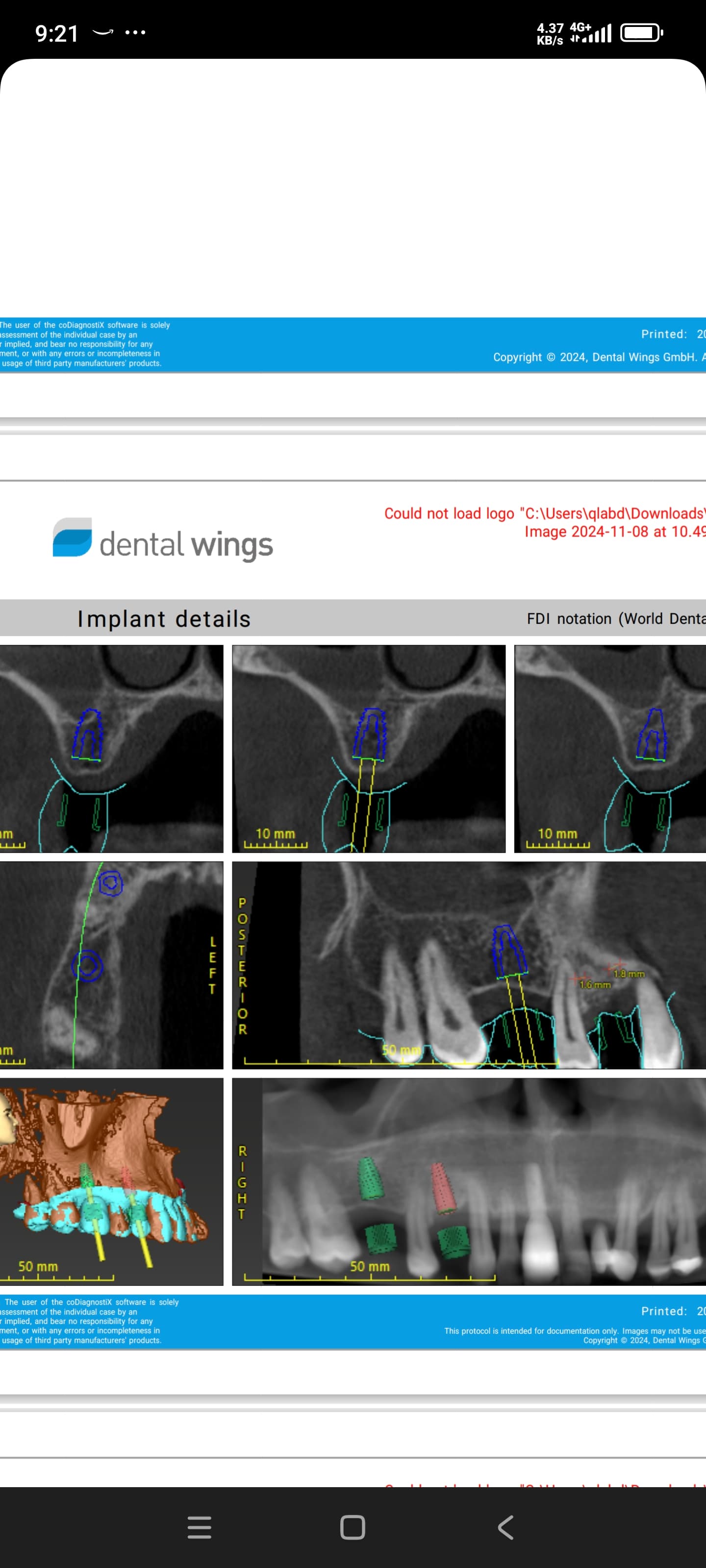
Systemic dentistry-based drill guides, based on CT data that shows all necessary prosthetic elements to plan in 3D, are not as prohibitive as you may think (app. $300 / guide)and the increasing ease with which a patient can find a scan site for iCAT or NewTom scanners, for instance, is bringing prices down.
Jin Kim, DDS, MPH, MS
5/30/2006
Look into Poitres surgical guide. It is not a product. It is a concept, and it is based on retention in opposing arch, and having th epatient go into RP for reproducable closure. I think there is a example in Misch textbook.
David C. Garrison DMD
5/30/2006
In my practice I usually find CT scans to be cost prohibitive. Try this. Make the tissue-born surgical guide showing the ideal implant locations and as many alternative locations as you can.(Make sure you have an accurate panoramic to be sure your mesio-distal measurements are accurate.) Using your guide, mark the locations on the tissue with a pilot drill or a surgical pen. Flap the tissue! Don't try this flapless. After visualizing all the bone, pick the best locations by re-opposing the flaps and using your tissue marks. Use paralling pens to get the implants as parallel as possible. With 6 or 7 implants, it is highly improbable that they will all be parallel whether you have a CT scan or not. If you are doing the prosthetics yourself, use a lab that is familiar with all implant systems. Send them the impression and the impression posts and let them do the rest.
sean meitner DDS
5/31/2006
I use a surgical guide mase from a wax set up, the components are available from Guide Right. A surgical guide made in the office takes 3 minutes. and must be evaluated radiographically be fore using using PA and or Panorex or cone beam. cost is $ 25 once you are set up.
Robert J. Miller, DDS
5/31/2006
You can solve your problem by fanricating an idealized occlusion rim with 50/50 barium sulfate/acrylic on the teeth only. Place 3 transitional implants and fix the appliance to them. Take a focused cone beam DVT scan to determine placement and bone volume. Then convert the appliance to a surgical guide by milling ou the occlusals and fixing it to the transitional implants during surgery. The guide will not move, you will get better implant placement, and you have the ability to modify trajectory within the confines of the guide.
michael williams, dmd
5/31/2006
Who makes Guide Right?
hersheydmd
4/29/2007
This thread is pretty old, so I am sure Dr. Tsanis' case is completed by now.
Nonetheless, Dr. Miller's suggestion is right on target.
Check out implantlogic.com and view their video demos. Their software and guides are very simple to use.
alvaro ordonez
6/23/2008
I will be happy to send you by email a step by step sequence that you can use.
You will need an impression of your patient with a nice set up of where and how your prosthetic will be (DX wax up or denture teeth), make an impression of it and pour it, trim the edges of the model and put it on the vacuum machine using a 0.6 mm hard disk of acetate (as if you were making a hard splint for TMJ), then you will cut the thinnest part of an old radio or TV antenna (i got the antenna trick from my friend ahmed osman in cairo), mix the acrylic with barium sulfate (4 ounces is only 11 dollars and most dental supplies have it), do the mix ratio the way Dr Miller proposed it (50-50), you have to seat it on the edentulous original model or on the patient mouth to get the shape of the ridge and make sure it will fit, the retention during single teeth will be friction just like with tmj splints, then make a perforation in the center of the occlusal of the area of the wax up teeth that were indexed with the stent, and make a scan (if you have CBCT available) or a panorex.
if you leave space in the gingival area of the stent you will be able to position the flap on the sides and the reference to the bone will be very nice. Do your analysis in your CBCT software (you can easily and cheaply do it with NNT which you can buy from a Newtom center below $300 us
then you do the angulation adjustments in a possible second scan or rx.
Newtom will alow you to do cuts and measurements, it doesnt have implant models but you can still very precisely do it without it if you do the slices carefully
I hope this helps, by the way, not even 10 dollars in materials and very precise.
alvaro ordonez


